Journal of Human Hypertension (2010) 24, 447–457
& 2010 Macmillan Publishers Limited All rights reserved 0950-9240/10
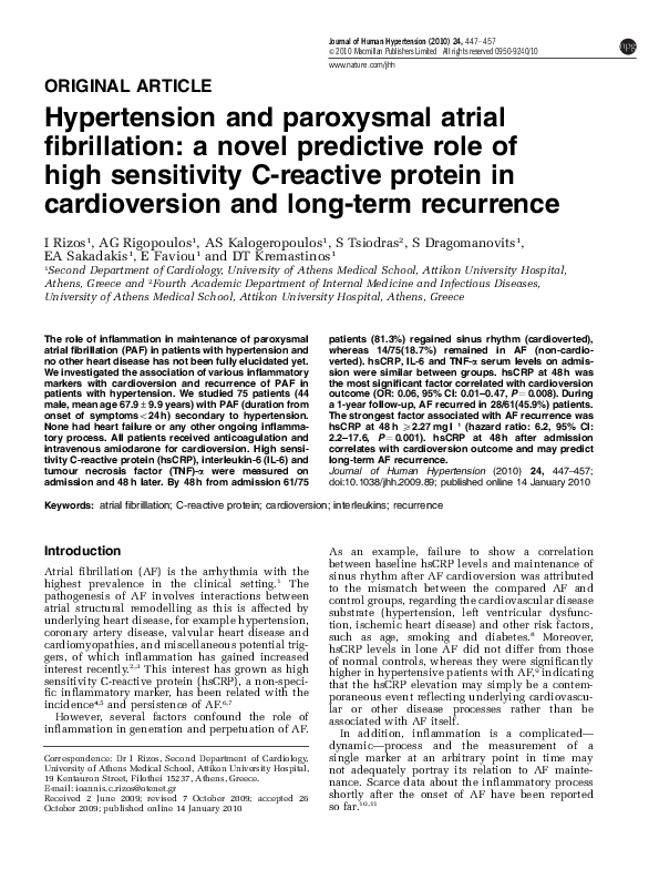
www.nature.com/jhh
ORIGINAL ARTICLE
Hypertension and paroxysmal atrial
fibrillation: a novel predictive role of
high sensitivity C-reactive protein in
cardioversion and long-term recurrence
I Rizos1, AG Rigopoulos1, AS Kalogeropoulos1, S Tsiodras2, S Dragomanovits1,
EA Sakadakis1, E Faviou1 and DT Kremastinos1
1
Second Department of Cardiology, University of Athens Medical School, Attikon University Hospital,
Athens, Greece and 2Fourth Academic Department of Internal Medicine and Infectious Diseases,
University of Athens Medical School, Attikon University Hospital, Athens, Greece
The role of inflammation in maintenance of paroxysmal
atrial fibrillation (PAF) in patients with hypertension and
no other heart disease has not been fully elucidated yet.
We investigated the association of various inflammatory
markers with cardioversion and recurrence of PAF in
patients with hypertension. We studied 75 patients (44
male, mean age 67.9±9.9 years) with PAF (duration from
onset of symptomso24 h) secondary to hypertension.
None had heart failure or any other ongoing inflammatory process. All patients received anticoagulation and
intravenous amiodarone for cardioversion. High sensitivity C-reactive protein (hsCRP), interleukin-6 (IL-6) and
tumour necrosis factor (TNF)-a were measured on
admission and 48 h later. By 48 h from admission 61/75
patients (81.3%) regained sinus rhythm (cardioverted),
whereas 14/75(18.7%) remained in AF (non-cardioverted). hsCRP, IL-6 and TNF-a serum levels on admission were similar between groups. hsCRP at 48 h was
the most significant factor correlated with cardioversion
outcome (OR: 0.06, 95% CI: 0.01–0.47, P ¼ 0.008). During
a 1-year follow-up, AF recurred in 28/61(45.9%) patients.
The strongest factor associated with AF recurrence was
hsCRP at 48 h X2.27 mg l 1 (hazard ratio: 6.2, 95% CI:
2.2–17.6, P ¼ 0.001). hsCRP at 48 h after admission
correlates with cardioversion outcome and may predict
long-term AF recurrence.
Journal of Human Hypertension (2010) 24, 447–457;
doi:10.1038/jhh.2009.89; published online 14 January 2010
Keywords: atrial fibrillation; C-reactive protein; cardioversion; interleukins; recurrence
Introduction
Atrial fibrillation (AF) is the arrhythmia with the
highest prevalence in the clinical setting.1 The
pathogenesis of AF involves interactions between
atrial structural remodelling as this is affected by
underlying heart disease, for example hypertension,
coronary artery disease, valvular heart disease and
cardiomyopathies, and miscellaneous potential triggers, of which inflammation has gained increased
interest recently.2,3 This interest has grown as high
sensitivity C-reactive protein (hsCRP), a non-specific inflammatory marker, has been related with the
incidence4,5 and persistence of AF.6,7
However, several factors confound the role of
inflammation in generation and perpetuation of AF.
Correspondence: Dr I Rizos, Second Department of Cardiology,
University of Athens Medical School, Attikon University Hospital,
19 Kentauron Street, Filothei 15237, Athens, Greece.
E-mail: ioannis.c.rizos@otenet.gr
Received 2 June 2009; revised 7 October 2009; accepted 26
October 2009; published online 14 January 2010
As an example, failure to show a correlation
between baseline hsCRP levels and maintenance of
sinus rhythm after AF cardioversion was attributed
to the mismatch between the compared AF and
control groups, regarding the cardiovascular disease
substrate (hypertension, left ventricular dysfunction, ischemic heart disease) and other risk factors,
such as age, smoking and diabetes.8 Moreover,
hsCRP levels in lone AF did not differ from those
of normal controls, whereas they were significantly
higher in hypertensive patients with AF,9 indicating
that the hsCRP elevation may simply be a contemporaneous event reflecting underlying cardiovascular or other disease processes rather than be
associated with AF itself.
In addition, inflammation is a complicated—
dynamic—process and the measurement of a
single marker at an arbitrary point in time may
not adequately portray its relation to AF maintenance. Scarce data about the inflammatory process
shortly after the onset of AF have been reported
so far.10,11
�Inflammation in atrial fibrillation due to hypertension
I Rizos et al
448
In this study, we sought to investigate whether the
outcome of cardioversion as well as the likelihood of
AF recurrence, in a select group of patients with
essential hypertension12 and paroxysmal AF (PAF),
relate to baseline levels of important inflammatory
markers such as hsCRP, interleukin-6 (IL-6) and
tumour necrosis factor (TNF)-a and with their levels
48 h after admission. Additionally, we examined if
there was an association between the baseline
inflammatory load and the level of atrial structural
remodelling.
Methods
Study population
This was an observational case–control study of 133
consecutive patients admitted to our department
with symptomatic PAF (duration from onset of
symptoms o24 h) secondary to hypertension from
January 2005 until January 2006. However, 58
subjects were excluded because of the presence of
additional comorbidities that complicated the origin
of PAF, namely, coronary artery disease, heart failure
(left and/or right ventricular dilatation and/or
systolic dysfunction), acute pulmonary edema,
acute coronary syndromes, lone AF, rheumatic valve
disease, sick sinus syndrome, autoimmune diseases,
chronic obstructive pulmonary disease, thyrotoxicosis, recent trauma or surgery, known neoplasm,
recent or ongoing exposure to non-steroidal antiinflammatory drugs or corticosteroids, renal failure.
All remaining patients were under intensive
antihypertensive treatment for at least a year with
well-controlled arterial blood pressure levels. The
diagnosis of AF was accomplished by at least one
12-lead electrocardiogram. Onset of AF was defined
precisely by considering the onset of symptoms
such as palpitations, shortness of breath and chest
discomfort.
Study protocol
The study protocol was approved by the hospital’s
ethics committee and conforms to the principles of
the Declaration of Helsinki. Written informed consent was obtained by all study participants and
eventually, each one underwent evaluation and
treatment according to a standardized protocol
described in detail below.
After history taking and physical examination on
admission, patients had a 12-lead electrocardiogram, an anteroposterior and lateral chest X-ray,
and routine laboratory evaluation and biochemical
testing. Patients received intravenous amiodarone at
a dosing regimen of 5 mg kg 1 bolus in 20 min and
then 500–900 mg intravenously for 12 h. For prevention of thromboembolism, anticoagulation with
enoxaparin 1 mg kg 1 subcutaneously twice daily
was initiated on admission and then acenocoumarol
was administered according to ACC/AHA guideJournal of Human Hypertension
lines.13 Serum levels of hsCRP, IL-6 and TNF-a
were measured on admission and at 48 h after
the beginning of intravenous administration of
amiodarone.
A standard two-dimensional echocardiogram was
performed before discharge, using Hewlett Packard
Sonos 2000 echocardiographic equipment (Andover,
MA, USA). Estimation of maximal left atrial volume
was performed as follows: from the apical four- and
two-chambers view, left atrial borders were manually traced at end-systole. The biplane and single
plane modified Simpson’s formula was applied for
maximal left atrial volume measurement.14,15 Images
were stored in VHS magnetic tape and two independent observers performed the final estimation of
atrial volume off-line. The mean value of three
consecutive cardiac cycles was derived as the final
atrial volume.
All patients were constantly monitored for 48 h
and the time of cardioversion to sinus rhythm was
recorded. Patients who regained sinus rhythm were
advised to seek medical care in the case of any
symptom recurrence and were followed regularly on
a monthly basis after discharge until the completion
of 1 year after cardioversion. Any sustained recurrence (electrocardiographically confirmed and lasting longer than 1 min) of AF was recorded. If
necessary, the patient’s physician was also contacted and all the essential information for the AF
recurrence was obtained. In addition, information
concerning any concurrent illness that might interfere with the probability of recurrence was also
sought. During follow-up, any anti-arrhythmic treatment was decided on by the patients’ attending
cardiologist at that time.
Laboratory methods and blood sampling
Blood samples for inflammatory markers measurement were obtained by a peripheral vein on
admission and 48 h later and were drawn without
stasis into serum tubes. Within an hour after
collection the samples were centrifuged at 3.200 g
for 10 min at a temperature of about 4 1C. The serum
was separated into aliquots and was stored in 80 1C
until the assay analysis was performed. Serum
levels of TNF-a and of IL-6 were determined by
commercial standardized solid phase sandwich
enzyme-linked immunosorbent assay methods according to the manufacturer’s instructions (BenderMed
Systems, Vienna, Austria and Biosource International, Camarillo, CA, USA). These assays detected only human cytokines with a sensitivity of
2.3 pg ml 1 for TNF-a and of 2 pg ml 1 for IL-6. The
clinical samples were kept at 2–8 1C and separated
rapidly before storing at 80 1C to avoid loss of
bioactive cytokine. hsCRP was measured by microparticle-enhanced immunonephelometric assay
(Cardiophase* hsCRP, Dade Behring, Siemens
Healthcare Diagnostics, Eschborn, Germany) on a
Behring BN ProSpec clinical chemistry analyzer,
�Inflammation in atrial fibrillation due to hypertension
I Rizos et al
449
for which the measuring range is 0.16–10.2 mg l 1,
the typical analytical sensitivity is 0.175 for the
measurements performed using a sample dilution of
1:20, and the total median intra- and inter-assay
coefficient of variation is less than 6% for values
below 1.0 mg l 1.
Statistical analysis
All quantitative variables were tested for normal
distribution by Kolmogorov–Smirnov test. Data nonnormally distributed are presented as median and
inter-quartile range values (IQR; 25th–75th percentile). Normally distributed variables are expressed as
mean and standard deviation values.
Univariate analysis for factors significantly
associated with the cardioversion and recurrence
outcome was performed for all patients. Differences
in continuous variables were estimated by Mann–
Whitney U-test and two-sample t-test for nonnormally distributed and normally distributed
variables, respectively. Differences in categorical
data were evaluated by w2-test for normally distributed and Fisher’s exact test for non-normally
distributed variables. Correlations were estimated
using Spearman’s rank correlation method. Values of
Po0.05 were considered statistically significant.
Stepwise multivariate logistic regression analysis
was performed to define factors significantly
associated with the cardioversion and recurrence
outcome. All factors with a significance level of
Pp0.1, during the univariate analysis, were included in the model. Covariates that were clinically
known to be important in AF persistence (age,
gender, left ventricular ejection fraction, left atrial
volume, diabetes and smoking) were included in the
analysis as well. Serum levels of hsCRP and IL-6
were each log transformed (log hsCRP and log IL-6,
respectively) for the regression analysis to improve
linear fitting. The probability value that was used for
entering or removing a variable was P ¼ 0.05 and 0.1,
respectively. To assess the most favourable cut-off
value, for the factor that correlated most significantly with AF relapse and best predicted the end
point of AF recurrence, a receiver operating characteristic curve analysis was applied. The optimal
cut-off value corresponded to the value with the best
sensitivity and specificity according to the maximal
positive and minimal negative likelihood ratios
(PLR and NLR). Further analysis of independent
predictors for AF recurrence including the optimal
cut-off value was performed by means of a Cox
proportional hazards regression model. The cumulative risk for AF recurrence, according to the cut-off
value of the factor that most significantly associated
with AF recurrence, was evaluated by Kaplan–Meier
analysis and the difference between patients with
and without recurrence of AF was assessed with the
log-rank test. Additionally, PLR and NLR were
assessed and the exact post-test probability of AF
relapse was calculated by Bayes’ Theorem method.16
The pre-test probability corresponded to the likelihood of AF recurrence in 1 year after successful
cardioversion as this is observed commonly in
clinical practice. Likelihood ratios have several
advantages in interpreting the diagnostic value
of diagnostic tests. They are independent of disease
prevalence and they do not vary in different populations and settings. PLR (sensitivity/(1 specificity))
corrects the true positive rate of a test by the false
positive rate, whereas NLR ((1 sensitivity)/specificity) compares the probability of a negative test in
diseased subjects, in comparison to the probability
of a negative test in those without the disease.
Power calculations
Formal sample size calculations were performed by
analysing data from earlier work by our group.17
Earlier reports showed that the success rate of
cardioversion after amiodarone administration in
patients with PAF is almost 80% in the first 24 h (1:4
case–control ratio)18 and the recurrence rate of PAF
after successful cardioversion with amiodarone is
about 50% (1:1 case–control ratio).19 Hence, it was
estimated, that to detect a 60% difference in hsCRP
levels between patients with a positive and negative
cardioversion outcome, a power of 80%, and a
two-tailed a of 0.05, 13 non-cardioverted and 50
cardioverted will be required for each arm of the
study (1:4 case–control ratio). Likewise, according
to the aforementioned criteria, 27 patients with AF
recurrence and 27 without AF recurrence will have
to be involved, in each arm of the study (1:1
case–control ratio).
Results
AF cardioversion
Seventy-five hypertensive patients with PAF of
o24 h duration were studied. Sixty-one patients
(81.3%) successfully cardioverted to sinus rhythm
(cardioverted), whereas 14 (18.7%) remained in AF
48 h after admission (non-cardioverted). All noncardioverted patients remained in AF during the
first week after admission and were excluded from
further study. The clinical profiles of the patients are
listed in Table 1.
Patients who failed to restore sinus rhythm had
significantly higher left atrial volume and longer
duration of AF from onset of symptoms until
admission than those who cardioverted successfully. In addition, the former used thiazide diuretics
(in combination with an ACE inhibitor or an
angiotensin receptor blocker) in a higher proportion
than cardioverted patients. On the contrary, cardioverted and non-cardioverted individuals had no
significant difference in both heart rate and blood
pressure levels on admission (Table 1).
The levels of the examined inflammatory markers
on admission were not significantly different beJournal of Human Hypertension
�Inflammation in atrial fibrillation due to hypertension
I Rizos et al
450
Table 1 Clinical characteristics according to cardioversion outcome
All patients
n ¼ 75
Cardioverted
n ¼ 61
Non-cardioverted
n ¼ 14
Pa
Age, mean±s.d. (years)
67.9±9.9
67.57±9.66
68.71±11.54
Male/female
44/31
34/27
10/4
Smoking, n (%)
16.0 (21.3)
14.0 (23.0)
2.0 (14.3)
Diabetes, n (%)
7.0 (10.4)
7.0 (11.5)
0.0 (0.0)
AF episodes in the last year, median (IQR)
2.00 (1.0–3.00)
2.00 (1.00–3.00)
1.50 (1.00–3.25)
AF duration from onset of symptoms till admission
5.0 (2.0–12.0)
4.0 (2.0–8.0)
11.0 (4.0–15.5)
(h), median (IQR)
Systolic blood pressure on admission (mm Hg), median (IQR) 135.0 (120.0–150.0) 130.0 (120.0–150.0) 137.5 (127.5–160.0)
Diastolic blood pressure on admission (mm Hg), median (IQR)
80.0 (75.0–82.5)
80 (75.0–81.0)
80 (77.0–83.0)
Heart rate on admission (beats per minute), mean±s.d.
120.97±23.8
123.4±23.6
111.71±23.0
0.17
0.48
0.11
Previous drug treatment on admission
ACE inhibitors, n (%)
Angiotensin receptor blockers, n (%)
Statins, n (%)
Aspirin, n (%)
b-adrenergic receptor blockers, n (%)
Calcium channel antagonists, n (%)
Diuretics, n (%)
0.33
1.00
0.44
1.00
0.54
0.20
0.014
22.0
14.0
12.0
24.0
26.0
21.0
26.0
(29.3)
(18.7)
(16.0)
(32.0)
(34.7)
(28.0)
(34.7)
16.0
12.0
11.0
20.0
20.0
15.0
17.0
(26.2)
(19.7)
(18.0)
(32.8)
(32.8)
(24.6)
(27.9)
6.0
2.0
1.0
4.0
6.0
6.0
9.0
(42.9)
(14.3)
(7.1)
(28.6)
(42.9)
(42.9)
(64.3)
0.73
0.28
0.72
0.34
0.53
0.016
Echocardiographic parameters
Left ventricular ejection fraction (%), median (IQR)
Left atrial volume (ml), median (IQR)
60.0 (55.0–63.0)
60.0 (56.5–64.5)
50.00 (45.00–70.84) 48.50 (42.25–63.00)
57.0 (50.0–60.0)
0.07
70.00 (49.50–90.25) 0.012
Inflammatory markers
Admission hsCRP (mg l 1), median (IQR)
Admission IL-6 (pg ml 1), median (IQR)
Admission TNF-a (pg ml 1), median (IQR)
hsCRP at 48 h (mg l 1), median (IQR)
IL-6 (pg ml 1) at 48 h, median (IQR)
TNF-a (pg ml 1) at 48 h, median (IQR)
2.10
4.35
13.00
3.24
5.44
12.90
3.01
3.86
13.91
16.29
15.09
13.73
(1.30–5.80)
(3.20–5.60)
(7.04–17.00)
(1.41–9.95)
(3.32–12.80)
(8.82–20.35)
1.97
4.30
13.80
2.27
4.62
12.38
(1.22–5.56)
(3.40–5.60)
(7.13–16.98)
(1.32–6.53)
(3.20–8.01)
(8.82–20.65)
(1.23–6.58)
(1.97–6.09)
(9.24–17.60)
(4.76–25.63)
(5.11–39.24)
(10.00–21.65)
0.84
0.35
0.78
0.001
0.011
0.65
Abbreviations: AF, atrial fibrillation; hsCRP, high sensitivity C-reactive protein; IL-6, interleukin-6; IQR, inter-quartile range; TNF, tumour
necrosis factor.
Bold values stand for statistically significant differences.
a
P for the comparison between cardioverted and not cardioverted patients.
Figure 1 Serum levels of hsCRP, IL-6, and TNF-a in non-cardioverted and cardioverted patients on admission and at 48 h. The black
and white bars show median values. The P-value is shown only for statistically significant comparisons.
tween non-cardioverted and cardioverted patients.
However, patients who failed to cardiovert exhibited
significantly higher levels of hsCRP and IL-6 at 48 h
after admission in comparison to the corresponding
values of cardioverted patients (Figure 1). Values of
hsCRP and IL-6 in most non-cardioverted patients
Journal of Human Hypertension
increased at 48 h in comparison to the corresponding values on admission, whereas they remained
relatively stable in most cardioverted patients
(Figure 2).
In all studied patients, we did not observe any
correlation between left atrial size and the examined
�Inflammation in atrial fibrillation due to hypertension
I Rizos et al
451
Figure 2 Serum hsCRP levels on admission and at 48 h in cardioverted and non-cardioverted patients. The interpolation lines show the
trend of each case for the hsCRP serum levels between the two points in time (admission and 48 h).
inflammatory markers: hsCRP on admission
(r ¼ 0.01, P ¼ 0.9); hsCRP at 48 h (r ¼ 0.1, P ¼ 0.4);
IL-6 on admission (r ¼ 0.1, P ¼ 0.4); IL-6 at 48 h
(r ¼ 0.2, P ¼ 0.09); TNF-a on admission (r ¼ 0.01,
P ¼ 0.9); TNF-a at 48 h (r ¼ 0.1, P ¼ 0.3). On the other
hand, hsCRP and IL-6 values at 48 h significantly
correlated with the total duration of AF (from
onset until cardioversion) (r ¼ 0.399, P ¼ 0.0004
and r ¼ 0.345, P ¼ 0.002, respectively). This correlation remained stable even after adjustment for left
atrial volume (r ¼ 0.343, P ¼ 0.003 and r ¼ 0.345,
P ¼ 0.003, respectively).
There was no significant association between
arterial systolic and diastolic blood pressure levels
on admission and the examined inflammatory
markers, with the only exception being TNF-a
serum levels at 48 h, which were positively correlated with baseline systolic (r ¼ 0.234, P ¼ 0.044)
and diastolic blood pressure (r ¼ 0.271, P ¼ 0.019).
Furthermore, patients with higher heart rate on
admission had also higher levels of baseline IL-6
(r ¼ 0.329, P ¼ 0.006).
The most significant factor independently associated with cardioversion outcome after adjustment
for age, gender, AF duration from onset of symptoms
until admission, left ventricular ejection fraction,
smoking, diabetes mellitus, thiazide diuretics and
left atrial volume was log hsCRP at 48 h (OR: 0.06,
95% CI: 0.01–0.47, P ¼ 0.008). In addition, log IL-6
at 48 h significantly correlated with the cardioversion outcome (OR: 0.16, 95% CI: 0.03–0.92,
P ¼ 0.04).
AF recurrence
Throughout the 1-year follow-up period, 28 (45.9%)
of the cardioverted patients had at least one episode
of sustained AF recurrence. None of them had any of
the conditions included in the exclusion criteria of
the study at the time of recurrence.
Patients who had a recurrence also reported more
AF episodes during the year before admission. In
addition, the total time from onset of symptoms
until cardioversion was slightly longer in patients
with AF recurrence, but this difference was not
statistically significant. There was no significant
difference between patients with AF recurrence and
those who remained in sinus rhythm during the
1-year follow-up regarding systolic and diastolic
blood pressure levels and heart rate values on
admission (Table 2).
Levels of hsCRP on admission and at 48 h as well
as IL-6 levels on admission were higher in patients
with AF recurrence, whereas no difference was
observed in IL-6 at 48 h and in TNF-a serum levels
on admission and at 48 h between the two groups
(Figure 3).
The number of relapses during the 1-year followup correlated positively with hsCRP levels at
48 h (r ¼ 0.425, P ¼ 0.001), hsCRP on admission
(r ¼ 0.326, P ¼ 0.01) and the number of AF episodes
during the last year before hospital admission
(r ¼ 0.353, P ¼ 0.005). Of all the examined inflammatory markers, only hsCRP at 48 h had a strong
negative correlation with the time that patients
remained free of AF recurrence (r ¼ 0.374, P ¼
0.003). Additionally, the only two cardioverted
patients that had a marked increase in hsCRP at
48 h (Figure 2) also had early recurrence of AF in the
first and second month of follow-up, respectively.
Binary logistic regression analysis showed that
after adjustment for age, gender, left atrial volume,
left ventricular ejection fraction, diabetes, smoking
and the number of AF episodes during the last year,
log hsCRP at 48 h was the most significant factor
associated with AF recurrence during the 1-year
follow-up (OR: 5.96, CI: 1.65–21.52, P ¼ 0.006). Log
hsCRP on admission was also associated with AF
relapse after adjustment for the same confounders
(OR: 4.58, CI: 1.23–16.99, P ¼ 0.02).
The optimal cut-off value of hsCRP at 48 h was
determined by the receiver operating characteristic
curve analysis (Figure 4) at 2.27 mg l 1 with a
sensitivity of 72% and a specificity of 68%. Cox
proportional hazards regression model after adjusting for hsCRP on admission, IL-6 on admission, age,
Journal of Human Hypertension
�Inflammation in atrial fibrillation due to hypertension
I Rizos et al
452
Table 2 Clinical characteristics of cardioverted patients (n ¼ 61) according to AF recurrence during the 1-year follow-up
No recurrence
Recurrence
Pa
Number of patients (%)
Age, mean±s.d. (years)
Male/female
Smoking, n (%)
Diabetes, n (%)
AF episodes in the last year, median (IQR)
AF duration from onset of symptoms until admission (h), median (IQR)
Time elapsed from admission until cardioversion (h), median (IQR)
Total time from onset of symptoms until cardioversion (h), median (IQR)
Systolic blood pressure on admission (mm Hg), median (IQR)
Diastolic blood pressure on admission (mm Hg), median (IQR)
Heart rate on admission (beats per minute), mean±s.d.
33.0 (54.1)
66.00±9.02
22/11
9.0 (27.3)
3.0 (9.1)
1.0 (1.0–2.0)
3.0 (2.0–6.0)
10.0 (6.0–13.5)
14.0 (10.5–24.5)
135.0 (120.0–150.0)
80.0 (75.0–80.0)
120.89±28.51
28.0 (45.9)
69.48±10.23
12/16
5.0 (17.9)
4.0 (14.3)
3.0 (2.0–4.0)
4.0 (2.0–10.0)
12.0 (5.0–24.0)
21.0 (10.75–31.25)
120 (114.8–148.8)
80.0(75.0–84.38)
126.04±17.32
—
0.09
0.06
0.38
0.46
0.006
0.18
0.35
0.132
0.68
0.37
0.43
Echocardiographic parameters
Left ventricular ejection fraction (%), median (IQR)
Left atrial volume (ml), median (IQR)
60.0 (57.0–63.5)
49.00 (40.65–62.00)
60.0 (55.0–65.0)
48.00 (45.00–67.00)
0.70
0.54
Inflammatory markers
Admission hsCRP (mg l 1), median (IQR)
Admission IL-6 (pg ml 1), median (IQR)
Admission TNF-a (pg ml 1), median (IQR
hsCRP at 48 h (mg l 1), median (IQR)
IL-6 (pg ml 1) at 48 h, median (IQR)
TNF-a (pg ml 1) at 48 h, median (IQR)
1.71
4.21
13.00
1.72
4.69
10.70
4.13
4.80
12.60
3.50
4.44
14.70
0.02
0.04
0.81
0.004
0.70
0.33
(1.06–2.60)
(2.71–5.04)
(7.52–15.92)
(1.07–3.64)
(3.86–7.96)
(8.63–16.45)
(1.33–13.90)
(3.78–10.40)
(5.29–24.00)
(2.03–9.95)
(3.18–8.15)
(9.07–20.83)
Abbreviations: AF, atrial fibrillation; hsCRP, high sensitivity C-reactive protein; IL-6, interleukin-6; IQR, inter-quartile range; TNF, tumour
necrosis factor.
Bold values stand for statistically significant differences.
a
P for the comparison between patients with and without AF recurrence.
Figure 3 Serum levels of hsCRP, IL-6 and TNF-a on admission and at 48 h in patients with and patients without recurrence of AF during
follow-up. The black and white bars show median values. The P-value is shown only for statistically significant comparisons.
gender, PAF episodes in the last year, left atrial
volume, ejection fraction, diabetes, smoking and left
atrial volume showed that this cut-off value of
hsCRP at 48 h was the most powerful marker
associated with AF recurrence (hazard ratio: 6.2,
CI: 2.2–17.6, P ¼ 0.001). Kaplan–Meier survival
analysis revealed that 74% of patients with hsCRP
at 48 h o2.27 mg l 1 maintained sinus rhythm at the
end of 1-year follow-up, whereas the corresponding
percentage for patients with hsCRP at 48 h
X2.27 mg l 1 value was only 33.3%, that is 2.2 times
lower (Figure 5). The PLR and NLR for this cut-off
value were 2.14 and 0.42, respectively, and by
Journal of Human Hypertension
considering that pre-test recurrence probability for
1 year after cardioversion is up to 75%,20 the posttest probability for PAF relapse in 1 year after
cardioversion for patients with hsCRP at 48 h
X2.27 mg l 1 is about 88%.
Discussion
The relationship between inflammation and maintenance of AF in a population restricted to subjects
with hypertension is, for the first time in the
literature, presented in this study. Several studies
�Inflammation in atrial fibrillation due to hypertension
I Rizos et al
453
Figure 4 Receiver operating characteristic (ROC) curve for
hsCRP serum levels at 48 h as predictors to AF recurrence within
1 year after admission.
Figure 5 Kaplan–Meier survival estimates for the cumulative
risk of AF recurrence during follow-up in two groups of patients
divided by the optimal cut-off value for hsCRP at 48 h.
have shown that higher baseline levels of hsCRP are
associated with failure of cardioversion and with
higher AF relapse rates.6,7,10,19,21–23 Others, however,
do not prove that hsCRP is a potent determinant of
either cardioversion outcome or AF recurrence.8,24,25
The divergence in these results could be attributed,
at least in part, to the significant variability of
the precipitating cardiovascular substrate (including
heart failure, valvular heart disease, cardiomyopathies) and AF type (paroxysmal, persistent or
permanent). In fact, it appears that AF duration and
the type of underlying heart disease are significant
confounding factors that may alter the factual
relation of inflammatory markers with AF cardioversion and recurrence likelihood. Acknowledging
the above, we attempted to assemble a more
homogeneous population, by enroling only patients
with hypertension and preserved cardiac function.
In addition, our study is the first to show
that baseline hsCRP failed to predict cardioversion
outcome in patients with PAF and essential hypertension. Rather, increased levels of hsCRP and IL-6
measured 48 h after admission strongly correlated
with failure of cardioversion. This suggests that
people sustaining an inflammatory process for a
more prolonged period have increased likelihood of
unsuccessful cardioversion.
On the other hand, levels of hsCRP—both on
admission and at 48 h—and IL-6 baseline levels
were significantly higher in cardioverted patients
who had PAF recurrence during 1-year follow-up.
Moreover, in the multivariate analysis, hsCRP at
48 h after admission was the most powerful factor
associated with PAF relapse. Our results suggest that
hsCRP levels shortly after cardioversion with amiodarone could be useful in identifying patients with
higher propensity for early AF recurrence. Recently,
other similar studies that included patients with
PAF have shown that pre-cardioversion CRP is an
important predictor for cardioversion outcome
and maintenance of sinus rhythm after successful
cardioversion.10,19 The usage of a non-ultrasensitive
assay to measure CRP serum levels in these studies
may have led to an overstatement of the importance
of these associations. To further corroborate
this, a recent meta-analysis investigating the role of
CRP in restoration of sinus rhythm and AF
recurrence revealed a great heterogeneity in the
analysed studies and regarded differences in the
used CRP assay as an important source for this
heterogeneity.26
CRP is a circulating acute-phase reactant considered to be the prototypic downstream marker of
inflammation. CRP is synthesized by the liver
primarily in response to IL-6 and reaches its highest
levels 48–72 h after the initiation of the inflammatory process.27 Regarding the relation of CRP with
AF, it is unclear whether acute CRP elevations may
be related to the AF incidence or maintenance.
The former appears to be true in cardiac surgeryinduced AF, in which the peak increase of hsCRP
concentration is noticed 48–72 h after cardiac
surgery and coincides with the peak AF occurrence.28 The additional second measurement of the
inflammatory markers, 48 h after the initial assessment on admission, was used in our study in an
attempt to elucidate the dynamics of such complex
inflammatory associations. Provided that our
patients had AF duration of up to 24 h on admission,
it can be ascertained that the second measurement at
48 h after admission was actually taken in all
patients inside the interval of 48–72 h after the
onset of AF. Thus, hsCRP measured at 48 h after
admission for PAF may offer a more explicit
expression of the dynamic progress of the suspected
initial inflammatory event compared with its corresponding value on admission. We have already
shown that IL-2 represents a potent trigger of this
process, as it depicts its highest concentration closer
Journal of Human Hypertension
�Inflammation in atrial fibrillation due to hypertension
I Rizos et al
454
to AF onset and mainly in AF subjects with
increased hsCRP values at 48 h.17 Interestingly,
hsCRP exhibits a rising tendency in the overwhelming majority of the non-cardioverted patients in the
first 48 h, whereas it remains almost stable in the
other group (Figure 2). The concurrent progression
of acute inflammatory response and the propensity
of AF to persist were probably further reinforced by
the positive association of hsCRP at 48 h with the
total duration of AF. Furthermore, the two cases in
the cardioverted group, in which a marked rise in
hsCRP levels after 48 h was noted, also showed
quick AF relapse into the first 2 months after
successful cardioversion. In support to our observations, a recent study of patients with persistent, nonvalvular AF and a homogeneous disease substrate
has shown a gradual decrease in serum hsCRP
within 1 month after successful cardioversion,
probably conveying the waning of the initial
inflammatory activity.23
Our study provides an hsCRP cut-off value of
2.27 mg l 1, assessed at 48–72 h after AF onset, as a
practical measure for the clinician treating PAF.
Higher values indicated an approximately 6.2 times
greater risk for arrhythmia recurrence in the first
year after successful cardioversion, whereas 74% of
patients, which had lower values, remained in sinus
rhythm during the 1-year follow-up. Moreover, after
analysis with the likelihood ratio form of the Bayes’
theorem, the post-test probability for AF recurrence
in patients with hsCRP at 48 h X2.27 was approximately 88% within 1 year after successful cardioversion. The observed cut-off value is remarkably
higher in comparison to the one referred by
Watanabe et al.22 (0.6 mg l 1), whereas the recurrence
rate is lower in our study (46 versus 76%). It is
likely, that the enrolment of patients with more
severe and variable cardiovascular disease substrate
(that involved heart failure, coronary artery disease,
valvular heart disease and cardiomyopathies), might
have significantly increased the propensity for AF
relapse in the study by Watanabe et al. Furthermore,
the probable increased structural and electrophysiological deterioration of the heart under such conditions might require a less intense inflammatory
trigger for AF to recur. Finally, the immense
variability regarding the duration of AF (range
1–265 days) in that study could also justify the
aforementioned differences, as increased AF duration promotes structural and electrophysiological
changes that potentially contribute to the persistence of AF.29
The impact of IL-6 and TNF-a on AF also remains
questionable. An earlier study of patients with
persistent AF who underwent direct current cardioversion failed to show any significant relation
between baseline IL-6 and the incidence or cardioversion outcome of AF.21 On the contrary, another
study revealed increased IL-6 serum levels in
patients with AF.30 Given that IL-6 is considered to
be a representative mediator of endothelial inflamJournal of Human Hypertension
mation, this result could be explained in part by the
effect of underlying comorbidities, such as coronary
artery disease, rather than a direct association with
AF.30 Another study showed higher levels of IL-6
and TNF-a in patients with PAF compared to
healthy volunteers;11 however, the population
sample was too small to substantiate any safe
conclusions. In our study, pre-cardioversion serum
levels of IL-6 were positively correlated with heart
rate on admission, indicating that higher states of
sympathetic activity may promote endothelial
inflammation and thus elevated levels of IL-6. In
addition, baseline IL-6 was also significantly associated with long-term AF recurrence in the univariate analysis. Nonetheless, after controlling for
other covariates, baseline IL-6 lost the significance
of the association with AF relapse, which may be
attributed to the small sample size of our study.
With regards to TNF-a, we did not find any
important associations with the cardioversion outcome and long-term maintenance of sinus rhythm
except for a weak correlation of TNF-a at 48 h with
baseline systolic and diastolic blood pressure
values. These findings may be compatible with a
focal inflammatory progression in fibrillating atria
that is not capable of inducing significant changes in
serum TNF-a. Indeed, TNF-a represents a mediator
of systemic inflammation, especially in conditions
with extensive immune disorders such as autoimmune diseases, sepsis, heart failure, ischemiareperfusion injury and cardiac allograft rejection.31
All such conditions, however, met the exclusion
criteria of our study.
Earlier clinical and experimental studies have
linked atrial structural and electrical remodelling to
the maintenance of AF.32,33 Inflammatory mediators
(IL-1b, IL-6 and TNF-a) can directly affect the
cardiac structural integrity by decreasing collagen
synthesis and procollagen mRNA expression in
cardiomyocytes and increasing the breakdown of
collagen by enhanced matrix metalloproteinases
activity.34–36 It has been recently shown that atrial
structural remodelling, assessed by left atrial diameter, correlated positively with hsCRP levels in
patients with PAF.23,37,38 Such a relation, however,
was not supported by our results. A possible
explanation could be the difference in the way we
assessed left atrial size, by measuring left atrial
volume instead of atrial diameter. Second, even
though the positive relation between hsCRP and left
atrial diameter was clearly illustrated in the univariate analysis, the above studies failed to reproduce this association in the multivariate model.
Heart rate and blood pressure levels on admission
had no association with cardioversion or recurrence
outcome. However, any such conclusion regarding
the aforementioned relations would be precarious,
as AF with high ventricular response, observed
commonly in PAF and in our study cohort, may
significantly alter sympathetic nerve activity and
thus affect blood pressure and heart rate levels.39
�Inflammation in atrial fibrillation due to hypertension
I Rizos et al
455
Study limitations
This study shows a dynamic progression of the
inflammatory process in PAF patients with hypertension who failed to restore sinus rhythm; however,
it uses only two early time points for the assessment
of this effect. The alteration in the levels of
inflammatory markers could be illustrated more
obviously with further measurements in more
instances or before the onset of PAF. Unfortunately,
the latter could not be assessed because our patients
were urgently admitted to the hospital and the time
of AF onset could not be predicted. As a result of
this, we were not able to have measurements of
inflammatory markers before the onset of AF in
subjects with such previous history.
Furthermore, the small number of evaluated
patients did not allow us to clearly elucidate the
effect of other factors such as gender or age in PAF
recurrences, for example female and older patients
were more likely to recur. The study, however, was
restricted to a homogeneous group of subjects with
hypertension and no evidence of other heart disease,
thus limiting the confounding effects of factors such
as left ventricular dysfunction, known to affect atrial
integrity and inflammatory indices.
Finally, the majority of the subjects with sinus
rhythm restoration were not under a constant
treatment protocol after hospital discharge. Thus,
we were not able to evaluate any effect of antiarrhythmic and/or other drug treatment on AF
recurrence likelihood.
Until further studies confirm these data, it is
premature to extrapolate our results in patients with
other forms of underlying cardiac diseases or other
forms of the arrhythmia, like ‘lone’ AF or persistent
AF. Finally, the pathophysiologic linkage between
the development of AF and the elevation of
inflammatory indices, especially hsCRP, as well as
the source of these agents, has to be clarified. It is
still ambiguous whether the elevation of inflammatory markers has a causal or a causative relation with
AF, or, eventually, if it is the result of the underlying
cardiac disease or another concurrent inflammatory
process.
In conclusion, our results suggest that in patients
with preserved left ventricular injection fraction,
essential hypertension and PAF, baseline hsCRP was
not associated with cardioversion outcome. On the
contrary, the corresponding levels 48 h after admission were the most influential factor correlated with
cardioversion outcome and AF recurrence in 1-year
follow-up. Thus, measurement of hsCRP shortly
after cardioversion could serve as a potent marker of
probable recurrence within the first year in hypertensive patients with PAF. This is indicative of an
important role of sustained inflammatory process
and validation of these observations in patients with
distinct and homogeneous cardiac substrates is
necessary. Furthermore, our findings are suggestive
of a potential therapeutic benefit, after an aggressive
anti-inflammatory treatment, for example with
anti-inflammatory agents,19 involving the improvement of AF cardioversion outcome and the reduction of AF recurrence rate in patients with arterial
hypertension. The risks and benefits of such an
approach should be further studied in carefully
designed clinical trials.
What is known about this topic
K Baseline levels of serum high sensitivity C-reactive protein
(hsCRP) have been correlated with incidence and
maintenance of AF.
K Several reports failed to show such a relation because of the
confounding effect of numerous, underlying cardiovascular
disorders (coronary artery disease, heart failure,
cardiomyopathies, valvular heart disease).
K In the operating setting, early incidence of atrial fibrillation
(AF) after heart surgery was accompanied by acute increase
of postoperative hsCRP serum levels.
What this study adds
K This study is focused on the likelihood assessment of the
cardioversion and recurrence of paroxysmal AF (PAF) in a
strictly select group of patients, in which the only trigger of
the arrhythmia is hypertension.
K Baseline hsCRP levels failed to predict cardioversion
outcome of PAF. A sustained inflammatory process, as this
is mainly reflected by the acute elevation of hsCRP 48 after
admission, appears to be the most important factor in
determining the cardioversion outcome of PAF.
K hsCRP levels, shortly after successful cardioversion of PAF,
appear to be a better predictor of long-term AF recurrence
compared with baseline levels, in patients with
hypertension.
Conflict of interest
The authors declare no conflict of interest.
References
1 Go AS, Hylek EM, Phillips KA, Chang Y, Henault LE,
Selby JV et al. Prevalence of diagnosed atrial fibrillation in adults: national implications for rhythm
management and stroke prevention: the AnTicoagulation and Risk Factors in Atrial Fibrillation (ATRIA)
Study. JAMA 2001; 285(18): 2370–2375.
2 Engelmann MD, Svendsen JH. Inflammation in the
genesis and perpetuation of atrial fibrillation. Eur
Heart J 2005; 26(20): 2083–2092.
3 Boos CJ, Anderson RA, Lip GY. Is atrial fibrillation
an inflammatory disorder? Eur Heart J 2006; 27(2):
136–149.
4 Chung MK, Martin DO, Sprecher D, Wazni O, Kanderian A, Carnes CA et al. C-reactive protein elevation
in patients with atrial arrhythmias: inflammatory
mechanisms and persistence of atrial fibrillation.
Circulation 2001; 104(24): 2886–2891.
5 Aviles RJ, Martin DO, Apperson-Hansen C, Houghtaling PL, Rautaharju P, Kronmal RA et al. Inflammation
as a risk factor for atrial fibrillation. Circulation 2003;
108(24): 3006–3010.
6 Malouf JF, Kanagala R, Al Atawi FO, Rosales AG,
Davison DE, Murali NS et al. High sensitivity
C-reactive protein: a novel predictor for recurrence
Journal of Human Hypertension
�Inflammation in atrial fibrillation due to hypertension
I Rizos et al
456
7
8
9
10
11
12
13
14
15
16
17
18
19
20
of atrial fibrillation after successful cardioversion.
J Am Coll Cardiol 2005; 46(7): 1284–1287.
Wazni O, Martin DO, Marrouche NF, Shaaraoui M,
Chung MK, Almahameed S et al. C reactive protein
concentration and recurrence of atrial fibrillation
after electrical cardioversion. Heart 2005; 91(10):
1303–1305.
Cosgrave J, Foley JB, Bahadur K, Bennett K, Crean P,
Walsh MJ. Inflammatory markers are not associated
with outcomes following elective external cardioversion. Int J Cardiol 2006; 110(3): 373–377.
Ellinor PT, Low A, Patton KK, Shea MA, MacRae CA.
C-Reactive protein in lone atrial fibrillation. Am J
Cardiol 2006; 97(9): 1346–1350.
Dernellis J, Panaretou M. C-reactive protein and
paroxysmal atrial fibrillation: evidence of the implication of an inflammatory process in paroxysmal atrial
fibrillation. Acta Cardiol 2001; 56(6): 375–380.
Sata N, Hamada N, Horinouchi T, Amitani S, Yamashita T, Moriyama Y et al. C-reactive protein and atrial
fibrillation. Is inflammation a consequence or a cause
of atrial fibrillation? Jpn Heart J 2004; 45(3): 441–445.
Lip GY, Felmeden DC, Li-Saw-Hee FL, Beevers DG.
Hypertensive heart disease. A complex syndrome or a
hypertensive ‘cardiomyopathy’? Eur Heart J 2000;
21(20): 1653–1665.
Fuster V, Ryden LE, Asinger RW, Cannom DS, Crijns
HJ, Frye RL et al. ACC/AHA/ESC guidelines for the
management of patients with atrial fibrillation: executive summary. A report of the American College of
Cardiology/American Heart Association Task Force on
Practice Guidelines and the European Society of
Cardiology Committee for Practice Guidelines and
Policy Conferences (committee to develop guidelines
for the management of patients with atrial fibrillation):
developed in collaboration with the North American
Society of Pacing and Electrophysiology. J Am Coll
Cardiol 2001; 38(4): 1231–1266.
Pritchett AM, Jacobsen SJ, Mahoney DW, Rodeheffer
RJ, Bailey KR, Redfield MM. Left atrial volume as an
index of left atrial size: a population-based study. J Am
Coll Cardiol 2003; 41(6): 1036–1043.
Wang Y, Gutman JM, Heilbron D, Wahr D, Schiller NB.
Atrial volume in a normal adult population by twodimensional echocardiography. Chest 1984; 86(4):
595–601.
Sookoian S, Castano G, Burgueno AL, Gianotti TF,
Rosselli MS, Pirola CJ. A diagnostic model to differentiate simple steatosis from nonalcoholic steatohepatitis based on the likelihood ratio form of Bayes
theorem. Clin Biochem 2009; 42(7–8): 624–629.
Rizos I, Tsiodras S, Rigopoulos AG, Dragomanovits S,
Kalogeropoulos AS, Papathanasiou S et al. Interleukin2 serum levels variations in recent onset atrial
fibrillation are related with cardioversion outcome.
Cytokine 2007; 40(3): 157–164.
Chevalier P, Durand-Dubief A, Burri H, Cucherat M,
Kirkorian G, Touboul P. Amiodarone versus placebo
and classic drugs for cardioversion of recent-onset
atrial fibrillation: a meta-analysis. J Am Coll Cardiol
2003; 41(2): 255–262.
Dernellis J, Panaretou M. Relationship between Creactive protein concentrations during glucocorticoid
therapy and recurrent atrial fibrillation. Eur Heart J
2004; 25(13): 1100–1107.
Naccarelli GV, Wolbrette DL, Khan M, Bhatta L, Hynes
J, Samii S et al. Old and new antiarrhythmic drugs for
Journal of Human Hypertension
21
22
23
24
25
26
27
28
29
30
31
32
33
34
35
converting and maintaining sinus rhythm in atrial
fibrillation: comparative efficacy and results of trials.
Am J Cardiol 2003; 91(6A): 15D–26D.
Conway DS, Buggins P, Hughes E, Lip GY. Predictive
value of indexes of inflammation and hypercoagulability on success of cardioversion of persistent atrial
fibrillation. Am J Cardiol 2004; 94(4): 508–510.
Watanabe E, Arakawa T, Uchiyama T, Kodama I,
Hishida H. High-sensitivity C-reactive protein is
predictive of successful cardioversion for atrial fibrillation and maintenance of sinus rhythm after conversion. Int J Cardiol 2006; 108(3): 346–353.
Kallergis EM, Manios EG, Kanoupakis EM, Mavrakis
HE, Kolyvaki SG, Lyrarakis GM et al. The role of the
post-cardioversion time course of hs-CRP levels in
clarifying the relationship between inflammation and
persistence of atrial fibrillation. Heart 2008; 94(2):
200–204.
Korantzopoulos P, Kolettis TM, Kountouris E, Siogas
K, Goudevenos JA. Variation of inflammatory indexes
after electrical cardioversion of persistent atrial fibrillation. Is there an association with early recurrence
rates? Int J Clin Pract 2005; 59(8): 881–885.
Aguiar-Souto P, Toquero-Ramos J, Fernandez-Lozano I.
Discordant regulation of CRP and NT-proBNP plasma
levels after electrical cardioversion of persistent
atrial fibrillation. Pacing Clin Electrophysiol 2006;
29(12): 1452.
Liu T, Li G, Li L, Korantzopoulos P. Association
between C-reactive protein and recurrence of
atrial fibrillation after successful electrical cardioversion: a meta-analysis. J Am Coll Cardiol 2007; 49(15):
1642–1648.
Gabay C, Kushner I. Acute-phase proteins and other
systemic responses to inflammation. N Engl J Med
1999; 340(6): 448–454.
Bruins P, te Velthuis H, Yazdanbakhsh AP, Jansen PG,
van Hardevelt FW, de Beaumont EM et al. Activation
of the complement system during and after cardiopulmonary bypass surgery: postsurgery activation
involves C-reactive protein and is associated with
postoperative arrhythmia. Circulation 1997; 96(10):
3542–3548.
Allessie MA, Konings K, Kirchhof CJ, Wijffels M.
Electrophysiologic mechanisms of perpetuation of
atrial fibrillation. Am J Cardiol 1996; 77(3): 10A–23A.
Roldan V, Marin F, Blann AD, Garcia A, Marco P,
Sogorb F et al. Interleukin-6, endothelial activation
and thrombogenesis in chronic atrial fibrillation. Eur
Heart J 2003; 24(14): 1373–1380.
Meldrum DR. Tumor necrosis factor in the heart. Am J
Physiol 1998; 274(3 Pt 2): R577–R595.
Allessie MA, Boyden PA, Camm AJ, Kleber AG,
Lab MJ, Legato MJ et al. Pathophysiology and
prevention of atrial fibrillation. Circulation 2001;
103(5): 769–777.
Wijffels MC, Kirchhof CJ, Dorland R, Allessie MA.
Atrial fibrillation begets atrial fibrillation. A study in
awake chronically instrumented goats. Circulation
1995; 92(7): 1954–1968.
Siwik DA, Chang DL, Colucci WS. Interleukin-1beta
and tumor necrosis factor-alpha decrease collagen
synthesis and increase matrix metalloproteinase activity in cardiac fibroblasts in vitro. Circ Res 2000; 86(12):
1259–1265.
Sarkar S, Vellaichamy E, Young D, Sen S. Influence of
cytokines and growth factors in ANG II-mediated
�Inflammation in atrial fibrillation due to hypertension
I Rizos et al
457
collagen upregulation by fibroblasts in rats: role of
myocytes. Am J Physiol 2004; 287(1): H107–H117.
36 Li YY, McTiernan CF, Feldman AM. Proinflammatory
cytokines regulate tissue inhibitors of metalloproteinases and disintegrin metalloproteinase in cardiac
cells. Cardiovasc Res 1999; 42(1): 162–172.
37 Watanabe T, Takeishi Y, Hirono O, Itoh M, Matsui M,
Nakamura K et al. C-Reactive protein elevation
predicts the occurrence of atrial structural remodeling
in patients with paroxysmal atrial fibrillation. Heart
Vessels 2005; 20(2): 45–49.
38 Psychari SN, Apostolou TS, Sinos L, Hamodraka E,
Liakos G, Kremastinos DT. Relation of elevated
C-reactive protein and interleukin-6 levels to left
atrial size and duration of episodes in patients
with atrial fibrillation. Am J Cardiol 2005; 95(6):
764–767.
39 Wasmund SL, Li JM, Page RL, Joglar JA, Kowal RC,
Smith ML et al. Effect of atrial fibrillation and an
irregular ventricular response on sympathetic nerve
activity in human subjects. Circulation 2003; 107(15):
2011–2015.
Journal of Human Hypertension
�
 Ioannis Rizos