[Palaeontology, Vol. 53, Part 3, 2010, pp. 533–569]
OSTEOLOGY OF BARAPASAURUS TAGOREI
(DINOSAURIA: SAUROPODA) FROM THE EARLY
JURASSIC OF INDIA
by SASWATI BANDYOPADHYAY*, DAVID D. GILLETTE ,
SANGHAMITRA RAYà and DHURJATI P. SENGUPTA*
*Geological Studies Unit, Indian Statistical Institute, 203 Barrackpore Trunk Road, Kolkata 700108, India; e-mails saswati@isical.ac.in and dhurjati@isical.ac.in
Museum of Northern Arizona, 3101 N. Fort Valley Road, Flagstaff, Arizona 86001, USA; e-mail dgillette@mna.mus.az.us,
àDepartment of Geology & Geophysics, Indian Institute of Technology, Kharagpur 721302, India; e-mail sray@gg.iitkgp.ernet.in
Typescript received 15 October 2007; accepted in revised form 25 February 2009
The sauropod dinosaur, Barapasaurus tagorei, is
known from the Early Jurassic Kota Formation (Sinemurian
to Pliensbachian) of India. The taxon is represented by c. 300
bones that were found associated with large fossilized tree
trunks and were collected from the interface of sandstone
and mudstone units covering an area of c. 276 m2. The collection includes one partial skeleton; most of the remainder
of the bones were disarticulated, disassociated and dispersed,
but taphonomic analysis permits recognition of associated
elements comprising several individuals. Skeletal anatomy of
Barapasaurus includes several teeth, vertebrae from the caudal
cervicals rearward to the terminal caudals, and most elements
of the appendicular skeleton. Barapasaurus is characterized by
spoon-shaped teeth with bulbous bases and grooves on the
anterolabial and posterolingual sides of the crown, coarse
tubercles on the carina, acamerate cranial and dorsal vertebrae, lateral laminae of the middle and caudal dorsal neural
spines composed of spinodiapophyseal and spinopostzygapophyseal laminae, neural canal of the mid-dorsal vertebrae
opens dorsally through a narrow slit into a large cavity and
sacrum with four co-ossified vertebrae. Phylogenetic analysis
reveals that Barapasaurus is basal in comparison with Vulcanodon and is removed from Eusauropoda.
Sauropod dinosaurs appeared during the Late Triassic
and diversified successfully in both the number of genera
and species throughout the Mesozoic but became extinct
by the end of Late Cretaceous. Fossil remains of these
gigantic, terrestrial, herbivorous dinosaurs with small
skull, simple teeth, long neck, long tail, quadrupedal gait
and upright limbs are found in almost all the continents
(McIntosh 1990; Upchurch et al. 2004). Nevertheless,
records of early sauropods of Late Triassic or Early
Jurassic age are still quite rare in comparison with their
Cretaceous successors and are known from a very few
places such as India, Thailand, China and Africa.
In India, early sauropod dinosaurs were first recorded
in 1962 with the discovery of about 300 bones occurring
just above the interface of sandstone and mudstone units
of the Kota Formation of peninsular India (Jain et al.
1962). The Early Jurassic Kota Formation occurs in the
Pranhita-Godavari basin, a Gondwana basin in Deccan,
India (Text-fig. 1). This is the only continental Early
Jurassic horizon in India and also one of the few in the
world producing rich terrestrial fauna including fishes,
reptiles, mammals, freshwater ostracodes, conchostrachans
and land insects (Jain 1980). After the initial discovery,
excavations in the following years produced a large number of bones including at least six skeletons in partial
association. Study of these collections led to the establishment of a new sauropod dinosaur, Barapasaurus tagorei
(Jain et al. 1975, 1979), one of the oldest sauropod dinosaurs of the world. The material included about six
individuals ranging in ontogenetic development from
juvenile to adult. Since the age of the Kota Formation is
generally considered as Early to early Middle Jurassic
(Bandyopadhyay and RoyChowdhury 1996; Bandyopadhyay and Sengupta 2006), this material constitutes an
early population of this sauropod in India. Jain et al.
(1975, 1979) presented preliminary osteological descriptions of some of the diagnostic elements in the hypodigm,
concluding that certain aspects of the anatomy are primitive for a sauropod, while other aspects resembled those
of the prosauropods.
According to the original diagnosis by Jain et al.
(1975), Barapasaurus tagorei is a large sauropod with
Abstract:
ª The Palaeontological Association
Key words: Dinosaur, Sauropoda, Jurassic, Pranhita-Godavari Basin, India.
doi: 10.1111/j.1475-4983.2010.00933.x
533
�534
PALAEONTOLOGY, VOLUME 53
T E X T - F I G . 1 . Type localities of Kota
sauropods Barapasaurus tagorei and
Kotasaurus yamanpalliensis, and the
geological map of the surrounding area
(modified after Rudra 1982;
Bandyopadhyay and Rudra 1985; Ghosh
1994; T. RoyChowdhury, pers. comm.
2003). Inset: Location of PranhitaGodavari basin in India.
slender limbs, spoon-shaped teeth with coarse denticles
on posterior and anterior keels, opisthocoelous cranial
dorsal vertebrae, sacrum consisting of four co-ossified
vertebrae and narrow width between the sacricostal yokes,
deep medial wall of the ilium, and shallow curvature of
the proximal part of the anterior border of the scapula. It
bears similarity with the prosauropod dinosaurs based on
slender limbs, distinctive fourth trochanter of the femur
and small pelvic basin (Jain et al. 1979).
Earlier description and diagnosis of Barapasaurus was
by Jain et al. (1975, 1979) during the time when sauropods were much less well known. Subsequently, information on several new basal sauropods came to light from
different parts of the world including two more basal
sauropods from the Pranhita-Godavari basin of India. Of
them, Kotasaurus yamanpalliensis Yadagiri, 1988 was also
recovered from the Early Jurassic Kota Formation of the
Pranhita-Godavari basin (Table 1) from a locality c.
40 km north of the type locality of B. tagorei (Text-
fig. 1). The other basal sauropod Lamplughsaura dharmaramensis Kutty, Chatterjee, Galton and Upchurch, 2004
has been recovered from the upper part of the early Early
Jurassic (Hettangian) Dharmaram Formation of the
Pranhita-Godavari basin (Table 1) and has recently been
described on the basis of a partially associated skeleton of
a nearly adult individual (Kutty et al. 2007).
The other known basal sauropods include Vulcanodon
karibaensis Raath, 1972 from the Early Jurassic of Zimbabwe, Isanosaurus attavipachi Buffetaut, Suteethorn, Cuny,
Tong, Le Loeuff, Khansubha, and Jongautchariyakul, 2000
from the northeastern Thailand, Antetonitrus ingenipes
Yates and Kitching, 2003 and Blikanasaurus cromptoni
Galton and Van Heerden, 1985 from the Late Triassic of
South Africa, Tazoudasaurus naimi Allain, Aquesbi, Dejax,
Meyer, Monbaron, Montenat, Richir, Rochid, Russell and
Taquet, 2004 from the Early Jurassic of the Moroccan
High Atlas, and ‘melanorosaurids’ from Africa and China
(Table 1). Apart from these Gondwanan basal sauropods,
�TABLE 1.
A synopsis of the known basal sauropods.
Taxon
Stratigraphic occurrences
Age
Material, characteristic features
References
Kotasaurus
yamanpalliensis
Lower part of the Kota
Formation, P-G basin, India
Early Jurassic
(Sinemurian
to Pliensbachian)
Yadagiri
(1988, 2001)
Lamplughsaura
dharmaramensis
Upper part of the
Dharmaram Formation,
P-G basin, India
early Early
Jurassic (Hettangian)
Vulcanodon karibiensis
Vulcanodon Beds
(Mashobaland North),
Zimbabwe
Early Jurassic
Isanosaurus attavipach
Nam Phong Formation
of northeastern Thailand
Late Triassic
(Norian-Rhaetian)
Antetonitrus ingenipes
Lower Elliot Formation
of South Africa
Late Triassic (Norian)
Chinshakiangosaurus
chunghoensis
Lower part of the
Fenghahe Formation of
Zhonghe, Yunnan Province
of Republic of China
Lower Lufeng Series
(Yunnan) of People’s
Republic of China
Early Jurassic
About 12 individuals; except teeth no skull material; simple dorsal
vertebrae without elaborated spinal laminae; absence of pneumatocoel on
the base of neural arch opening into neural canal; long but low iliac blade
with straight dorsal border; scapula with narrow proximal surface; limb
bones relatively slender; femur with lesser trochanter, v-shaped chevrons
with well-developed articular facets on the dorsolateral corners.
A heavily built quadrupedal taxon with a body length of 10 m; teeth with
strongly emarginated distal edge; caudal cervical neural spines bearing a
transversely expanded spine table and a vertically oriented ligamentous
furrow on cranial and caudal surfaces; craniodorsally directed spur on the
proximal caudal neural spines while a large process on midcaudal neural
spine; caudal neural spines shorter than transverse process and
consequently lost in tail; manual ungual I nontrenchant.
Sacrum with ?4 fused vertebrae; ischium longer than pubis, pubes forming
a prominent anterior facing pubic apron, ilium with long pubic peduncle,
amphicoelous caudals, straight columnar femur, forelimb 78% of the
length of hindlimb, proximal end of metatarsal II transversely narrower
than the other metatarsals, hallux ungual enlarged, distal nonungual pedal
phalanges of digit III and IV stubby and wider, proximal articular
surfaces of pedal digits unguals II and III dorsoventrally compressed
resulting in wider breadth than height.
Associated skeleton with a prominent process (?cranial process) projecting
laterally from the proximal end of the femur, making the lateral margin
of the femoral shaft strongly concave in cranial view and a very
prominent, acuminate, S-shaped fourth trochanter in the proximal half of
the robust straight femur.
Partial disarticulated skeleton; dorsal neural spines flared transversely at
their distal end; dorsal vertebrae with broad, triangular, hyposphene in
caudal view; ventral ridge on the hyposphene of the caudal dorsal
vertebrae; a deep sulcus adjacent to the lateral distal margin of the
deltopectoral crest; and an extremely short, broad metacarpal I.
One or more partial skeleton including a 12–13-m-long individual; the
holotype material is a partial skeleton consisting of a nearly complete left
dentary, one cervical vertebra, several dorsal vertebrae, cranial caudal
vertebrae, both scapulae, incomplete pelvic girdle and the hind limbs.
A nearly complete skull and postcranial skeleton of an adult individual.
early Early Jurassic
(Hettangian to
Pliensbachian)
Raath (1972);
Cooper (1984)
Buffetaut
et al. (2000)
Yates and
Kitching (2003)
Dong (1992);
Upchurch
et al. (2007b)
Zhang and
Yang (1994)
BANDYOPADHYAY ET AL.: OSTEOLOGY OF BARAPASAURUS TAGOREI
Jingshanosaurus xinwaensis
Kutty
et al. (2007)
535
�536
(Continued).
Taxon
Stratigraphic occurrences
Age
Material, characteristic features
References
Gongxianosaurus shibeinsis
Ziliujing Formation of
Sichuan Province of China.
Early Jurassic
He et al. (1998)
Yunnanosaurus robustus
Lower Lufeng Formation,
Yunan Province of China
Early Jurassic
Tazoudasaurus naimi
High Atlas of Morocco
Early Jurassic
Blikanasaurus cromptoni
Lower Elliot Formation
of South Africa
Late Triassic
(Early Norian)
Nearly complete skeleton except the skull; well-developed posterior process
in the premaxilla; large, spoon-shaped teeth without any serration but
with thick longitudinal striations on lingual and labial sides;
amphiplatyan to amphicoelous vertebrae; three fused sacrals; dense
internal structures in cranial sacral; caudal chevrons not bifurcated.
A dorsoventrally shallow maxilla with ten preserved teeth in various stages
of eruption in 16 alveoli which gradually decrease in size posteriorly;
spatulate teeth with wrinkled texture in the enamel; tooth crowns
lingually concave and labially convex.
A partly articulated skeleton; thin bony plate present on the posterodorsal
margin of the postorbital; 20 dentary teeth with denticulate crown
margins; anterior end of dentary slightly expanded in comparison to the
depth of dentary at midlength; quadrate without any posterior fossa;
forked chevron; prominent crest on the lateral surface of the proximal
end of the fibula; lesser trochanter occurring laterally on the femur; flat
pubic apron and flattened planter surface of pedal unguals II.
Partial stocky hindlimb consisting of associated left tibia, fibula, tarsus and
pes; the distal end of the fibula faces strongly ventromedially.
‘melanorosaurids’
Lower Elliot Formation
of South Africa
Lower Elliot Formation
of South Africa
Late Triassic
(Early Norian)
Late Triassic
(Early Norian)
Melanorosaurus readi
Melanorosaurus thabaensi
‘Melanorosaurids’
Upper Elliot Formation
of Lesotho
China
Early Jurassic
Early Jurassic
A nearly complete skeleton with a length of 7Æ5 m but without any skull
and two more partial skeletons
Vertebrae, two complete sacra, scapulae, a humerus, an ulna, most of the
pelvis and hindlimb
Skull and atlas-axis complex
Femur
Young (1951);
Simmons (1965);
Barrett (1999)
Allain et al. (2004);
Allain and Aquesbi
(2008)
Charig et al. (1965);
Galton and Van
Heerden (1985):
Galton et al. (1998)
Galton and Upchurch
(2004)
Haughton (1924)
Galton et al. (2005)
Yates (2007a)
Gauffre (1993)
Galton and Upchurch
(2004)
PALAEONTOLOGY, VOLUME 53
TABLE 1.
�BANDYOPADHYAY ET AL.: OSTEOLOGY OF BARAPASAURUS TAGOREI
descriptions of four sauropod taxa from the People’s
Republic of China establish a Laurasian distribution of
basal sauropods in the Early Jurassic. These are Chinshakiangosaurus chunghoensis Ye vide Dong, 1992, Jingshanosaurus xinwaensis Zhang and Yang, 1994, Gongxianosaurus
shibeinsis He, Wang, Liu, Zhou, Liu, Cai and Dai, 1998
from Sichuan and Yunnanosaurus robustus Young, 1951
from Yunnan Province (Table 1).
The above-mentioned early sauropod genera were
considered to belong to Eusauropoda (Upchurch 1995,
1998; Upchurch et al. 2004; Salgado et al. 1997; Wilson
2002; Wilson and Sereno 1998). Eusauropoda or the basal
Sauropoda are characterized by tall, broad dorsal neural
spines produced by bony laminae, columnar limbs with
reduced processes for muscle attachment, an elongated
forelimb, a deep radial fossa on proximal ulna, radius with
flat caudally facing ulnar facet at its distal end, semitubular
metacarpal, an elliptical femoral cross-section and a shortened, spreading pes (Upchurch 1998; Upchurch et al. 2004;
Wilson 2002; Wilson and Sereno 1998; Bonnan 2003).
However, Upchurch et al. (2007a) concluded that Barapasaurus, Kotasaurus and Lamplughsaura from India were
basal Sauropoda and removed from the Eusauropoda,
although Allain and Aquesbi (2008) considered Barapasaurus as a Eusauropod. It is evident that the relationships among all basal sauropods cannot be fully analysed
without additional elucidation of osteology in all these
genera. With the recent expansion of knowledge of early
sauropods (e.g. Vulcanodon, Kotasaurus, Chinshakiangosaurus, Antetonitrus, Blikanasaurus, Tazaoudasaurus and
Jingshanosaurus), a more extensive description of Barapasaurus became imperative. Hence, this article reviews the
osteology of B. tagorei, with emphasis on specimens that
have not been previously described.
GEOLOGICAL SETTING
The Gondwana succession of the Pranhita-Godavari basin
(Text-fig. 1) occurs as a narrow, rectilinear outcrop
trending NNW–SSE and is bordered on both sides by
Proterozoic and ⁄ or Archaean rocks; the succession is
overlain by Deccan Trap basalt of Cretaceous to Palaeocene age (69–63 Ma) (Pande 2002). The overall dip of the
succession is 5–12 degrees north and north-west, while
the general palaeocurrent direction is north (Sengupta
1970). The Gondwana sediments were deposited in glacial, fluvioglacial, fluviatile and lacustrine conditions and
range in age from Permian to Cretaceous. The Kota Formation in the Upper Gondwana of the Pranhita-Godavari
basin overlies the Dharmaram Formation and shows a
more or less uniform lithology throughout the valley
(Table 2). Rudra (1982) divided the Kota Formation into
two lithological units. The lower unit includes 15–25 m
537
of thick, hard, compact and coarse sandstone, which is
pebbly in places. This sandstone becomes finer and grades
both laterally and vertically into finer siltstone and mudstones. The upper part of the Kota Formation includes
marl and 1–2 m thick limestone beds. This is followed
upward by mudstone and ferruginous shale interbedded
with sandstone. Rudra and Maulik (1994) suggested that
a meandering river system deposited the Lower Kota,
while a braided river system formed the upper part; the
limestone facies was interpreted to be a lacustrine deposit.
The Kota Formation has quite a rich vertebrate fauna
whose remains come from two successive stratigraphic
levels (Table 3). The mudstone of the dominantly fluviatile lower unit has yielded two sauropod dinosaurs and
three mammals. Barapasaurus and Kotasaurus are the two
basal sauropod dinosaurs. The osteology of Barapasaurus
is treated in this article. Kotasaurus is characterized by
simple dorsal vertebrae (without spinal laminae), low iliac
blade, narrow proximal surface to the scapula, relatively
slender limb bones, femur with lesser trochanter and ‘v’shaped chevrons with well-developed articular facets on
the dorsolateral corners (Yadagiri 1988, 2001). Some theropod teeth have also been recovered from this horizon.
On the basis of isolated teeth, three mammals have been
identified from this horizon – a kuehneotherid (Kotatherium) (Datta 1981) and a probable amphilestid (Indotherium) (Yadagiri 1984). Datta and Das (2001) recently
described a molariform tooth Indozostrodon belonging to
Megazostrodontidae.
The fauna of the upper unit of the Kota Formation, a
limestone-dominated lacustrine deposit, includes three
semionontids (Lepidotes, Paradapedium and Tetragonolepis) (Jain 1959, 1973, 1983), a pholidophorid (Pholidophorous) (Yadagiri and Prasad 1977) and a coelacanth
(Indocoelacanthus) (Jain 1974a). Of the ‘holostean’ fishes,
Lepidotes is the most common semionotid. Paradapedium,
though very close to Dapedium, morphologically differs in
skull and body proportions. Among the reptiles, there are
a pterosaur (Campylognathoides) (Jain 1974b), a mesosuchian crocodylomorph (Bandyopadhyay and Roychowdhury 1996) and a cryptodiran turtle (Indochelys) (Datta
et al. 2000). The turtle, Indochelys, shows similarity with
the North American Kayentachelys. A scute and other
fragmentary limb bones indicate the presence of another
diapsid – a scelidosaurid dinosaur in this horizon (Bandyopadhyay and Roychowdhury 1996).
Yadagiri (1986) reported the presence of a micro-vertebrate assemblage comprising an elasmobranch (Lissodus),
a rhynchocephalian and fragments of a pleurodont dentition, identified as Paikasisaurus indicus of uncertain affinity. Later, Prasad and Arratia (2004) described two
elasmobranchs ?Polyacrodus and Lissodus along with some
other ‘holosteans’. Two sphenodontians, Rebbanasaurus
and Godavarisaurus, and three dentary fragments and a
�538
PALAEONTOLOGY, VOLUME 53
TABLE 2.
Formations
Gondwana stratigraphy of the Pranhita-Godavari basin.
Main Lithologies
Important Fossils
Age
UPPER GONDWANA
Chikiala
Highly ferruginous sandstone
?
?
and conglomerate
Gangapur
Coarse, gritty sandstone, greyish
Gleichenia, Pagiophyllum,
Early Cretaceous
white to pinkish mudstone with
Ptilophyllum, Elatocladus
interbedded ferruginous sandstone
and concretions
-------------------------------------------------------------------- Unconformity ------------------------------------------------------------------Kota
Sandstone, siltstone and mudstone
‘holosteans’, sauropods,
Early to Middle Jurassic
with limestone bands
sphenodontians, lepidosaurids,
crocodylomorphs, cryptodire,
‘symmetrodonts’
Dharmaram
Coarse sandstone and mudstone
Sauropods, theropods,
late Late Triassic to
sphenosuchians, phytosaurids,
early Early Jurassic
aetosaurids
early Late Triassic
Maleri
Mudstone, fine to medium
Metoposaurids, chigutisaurids,
sandstone and calcirudite
phytosaurids, rhynchosaur,
aetosaurids, theropods, cynodont,
prolacertid, therapsid, basal
saurischian, eosuchian
Bhimaram
Coarse to fine sandstone,
–
late Middle Triassic
ferruginous or calcareous at
places and mudstone
Yerrapalli
Red and violet mudstone
Capitosaurids, therapsids, cynodont,
early Middle Triassic
and calcirudite
rhynchosaur, prolacertid, rauisuchid,
erythrosuchid
Kamthi
Siltstone, ferruginous
Brachyopid, therapsid
Early Triassic
sandstone – pebbly at places
After Bandyopadhyay and Sengupta (2006) and Kutty et al. (2007).
partial maxilla of a probable pleurodont lepidosauromorph similar to basal rhynchocephalians have been
described by Evans et al. (2001). Subsequently, Evans
et al. (2002) described an acrodont lizard, Bharatagama
and two indeterminate agamid lizards. Among the micromammals, two docodonts, Gondtherium and Godavariodon (Prasad 2003; Prasad and Manhas 2007) and a small
lower molar of uncertain affinities, Dyskritodon (Prasad
and Manhas 2002), two dryolestids and a probable ‘amphilestid’ Paikasigudodon (Prasad and Manhas 1997,
2002) have been described from this horizon. Besides,
there are a therian (Trishulotherium) (Yadagiri 1984), and
a holotherian (Nakunodon) (Yadagiri 1985) of uncertain
familial affinities (Averianov 2002).
On the basis of fishes, the Kota Formation has long
been considered to be of Liassic age (King 1881; Robinson 1970). However, several workers noticed the similarity of Kota fishes with the European Toarcian fishes
(Schaeffer and Patterson 1984). Patterson and Owen
(1991) suggested that the major marine transgression
during the Toarcian might have been instrumental for the
invasion of the European Liassic fishes in the circum-
tethyean continents including parts of Indian subcontinent. Recovery of Darwinula, an early Middle Jurassic
ostracod, led Govindan (1975) to suggest a Middle Jurassic age for the Kota Formation. Analysing the faunas of
the underlying Dharmaram Formation and of the lower
and upper units of the Kota Formation and comparing
them with faunas from coeval horizons, Bandyopadhyay
and Roychowdhury (1996) and Bandyopadhyay and Sengupta (2006) suggested that the Lower Kota Formation has
an age ranging from Sinemurian to Pliensbachian, while
the age of the upper Kota is Toarcian and may even be
extended to Middle Jurassic (?Aalenian).
TAPHONOMY OF THE BARAPASAURUS
ASSEMBLAGE
A large number of dinosaur bones were discovered as surface finds from the Kota Formation of the P-G valley in
1958–59 (Jain et al. 1962). Proper excavation in 1960–61
at a site about 18 km south-east of Sironcha (18�51¢N,
79�58¢E) (Text-fig. 1), near a small village of Pochampalli
�BANDYOPADHYAY ET AL.: OSTEOLOGY OF BARAPASAURUS TAGOREI
TABLE 3.
539
The vertebrate fauna of the Kota Formation.
UPPER FAUNA
Fishes
Reptiles
Mammals
Semionotid
Pholidophorid
Coelacanthid
Kyantachelyid
Campylognathoidid
Sphenodontian
Lepidosaur
Docodontid
Morganucodontid
‘Amphilestid’
Incertae sedis
Incertae sedis
Incertae sedis
LOWER FAUNA
Reptiles
Mammals
Sauropods
Kuehneotheriid
Amphilestid
Morganucodontid
(18�44¢N, 80�05¢E) (Gadchiroli district, Maharashtra) led
to the recovery of a rich layer of sauropod bones, later
described as Barapasaurus tagorei (Jain et al. 1975, 1979).
These bones were found with fossilized large tree trunks
just above a sandstone–mudstone interface (Jain et al.
1962). The rich dinosaur bone layer occurred in an area
of about 276 m2. Only postcranial material and a few
teeth of adult individuals totalling c. 300 bones were
found from the site. On the basis of six left femora, it
is estimated that there were at least six individuals
(Text-fig. 2). Apart from one partially associated skeleton,
the bones were disarticulated, disassociated and dispersed.
However, the state of preservation of the majority of the
fossil bones is remarkably good. Although orientation of
the bones is polymodal (Bandyopadhyay et al. 2002,
fig. 14), two strong modes were identified. The majority
of the bones were oriented nearly NNW–SSE while
another set of bones along with logs were oriented NE–
SW. The disarticulated bones are all complete and well
preserved. The more or less good shape of the vertebrae
and the girdle bones indicate that the bones did not suffer
much distortion. Some of the bones show long, fine desiccation cracks indicating brief surface exposure before
burial, but do not show any other surface marks.
Bandyopadhyay et al. (2002) interpreted the bone
assemblage of B. tagorei as an accumulation from mass
mortality because of a catastrophic event. A flood might
have been the cause of death of this herd of Barapasaurus,
Lepidotes deccanensis
Paradapedon egertoni
Tetragonolepis oldhami
Pholidophorus kingii, P. indicus
Indocoelacanthus robustus
Indochelys spatulata
Campylognathoides indicus
Rebbanasaurus jaini
Godavarisaurus lateefi
Bharatagama rebbanensis
Gondtherium dattai,
Godavariodon denisei
Indotherium pranhitai
?Paikasigudodon yadagiri
Dyskritodon indicus
Nakunodon paikasiensis
Trishulotherium kotaensis
Toarcian to ?Aalenian
Barapasaurus tagorei,
Kotasaurus yamanpalliensis
Kotatherium haldanei
Indotherium pranhitai
Indozostrodon simpsoni
Sinemurian to
Pliensbachian
whose carcasses were transported by floodwater for a distance. The associated tree logs were uprooted in the flood
event and were transported together with Barapasaurus.
Subsequently, these carcasses became entangled with the
tree trunks, decomposed and disarticulated. The sauropod
skulls being fragile, light and with weak necks were fragmented and washed away, while the heavier postcranial
bones were left behind along with the tree trunks. This
bone assemblage was exposed on the surface for some
time and subsequently buried by silt and clay material.
MATERIAL
A major part of the material is in the mounted skeleton
of B. tagorei, displayed at the Geology Museum of ISI,
and the rest of the material is in the Palaeontological collection of the Geological Studies Unit, ISI. Most of the
material (Material A) had been collected from the area
near the village Pochampalli, about 18 km southeast of
Sironcha (18�51¢N, 79�58¢E), Gadchiroli district, Maharashtra, India.
Material A. ISIR 50, sacrum (four co-ossified sacral vertebrae
with sacricostal yoke); ISIR 51, right ilium (mounted); ISIR 52,
right ilium; ISIR 53, right ischium (mounted); ISIR 54, left
ischium; ISIR 55, right pubis (mounted); ISIR 56, left pubis
(mounted); ISIR 57, right pubis; ISIR 58, right femur in two
�540
PALAEONTOLOGY, VOLUME 53
T E X T - F I G . 2 . Bone distribution and bone association of Barapasaurus tagorei. The bones collected from the ‘Colbert excavation’ are
in the inset. The associated bones as occurred in the field are within the grey boundary and marked here as A, B, C, F, G, H and J. D
and E (with different shades) indicate two different associated vertebrae. This is not a complete plan; the overlapping and the surface
bones are omitted. Detailed information will be found in the text. Fossil tree logs are stippled.
pieces (mounted); ISIR 59, left femur (mounted); ISIR 60, left
femur in two pieces; ISIR 61, left tibia; ISIR 62, left tibia
(mounted); ISIR 63, only distal end of left tibia; ISIR 64, left fibula; ISIR 68, left scapula (mounted); ISIR 69, left coracoid
(mounted); ISIR 70, left humerus; ISIR 71, left radius; ISIR 72,
left ulna; ISIR 74, twelfth dorsal vertebra (mounted); ISIR 79,
first dorsal vertebra (mounted); ISIR 80 third cranial dorsal vertebra (mounted); ISIR 81, second dorsal vertebra (mounted);
ISIR 83, ungual phalanx (digit I) of right pes (mounted); ISIR
84, ungual phalanx of digit III of right pes, (mounted); ISIR 85,
right humerus; ISIR 86, proximal half of left humerus
(mounted); ISIR 87, left humerus; ISIR 88, right humerus
(mounted); ISIR 89, right radius; ISIR 90, right ulna; ISIR 91,
left ulna (mounted); ISIR 92, right scapulo-coracoid; ISIR 94,
metacarpal III; ISIR 95, metacarpal IV; ISIR 96, metacarpal V;
ISIR 97–98, left femora; ISIR 99–100, right femora; ISIR 101,
right tibia; ISIR 102, left tibia; ISIR 105, right fibula; ISIR 106,
left fibula; ISIR 108, left metacarpal of digit I; ISIR 110, ungual
phalanx of left digit I (juvenile); ISIR 111, large left ilium, ischiadic peduncle & iliac plate broken (mounted); ISIR 112, right
large ilium; ischiadic peduncle and iliac plate broken; ISIR 114,
left ischium (mounted); ISIR 115, right ischium; ISIR 116, left
ischium; ISIR 117, left pubis; ISIR 118, left pubis; ISIR 121,
tenth cervical vertebra (mounted); ISIR 122, fourth dorsal vertebra (mounted); ISIR 123, mid-dorsal vertebra (D5) (mounted);
ISIR 124, mid-dorsal vertebra (D6) (mounted); ISIR 125, middorsal vertebra (D7) (mounted); ISIR 126, mid-dorsal vertebra
(D8) (mounted); ISIR 127, mid-dorsal vertebra (D9) (mounted);
ISIR 128, mid-dorsal vertebra (D10) (mounted); ISIR 129, middorsal vertebra (D11) (mounted); ISIR 133, posterior caudal
vertebra: ISIR 134, posterior caudal (Ca43) (last in the mount);
ISIR 700, mid- to caudal dorsal vertebra; ISIR 701, third dorsal
vertebra; ISIR 702, isolated cervico-dorsal vertebra; ISIR 703,
second dorsal vertebra; ISIR 717, complete right upper tooth;
ISIR 718, small complete tooth; ISIR 719, tooth with complete
root but incomplete crown; ISIR 720, tooth crown only; ISIR
721, tooth crown only; ISIR 722, tooth crown only; ISIR 723,
distal chevron pair; ISIR 724, distal chevron pair; ISIR 725, single chevron co-ossified with the proximal part of the right femur
of the mounted skeleton; ISIR 726, almost complete dorsal vertebra (D13) (mounted); ISIR 727, almost complete mid- to caudal dorsal vertebra with slit-like neural canal; ISIR 728, most
elongate cervical vertebra (mounted); ISIR 733, distal caudal vertebra (Ca 34); ISIR 734, distal caudal vertebra (Ca 32); ISIR 737,
isolated sacral vertebra ISIR 739, distal caudal; ISIR 740, pubis
found near ISIR 739; ISIR 741, small right femur; ISIR 743, right
calcaneum; ISIR 745, anterior caudal vertebra (Ca 1 series); ISIR
746–747, mid-caudal (Ca 13–14); ISIR 748, distal caudal (Ca 33);
ISIR 749, large ungual phalanx of left digit I of manus; ISIR770,
almost complete posterior dorsal vertebra (D14) (mounted).
Besides the first excavation during 1960–61, another small
excavation was carried out in 1961–62 in north of the village
Krishnapur (19�13¢20¢¢N, 79�31¢18¢¢E), Adilabad district, Andhra
Pradesh, India (Material B).
Material B. ISIR 65, left metatarsal I (mounted); ISIR 66, left
metatarsal II (mounted); ISIR 67, left metatarsal IV (mounted);
ISIR 77, right tibia; ISIR 93, right humerus; ISIR 103, right tibia
(mounted); ISIR 104, right fibula (mounted); ISIR 107, right
astragalus.
A third excavation of very limited scope was attempted in
1964 in the same locality at Sironcha, and those collections
(Material C) are referred as ‘Colbert excavation’ in memory of
late Prof. Edwin H. Colbert’s participation (Colbert 1980, 1989).
�BANDYOPADHYAY ET AL.: OSTEOLOGY OF BARAPASAURUS TAGOREI
Material C. Colbert collection. ISIR 113, left ilium; ISIR 120, cervical vertebra; ISIR 135, right femur; ISIR 136–143, co-ossified
neural arches with spine; ISIR 144–149, caudal centra; ISIR 704,
caudal centrum.
There is no evidence that this material includes more than
one species.
Institutional Abbreviations. GSI, Geological Survey of India,
Kolkata, India; ISI, Indian Statistical Institute, Kolkata, India
(ISIR, prefix to specimen number indicating reptile collection).
Abbreviation of the vertebral laminae used in the text. cdl, anterior centrodiapophyseal lamina; acpl, anterior centroparapophyseal lamina; cpol, centropostzygapophyeal lamina; cprl,
centroprezygapophyseal lamina; pcdl, posterior centrodiapophyseal lamina; pcpl, posterior centroparapophyseal lamina; podl,
postzygodiapophyseal lamina; posl, postspinal lamina; ppdl,
paradiapophyseal lamina; prdl, prezygodiapophyseal lamina; prsl,
prespinal lamina; spdl, spinodiapophyseal lamina; spol, spinopostzygapophyseal lamina; sprl, spinoprezygapophyseal lamina;
tpol, intrapostzygapophyseal lamina; tprl, intraprezygapophyseal
lamina.
SYSTEMATIC PALAEONTOLOGY
DINOSAURIA Owen, 1842
SAURISCHIA Seeley, 1888
SAUROPODOMORHA von Huene, 1932
SAUROPODA Marsh, 1878
BARAPASAURUS Jain, Kutty, RoyChowdhury and Chatterjee,
1975
Barapasaurus tagorei Jain, Kutty, RoyChowdhury and
Chatterjee, 1975
Text-figures 3–16
Type specimen. Sacrum (ISIR 50).
Repository. Palaeontological collection, Geological Studies Unit,
Indian Statistical Institute.
541
Locality and horizon. Pochampalli (18�44¢N, 80�05¢E), Gadchiroli district, Maharashtra, India; Early to early Middle Jurassic
Kota Formation, Pranhita-Godavari basin, Deccan, India.
Remarks. The following amended diagnosis, representing a
unique combination of characters including at least one autapomorphy, the slit-like opening of the neural canal of the dorsal
vertebrae, distinguishes the species.
Amended diagnosis. Large sauropod with slender limbs; teeth
spoon-shaped with bulbous base and with grooves on anterolabial and posterolingual sides of the crown; coarse tubercles mostly
on posterior carina; cervical and cranial dorsal vertebrae opisthocoelous while others platycoelous; primitive acamerate vertebrae
morphology in cervical and cranial dorsal vertebrae; intraprezygapophyseal laminae joined the prezygapophyses on the
midline at the dorsal margin of the neural canal in the caudal
cervicals; hyposphene-hypantrum articulation well-developed in
middle and caudal dorsal vertebrae; lateral laminae of the middle
and caudal dorsal neural spines composed of spinodiapophyseal
and spinopostzygapophyseal laminae; dorsal neural spines flattened craniocaudally but wide transversely; neural canal in the
mid-dorsal vertebrae open dorsally through a narrow slit-like
opening into a large cavity; sacrum with four co-ossified vertebrae; sacral centra hour glass-shaped and amphiplatyan; sacral
neural spines high; sacricostal yoke set close together; distal caudals spool-shaped with caudally inclined neural spine; y-shaped
chevrons with fused cranial and caudal projections; scapula with
tall narrow blade; coracoid subcircular with coracoid foramen;
humerus with prominent deltopectoral crest, expanded at both
ends; ulna with triradiate proximal end stouter than radius but
slender in shaft; ilium with prominent preacetabular process;
medial wall of acetabulum quite deep; ischiadic peduncle of ilium
short, while pubic peduncle long and directed downward and a
little cranially; pubis and ischium almost of same length; pubis
with a large obturator foramen and the pubic apron articulated
with its partner along nearly the full length of the midline resulting a narrow pelvic basin; ischium slender, straight and distally
moderately expanded; the symphyseal contact narrow; femur
long and slender with hemispherical femur head set at right angle
to the straight, slender shaft; well-developed fourth trochanter
projecting caudally a ridge-like process with an acuminate and
declined tip; short, robust tibia with well-developed cnemial
T E X T - F I G . 3 . Schematic drawing of left side of mounted skeleton of Barapasaurus tagorei in modern pose with elevated tail at the
Indian Statistical Institute.
�542
PALAEONTOLOGY, VOLUME 53
tu
tu
tu
e
tu
E
tu
F
G
20 mm
D
B
A
H
C
I
J
K
L
M
N
O
T E X T - F I G . 4 . Teeth of Barapasaurus tagorei. A–E, complete tooth ISIR 717 with A, root and crown in side view. B, other side view.
C, buccal aspect. D, close-up view of crown. E, close-up, oblique view of occlusal surface. F–G, ISIR 722, an isolated crown in two
views, orientation uncertain. H–I, ISIR 721, isolated crown in H, lingual and I, buccal views. J–K, ISIR 720, isolated crown in two
views, orientation uncertain. L–M, ISIR 719, a medium-sized complete tooth with root and crown in L, lingual and M, buccal views.
N–O, ISIR 718, a small complete tooth with root and crown in two views, orientation uncertain. D and E enlarged, all others at 2-cm
scale. Abbreviations used in the text-figures: a, acetabulum; as, astragalus; c, caudal vertebrae; ce, cervical vertebra; co, coracoid; c-fi,
fibular facet of calcaneum; cf, wing of fan suspended from ceiling; cl, ceiling light; e, enamel ridge of occlusal surface; f, femur; f-l, left
femur; f-r, right femur; f4t, fourth trochanter; fi, fibula; gl, glenoid cavity; h, humerus; il-l, left ilium; il-r, right ilium; is, ischium; is-l,
left ischium; is-r, right ischium; Mt1, metatarsal 1; Mt 2, metatarsal 2; Mt 3, metatarsal 3; Mt 4, metatarsal 4; Mt 5, metatarsal 5; nc-i,
interior cavity of neural canal; p, pubis; p-l, left pubis; p-r, right pubis; pp, pubic peduncle; sc, scapula; s, sacral vertebra; s-1, first
sacral vertebra; t, tibia; tu, tubercle.
crest; fibula slender with weakly developed lateral trochanter;
tibia articulating with prominently raised elliptical rugosity on
the lateral side of the fibula; subtriangular astragalus bearing
proximally a prominent ascending process and a medial depression; calcaneum quadrangular but proximally semi-trapezoidal;
distal articular surface for the metatarsals with a fine mediolateral
ridge.
OSTEOLOGY
Most of the elements of Barapasaurus were not found in
direct association. While examining the nature of the
association of some specimens, partial associations of the
bones could be established. The holotype sacrum (ISIR
50) of B. tagorei is associated with right and left ilium
(ISIR 51 and ISIR 111) indicating the presence of a medium-sized individual (Text-fig. 2, Association A). One
partial skeleton (ISIR 113, ISIR 120, ISIR 135–149) consisting of limb bones, pelvic bones and vertebrae is a
nearly mature adult as indicated by ossification of neural
arches. These were excavated in a condition of disarticulation, but in close association from the ‘Colbert excavation’. From this skeleton, proportions can be established
for certain limb elements, and some of the vertebrae can
be identified by region (Text-fig. 2, Association J).
Skeletal elements of a small individual are so far the
most complete association in the collection. This includes
left scapula (ISIR 68) and complete coracoid (ISIR 69),
left humerus (ISIR 70) and left radius and ulna (ISIR 71
and ISIR 72). In the same association were found the
right ilium (ISIR 52), right and left ischium (ISIR 115
and ISIR 54), right and left pubis (ISIR 57 and ISIR 117),
left femur (ISIR 60), left tibia and fibula (ISIR 62 and
ISIR 64) (Text-fig. 2, Association C).
Published descriptions of B. tagorei include Jain et al.
(1975, 1979). The following osteological descriptions are
based on all available material in the ISI collections. The
side view of the skeleton of B. tagorei (Text-fig. 3) summarizes our present understanding of the anatomy of this
dinosaur in a contemporary interpretation of stance and
posture.
Skull and teeth
No skull bones are known. Several teeth were recovered with the
B. tagorei skeleton. Jain et al. (1975) described these teeth as
spoon-shaped, with anterior and posterior keels bearing coarse
denticles. This dentition (Text-fig. 4) includes three nearly complete teeth and three crowns without roots.
�BANDYOPADHYAY ET AL.: OSTEOLOGY OF BARAPASAURUS TAGOREI
Cervical and dorsal
vertebrae of Barapasaurus tagorei,
mounted skeleton. A, cervicals and
cranial dorsals, left side. B, middle and
caudal dorsals, left side. C, close-up of
caudal cervicals and cranial dorsals, left
side. D, close-up of caudal dorsals, left
side. E, caudal dorsals, cranial sacral and
cranial pelvis, ventral aspect. F, caudal
dorsals, postero-oblique aspect, numbers
placed on tips of neural spines; 1–14,
dorsal vertebrae.
TEXT-FIG. 5.
11 12 13 14 15
7 8 9 10
6
5
34
12
co
cf
sc
543
il-r
il-l
cl
f
h
A
is
p
B
2
1
4
3
11
12
13
14
15
il-l
co
C
C
C
D
11
8
12
9
10
13
11
12
14
13
15
il-r
14
il-l
S-1
il-l
pp
E
The following description for the spoon-shaped teeth adopts
the orientation terminology of Calvo (1994). Moreover, the
outer ‘labial’ surface of the crown is described below as the distal
surface, and the inner ‘lingual’ surfaces of the crown as the
mesial surface. Grooves are situated on the anterolabial and posterolingual sides of the crown. The largest tooth (ISIR 717, Textfig. 4A–E) consists of a nearly complete root, missing only its
tip, and nearly complete crown, missing parts of the apex. It is
58 mm tall (root tip to apex diameter). Crown dimensions are
15 mm maximum anterior-posterior (carina-carina) diameter,
and 24 mm maximum height. In overall shape, the tooth is only
slightly curved in anterior aspect and straight in lingual aspect
with slight asymmetrical bulge of the crown. The tapered root is
subcircular in cross-sectional aspect, and is slightly constricted at
its junction with the crown, which is also constricted at its base
and subcircular. By Calvo’s (1994) definition of anterior-posterior, this tooth is right upper or left lower; its straight profile
suggests this tooth is maxillary rather than mandibular.
il-r
15
F
Most of the enamel on the crown is weakly wrinkled, visible
only under magnification, as in almost all basal sauropods (Barrett and Upchurch 2007). Along its apicobasal axis (root tip to
apex), the labial surface of the crown is convex; the lingual surface
along the same axis is weakly sigmoid, producing a spatulate profile in lateral or mesial aspect. Its labial surface is convex in overall cross-sectional shape, but with a weakly developed groove on
the apical one-third of the crown near the anterior carina. The
lingual surface is likewise convex in cross-section, but with a
weakly developed groove near the opposite (posterior) carina.
These labial and lingual grooves are primitively present in eusauropods (Barrett and Upchurch 2007). This tooth is missing part
of the apex, but its bulbous base retains anterior and posterior
carinae, with three tubercles on the posterior carina. These tubercles are coarse, each with an overall scalloped, asymmetrical outline, shallow profile proximally and steep profile apically.
Upchurch et al. (2007a) reported in the discussion of their character state C86 that Barapasaurus possesses coarse denticles with a
�544
PALAEONTOLOGY, VOLUME 53
A
B
C
D
100 mm
F
E
K
nc-i
G
H
I
J
L
T E X T - F I G . 6 . Dorsal vertebrae of Barapasaurus tagorei, isolated elements. A–B, ISIR 701, posterior cervicodorsal in A, left and B,
posterior aspects. C–D, ISIR 703, dorsal vertebra in C, anterior and D, left aspects. E–F, ISIR 700, dorsal vertebra in E, anterior and F,
left aspects. G–H, ISIR 702, dorsal vertebra in G, anterior and H, left aspects. I–L, ISIR 727, dorsal vertebra in I, anterior; J, left; K,
dorsal and L, anterodorsal oblique aspects; tipped to provide perspective with respect to J and K.
45 degree orientation on the basis of a published figure (Kutty
et al. 2007, fig. 8). However, a close look at ISIR 717 reveals that
the denticles are actually set at higher angles with the long axis;
the angle of the individual denticle varies from c. 60–70 degrees
to the long axis (Text-fig. 4C–D). This tooth does not show evidence of an apical wear facet, perhaps because part of the apex is
missing. The crenulated enamel in the labial and lingual grooves
lacks any indication of occlusal wear.
The three crowns (ISIR 720–722) (Text-fig. 4F–K) are roughly
the same sizes as ISIR 717 and possess nearly identical anatomy,
with only slight variation. In all three, the carinae are incomplete. These teeth are spatulate, with bulbous base, and tapering
profile in lingual and labial aspect. ISIR 721 has two weakly
developed tubercles on its posterior carina. These tubercles,
which are slightly raised surfaces of the enamel, extend onto the
lingual surface as weakly expressed, linear wrinkles in the crenulations, but show no evidence of striation or wear. ISIR 720 has
a slightly narrowed crown, but nearly complete apex, demonstrating the complete shape of the crown; it lacks evidence of
wear facets, perhaps because the carinae are incomplete.
Tooth ISIR 719 (Text-fig. 4L–M) is approximately two-thirds
the size of the previously described teeth. Most of its root and
crown are intact, but the tooth is poorly preserved. A much
smaller tooth (ISIR 718) (Text-fig. 4N–O) is nearly complete,
with elongate root and reduced crown. In overall shape, this
tooth resembles a mammalian incisor. The bulbous crown is
truncated by a beveled surface, apparently a lingually inclined
wear facet. This is probably the tooth of a juvenile individual.
In the two teeth that have tubercles, they occur only on the
posterior carina. None have tubercles on the anterior carina, a
condition that might be attributed to incomplete preservation.
Presacral vertebrae
Partial vertebral associations and a number of isolated vertebrae
were collected. Three pairs of dorsal vertebrae, ISIR 123 and ISIR
124, ISIR 726 and ISIR 770 (Text-fig. 2, Association D) and ISIR
127 & ISIR 128 (Text-fig. 2, Association E), which are very likely
to be adjacent ones, belong to such associations. Of the other
�BANDYOPADHYAY ET AL.: OSTEOLOGY OF BARAPASAURUS TAGOREI
1 2 3
4
545
5
6
7
8
9
10
15
A
20
25
30
35
40
43
100 mm
C
B
8
16
15
14
13
12
11
10
9
D
Tail of Barapasaurus tagorei. A, tail of mounted skeleton, left side in photomosaic. B, photograph showing detail of
articulated mid-caudals, right side. C, isolated anterior chevron affixed to right femur on mounted skeleton, posteroproximal
extremity, in photograph and line drawing interpretation. D, ISIR 723, isolated posterior chevron in dorsal, lateral-oblique, and ventral
aspects, as photographs and line drawing interpretations. Numbers indicate caudal position.
TEXT-FIG. 7.
two, one consists of eight articulated neural arches (ISIR 136,
ISIR 137, ISIR 138, ISIR 139, ISIR 140, ISIR 141, ISIR 142 and
ISIR 143) of a small individual from ‘Colbert excavation’ and a
series of caudal centra (ISIR 144–ISIR 149) found detached from
the neural arches but lying close to them (Text-fig. 2, Association
J). These cover the region of the caudal cervicals and cranial dorsals. The third association includes five vertebrae (ISIR 728, ISIR
79, ISIR 81, ISIR 80 and ISIR 122) of a medium sized individual,
disarticulated but lying close together and belongs to the cervicodorsal region (Text-fig. 2, Association B).
Precise count of presacral vertebrae (cervicals and dorsals) is
not possible to determine, as there was no completely associated
vertebral column. However, it is assumed by analogy with other
related forms (sauropods and prosauropods) that there were 26
presacral vertebrae. Except for the associated caudal cervical and
the cranial dorsals, there are very few duplicate specimens in the
collection of the presacral vertebrae. The material in the collec-
tion suggests that B. tagorei had at least 14 dorsal vertebrae as
indicated by the cervico-dorsal transition determined on the
basis of the available associations of this region. Moreover, the
caudal cervicals show certain modification from the typical cervical structure. The available material does not allow a complete
cervical count and it is assumed that there were at least eight
cervical vertebrae posterior to the atlas-axis. Thus, this set of
eight, the atlas-axis, and the two caudal cervicals give a minimum count of 12. We identify the next vertebra as the first dorsal, D1 rather than C13. According to Wilson and Sereno (1998),
Upchurch (1995) and Upchurch et al. (2004) basal sauropods
had 13 cervicals, 13 dorsals, and a minimum count of 26 presacrals. Because this count is not inconsistent with the typical presacral count in basal sauropods, we assume that B. tagorei had
12 cervicals and 14 dorsals making up the presacral region.
The arranging of the presacrals in their proper order is essentially ordering of the dorsals. There is no atlas or axis in the
�546
PALAEONTOLOGY, VOLUME 53
co sc
co
A
sc
B
co
gf
D
sc
100 mm
sc
sc
sc
co
co
co
E
gf
C
Scapula and coracoid of Barapasaurus tagorei. A–C, mounted skeleton from three viewing angles of the left scapulacoracoid complex in overall dorsolateral aspect. A, a slightly anterior perspective. B, lateral aspect of scapula with lateral perspective of
the coracoid in articulation. C, anterodorsal aspect, cranial surface of coracoid and cranial edge of scapula in articulation. D–E, line
drawing interpretation based on stylized reconstruction with restoration of missing parts after left scapula on mounted skeleton; D, in
medial aspect, with internal surface of coracoid curving upward towards viewer from plane of scapula; E, line drawing of the
conjoined right scapula and coracoid in lateral aspect. 10-cm scale bar applies only to D and E.
TEXT-FIG. 8.
collection. The last cervical vertebra (C12) is ISIR 728 (Textfig. 5, labeled ce-12), and the first four dorsals (D1–D4) are ISIR
79, ISIR 81, ISIR 80 and ISIR 122, respectively (Text-fig. 5,
labeled 1–4). Lack of sufficient cranial cervical and mid-cervical
vertebrae precludes proper ordering in this region. Among the
collected cervicals, ISIR 121 has been separated as C10 from C12
(ISIR 728) but it is not possible to distinguish it from the other
cranial or mid-cervicals. For the ordering of dorsals D5–D14, vertebrae from this region were first placed into groups on the basis
of some broad characters. For example, on the basis of the structure of the neural canal and associated features on the neural
arch ISIR 126 (D8), ISIR 127 (D9), ISIR 128 (D10) and ISIR 129
(D11) are placed in one group and ISIR 74 (D12), ISIR 726
(D13), ISIR770 (D14) in another. The latter is placed as the caudal set of dorsals, i.e., D12–D14, on the basis of its similarities
with the vertebrae in the sacrum. ISIR123 and ISIR 124 are
placed as D5 and D6 as indicated by their similarities with D4,
especially in the position of the zygapophyses. Similarly, ISIR
125 (D7) based on its similarities with D6 on one hand and the
ISIR 126 (D8) group on the other. Further ordering within the
groups is based on details of individual vertebrae. In the follow-
ing description of the vertebrae, the abbreviated form of the
nomenclature of the vertebral laminae as proposed by Wilson
(1999) have been used (for details, please see above).
Cervical vertebrae. Jain et al. (1975, 1979) described the cervical
vertebrae as opisthocoelous, with centra probably a little less
than twice the length of dorsal centra. The caudal cervicals
increase in length rearward. Because only the caudal cervicals are
known, this proportion applies only to the caudal-most cervical
of Barapasaurus. (The exact positions of other cervical vertebrae
in the collection are not clear). On the mounted skeleton, the
caudal cervicals increase in length rearward.
Cervical vertebrae are deeply opisthocoelous with markedly
convex cranial facets. The centra are elongate, longer than tall.
They have a weakly developed double-keel construction ventrally, producing a flattened ventral surface and slightly squared
cranial ⁄ caudal profiles of the articular facets. In lateral aspect,
the vertebrae are constricted at mid-centrum, and a well-defined
ridge is present along the ventrolateral edges; the diapophysis
position is low. Facets for cervical ribs are indistinct.
�BANDYOPADHYAY ET AL.: OSTEOLOGY OF BARAPASAURUS TAGOREI
547
E
100 mm
F
B
A
D
C
Q
K
R
L
G
H
I
J
M
N
O
P
Forelimb of Barapasaurus tagorei. A–F, ISIR 70, left humerus in A, anterior, B, lateral, C, posterior, D, medial, E,
proximal, and F, distal aspects. G–L, ISIR 72, left ulna in G, anterior, H, lateral, I, posterior, J, medial, K, proximal, and L, distal
aspects. M–R, ISIR 71, left radius in M, anterior, N, lateral, O, posterior, P, medial, Q, proximal, and R, distal aspects.
TEXT-FIG. 9.
Jain et al. (1979, pl. 98A) illustrated the largest cervical vertebra (ISIR 728), which we assign to position C12. Its centrum is
approximately two times longer than tall. In lateral aspect, the
cranial and caudal facets are not parallel; instead their extended
outlines converge ventrally (Text-fig. 5C). This orientation indi-
cates an arched profile in the caudal cervical series. Cervicodorsals display a similar condition, described below. The acdl
extends from the diapophysis to the cranial part of neurocentral
suture, whereas the pcdl connects the diapophysis to the caudal
part of the neurocentral junction.
�PALAEONTOLOGY, VOLUME 53
548
D
H
100 mm
A
C
B
E
F
G
M
I
J
K
L
N
O
P
Q
R
Manus of Barapasaurus tagorei. A–D, ISIR 108, left metacarpal I in A, anterior, B, lateral, C, posterior, and D,
proximal aspects. E–H, ISIR 94, left metacarpal III in E, anterior, F, lateral, G, posterior, and H, proximal aspects. I–M, ISIR 96, left
metacarpal IV in I, anterior, J, lateral, K, posterior, L, medial, and M, proximal aspects. N–Q, ISIR 749, ungual phalanx, left digit I
in N, anterior, O, lateral, P, medial, and Q, proximal aspects. R, ISIR 110, ungual phalanx, left digit I of a young individual, in medial
aspect.
TEXT-FIG. 10.
The parapophyses of the caudal cervicals occur on an outward
and downward projecting buttress of the ventrolateral ridge just
behind the rim of the cranial face of the centrum. The parapophyses on C12 (ISIR 728) are larger, project downward and
outward and originate on the lateral sides of the dorsal to the
ventrolateral ridge. The prezygapophyseal facets are projected
more forward than outward from the cranial margin. The prezygapophyses are subcircular and fairly highly tilted. The cprl
extend from the anterolateral margin of the centrum to the prezygapophyses. The prdl (for details, please see above) connect
the lateral surfaces of the prezygapophyses with the cranial part
of the transverse process. The transverse processes originate from
anterior position low on the lateral surface of the neural arch.
The postzygapophyseal facets are shortened but not very distinct. The podl connect the diapophyses posterodorsally to the
postzygapophyses and are almost parallel to the sprl which connect the prezygapophysis with the neural spine.
Part of the neural spine and neural arch is preserved in the
tenth vertebra (ISIR 121). The neural spine appears to be short,
craniocaudally elongated and laterally compressed near its base.
At the mid-length, the spine expands upward, giving it a diamond-shaped outline for the top of the spine. The prespinal
cavities are deeper than postspinal cavities; however, the depth
reduces caudally. The cranial and caudal openings of the neural
canal are triangular and low in position, with a relatively broad
base in outline. The cranial and caudal faces of the neural arch
are concave on both sides.
Cranial dorsal vertebrae (D1–D3). Jain et al. (1979) identified
two cranial dorsal vertebrae as opisthocoelous. In the mounted
skeleton, these two vertebrae (ISIR 79 and ISIR 81) are immediately posterior to the longest cervical (Text-fig. 5A, C). The cranial dorsal centra are the longest in the dorsal series. Their rib
facets are situated high on the neural arch, and their neural
spines are transversely expanded, indicating without doubt that
they are dorsal vertebrae. Because their anatomy is distinctive,
these two vertebrae represent separate vertebral positions, D1
and D2 respectively, as mounted.
The ventral surface of the opisthocoelous centrum of the cranialmost of the two mounted cranial dorsal vertebrae (ISIR 79)
is craniocaudally concave, gently convex transversely, weakly
double-keeled like the condition in the caudal cervical described
above, and in contrast to succeeding dorsal vertebrae in which
the ventral surfaces of the centra are flat. In lateral aspect, the
projected planes of the centrum in this vertebra, like those of
the caudal cervical described above, converge ventrally, continuing the arch of the cranio-dorsal region. Accordingly, this vertebra is assigned to the cranialmost position (D1) among the
cranial dorsals. The centrum has deep cavities on the lateral surfaces; neither penetrates to the interior of the centrum; these
depressions are not pleurocoels. Britt (1993; cf. Wedel et al.
2000) termed these lateral excavations as pneumatic fossae.
Wedel et al. (2000) characterized these pneumatic fossae to be
broad in contour but not enclosed by ostial margin to form a
foramen. They suggested that Barapasaurus has primitive acamerate vertebrae morphology in which pneumatic fossae are
present but do not significantly invade the centrum.
The parapophyses are small and originate on the lateral face of
the centrum with the ventral border still on the ventrolateral
ridge as in the caudal cervicals. One sharp, well-defined dorsal
�BANDYOPADHYAY ET AL.: OSTEOLOGY OF BARAPASAURUS TAGOREI
549
B
A
C
100 mm
D
E
T E X T - F I G . 1 1 . Ilium of Barapasaurus tagorei. A, ISIR 51, left ilium as mounted, in articulation with pubis, ischium and femur,
lateral view. B–D, ISIR 52, right ilium in B, lateral and C, medial aspects, and D, oblique view looking into acetabulum, ventral aspect.
E, left ilium as mounted, with proximal femur, proximal ischium, sacral spines, and proximal caudal vertebrae, oblique posterolateral
aspect.
ridge extends cranially and another caudally on the neurocentral
suture. The rib facets are situated high on the neural arch, and
their neural spines are transversely expanded. From the diapophysis, a lateral lamina, the acdl and another lamina, the pcdl,
extend downward and join the cranial and the caudal part of the
neurocentral contact. The prezygapophyses are slightly elongated
craniocaudally and extend well beyond the plane of the cranial
centrum facet. The prezygapophyses are joined on the midline at
the dorsal margin of the neural canal by tprl. The prezygapophyseal process is supported by the prominent cprl connecting the
prezygapophyses ventrolaterally with the cranial part of the centrum and by the prdl extending laterally from the prezygapophysis to the cranial part of the diapophysis which occurs above the
neurocentral junction. The postzygapophyseal facets are large,
subcircular, moderately tilted and are set well apart. A pair of
laminae originates from the postzygapophysis; the podl connects
the postzygapophysis to the diapophysis, while cpol connects the
postzygapophysis with the caudal part of the neurocentral contact. The cranialmost dorsal vertebra (D1) has a coalesced neural
spine, which is short, transversely expanded, convex on its cranial
surface and concave on its rear surface. The neural arch arises
from the cranial half of the centrum. The sprl flares from the
caudal part of the prezygapophysis towards the cranial surface of
the neural spine; the spol extends from the postzygapophysis to
the caudal surface of the neural spine. On the ventral surface, a
median keel and sharp ridges on the ventrolateral edges are well
defined.
The second cranial dorsal vertebra (D2) (ISIR 81) in the
mounted skeleton is taller, but its centrum length is shorter
than the previous vertebra. An isolated vertebra (ISIR 703)
�550
PALAEONTOLOGY, VOLUME 53
c
c c
c
c
100 mm
f-l
f-r
is-l
is-r
p
I
il-l
C
B
A
il-r
D
c
c
c
c
f-l
is-l
is-r
J
il-r
il-l
s
ilpp-r
p
E
F
G
H
K
r-f
ilpp-l
p
l-f
T E X T - F I G . 1 2 . Pelvis of Barapasaurus tagorei. A–D, ISIR 54, right ischium in A, anterior, B, lateral, C, posterior and D, medial
aspects. E–H, ISIR 57, right pubis in E, lateral, F, posterior, G, medial, and H, distal aspects. I, posterior view of pelvis in mounted
skeleton, showing left and right ischia projecting rearward towards viewer with tail deflected to right side as mounted, obscured pubes
projecting forward, caudal vertebrae and left and right femora. J, oblique view of pelvis of mounted skeleton, left side, showing
articulation of left and right ischia, left femur, lateral surface of left ilium, medial surface of right ilium and anterior caudal vertebrae.
K, anterior view of pelvis in mounted skeleton from within ribcage, showing left and right ischia, pubic peduncle of ischia, left and
right pubes, sacral vertebrae and left and right femora.
(Text-fig. 6C–D) is similar to ISIR 81 with respect to its centrum proportions, but is somewhat smaller in overall dimensions. Because it is detached and can be examined from all sides,
this vertebra adds considerable knowledge of this position in the
vertebral column. These two vertebrae represent a position caudal to the first cranial dorsal, perhaps the next vertebra in suc-
cession and are assigned the position of second dorsal (D2). Like
the first cranial dorsal, D2 is opisthocoelous. The centrum is
deeply constricted, and the planes of the centrum facets are
inclined forward with respect to the long axis of the centrum,
the third vertebra in the cervicodorsal region to continue the
arched profile in lateral aspect. This vertebra has shallow depres-
�BANDYOPADHYAY ET AL.: OSTEOLOGY OF BARAPASAURUS TAGOREI
Femora of
Barapasaurus tagorei. A–D, ISIR 741
right femur in A, anterior, B, lateral, C,
posterior, and D, proximal aspects; E–F
ISIR 58, right femur in the mounted
skeleton in E, lateral and F, posterior
aspects.
551
TEXT-FIG. 13.
D
f4t
A
E
sions on the lateral surface of the centrum, but they are not
pleurocoels. Two laminae, the ppdl and the acpl originating
from the parapophysis (which has moved upward compared to
its position on D1), extend to the diapophysis and the cranial
part of the centrum, respectively. The diapophysis is lower than
on the succeeding dorsals. The pcdl extends from the diapophysis to the caudal part of the neurocentral junction.
The prezygapophysis is shortened, and the facets are more or
less subcircular, less tilted and project more outward than for-
100 mm
C
B
F
ward. Like the first cranial dorsal vertebra, the tprl form winglike expansions that buttress the prezygapophyseal process. The
prdl and the cprl extend from the prezygapophysis to the diapophysis occurring above the neurocentral junction and the cranial part of the centrum, respectively. The postzygapophysis is
shortened, and the facets are closer to the neural spine but away
from each other. The podl extends from the postzygapophysis to
the diapophysis. The prominent transverse processes are
supported by the tprl. The neural arch arises on the cranial
�552
PALAEONTOLOGY, VOLUME 53
f
t
A
D
C
B
Mt IV
Mt V
J
H
I
Mt III
Mt II
L
100 mm
G
as
Mt I
E
F
fi
K
Lower leg of Barapasaurus tagorei. A–E, ISI R62, left tibia in A, anterior, B, lateral, C, posterior, D, medial, and E,
distal aspects. F–K, ISIR 64, left fibula in F, anterior, G, lateral, H, posterior, I, medial, J, proximal and K, distal aspects. L,
photograph of lower limb, left rear leg on mounted skeleton, posterior aspect; astragalus in oblique posterodorsal aspect showing
articular surface for reception of femur and posterior margin. A–K, scale as shown; L, enlarged for details in articulation. The
calcaneum, which was not mounted in this skeleton, would fit between the distal extremity of the femur and metatarsal I–III.
TEXT-FIG. 14.
two-thirds of the centrum and is more erect than in the first
cranial dorsal vertebra, and the diapophysis is lower than on
succeeding dorsals. The rib facets, like those on the first cranial
dorsal, are situated high on the neural arch. The neural spine is
flat and transversely expanded dorsally. The sprl extends from
the prezygapophysis to the cranial surface of the neural spine;
the spol is not clearly preserved.
The third cranial dorsal vertebra (D3) in the mounted skeleton (ISIR 80) with its opisthocoelous centrum is quite similar to
the previous vertebra barring its tall height and lower centrum
length. The width of the centrum is narrow in the middle, less
than half of the cranial face. The large parapophyses face more
outward than downward. The acpl develops as a supporting
ridge extending from the parapophyses to the centrum. The ppdl
extends from the parapophysis to the diapophysis. The prezygapophyseal facets are narrow, less tilted and more transversely
elongate than the previous vertebra and are projected more outward than forward from the neural spine. The tprl meet just
dorsal to the neural canal opening and then descend as a single
lamina. The prdl connects the prezygapophysis to the diapophysis above the neurocentral junction. The postzygapophyseal facets, however, are closer together. Accordingly, the tpol meet
some way up from the neural canal opening and descend as a
single lamina. The transverse process arises on the neural arch to
�BANDYOPADHYAY ET AL.: OSTEOLOGY OF BARAPASAURUS TAGOREI
Tarsus of
Barapasaurus tagorei. A–C, right tarsus
in the mounted skeleton in A, posterior,
B, anterior, and C, lateral aspects. D–G,
ISI R743, right calcaneum in D, anterior,
E, proximal, F, distal, and G, medial
aspects. Calcaneum was not installed on
the mounted skeleton. The 10-cm scale
applies to the calcaneum only; scale for
A–C modified slightly to provide
perspective.
553
TEXT-FIG. 15.
t
fi
fi
t
a
a
Mt IV
Mt II
Mt IV
Mt V
Mt III
Mt III
Mt II
Mt I
Mt V
A
Mt I
B
fi
f
a
Mt
Mt V IV
E
Mt III
G
D
100 mm
C
a height roughly equalling the centrum height and it remains
more or less in the same position in the subsequent dorsal vertebrae. It is directed outward and very slightly upward and located
closer to the postzygapophyses than the cranial dorsals. The podl
extends from the postzygapophysis to the diapophysis. The neural canal is tabular, with an oval cross-section in the middle and
the height of the neural canal is relatively larger than its width.
The floor of the canal is slightly sunk into the body of the centrum. The neural spine is flattened craniocaudally and wide
transversely. The transverse width reduces considerably towards
the base, more on the cranial face than on the caudal.
The dorsal vertebrae of Barapasaurus have hollow neural
spines whose chambers communicate directly with the neural
canal (Jain et al. 1979; Britt 1993; Wedel 2003). Wilson (1999)
interpreted the vertebral laminae as primarily pneumatic in origin but also have a secondary function, and these laminae
evolved initially to partition pneumatic diverticula. Wedel
(2003) concluded that the presacral vertebrae of basal sauropods
were probably pneumatized by diverticula of cervical air sacs.
On the basis of presence of neural spine laminae and supramedullary chambers, he further suggested that the presacral vertebral pneumatic fossae of Barapasaurus are osteological correlates
of a system of pneumatic diverticula.
The anatomy of another isolated opisthocoelous dorsal vertebra (ISIR 701) (Text-fig. 6A–B) assigned to position D3 differs
from the previously described dorsals in several respects. The
planes of the articular facets of the centrum are orthogonal with
respect to the long axis of the centrum, and the neural arch is
simple and arises from nearly the full length of the centrum.
The depression on the dorsolateral surface of the centrum is
broad and poorly defined. The centrum shape in lateral aspect is
F
a modified spool, with broad ventrolateral ridges and a weak
ventral keel.
Another cranial dorsal vertebra with an indeterminate position
from the associated skeleton (ISIR 139) is a complete neural
arch and neural spine. The transversely expanded spine is weakly
convex cranially and weakly concave on its caudal surface. The
neural spine is constricted at its base and expands upward to
form a fan shape in cranial and caudal aspects.
Dorsal vertebrae (D4–D14). Jain et al. (1979) described the first
two dorsal vertebrae (D1–D2) as opisthocoelous; the remaining
dorsals (D3–D14) are essentially platycoelous. The mounted skeleton corresponds to the description of Jain et al. (1979) with 14
dorsal vertebrae (Text-fig. 5) of which the first two are opisthocoelous, and the rest are variously platycoelous or slightly biconcave and short. All the centra are real; neural arches and neural
spines of the few cranial and some caudal dorsal vertebrae in the
mounted skeleton are real; those in the middle are reconstructed. These dorsal vertebrae form an arch that rises gently
rearward, where the top of the arch is established several positions cranial to the sacral complex. These vertebrae articulate in
closed-pack condition in the mounted skeleton, and this arch
appears to be natural. The centrum length in these vertebrae is
remarkably uniform and shorter than the centra of the cranial
dorsals. All centra have deep dorsolateral depressions, but none
have pleurocoels. The centra are strongly constricted and spoolshaped.
The parapophyses in the fourth dorsal vertebra D4 (ISIR 122)
are shared by the neural arch although they are mainly on the
centrum. A vertical ridge supports each parapophysis ventrally
�554
PALAEONTOLOGY, VOLUME 53
a
a
a
Mt
I
Mt
II
Mt
IV
Mt II
IV
Mt I
I
II
100 mm
A
B
C
H
L
I
I
E
F
J
K
G
III
D
T E X T - F I G . 1 6 . Rear foot of Barapasaurus tagorei. A–C, mounted skeleton, left foot in A, anterior, B, medial, and C, posterior
aspects. I, II, IV, metatarsals, original bone; unlabelled elements are restored. D, mounted skeleton, right foot in anterior aspect. E–H,
ISIR 83, isolated right ungual I in E, lateral, F, dorsal, G, medial, and H, proximal aspects. I–L, ISIR 84, isolated right ungual III in I,
lateral, J, dorsal, K, medial, and L, proximal aspects. 10-cm scale identical in A and B slightly larger in C.
on the lateral face of the centrum. In the succeeding dorsals, the
position of the parapophyses gradually migrates upward and
arises almost entirely on the neural arch thereafter to the caudal
dorsals. The parapophyses from the mid-dorsals onwards are
supported below by the acpl, which joins the ventral end of the
cprl. A pcpl joins the parapophysis to the cranial and posterolateral part of the centrum. The ppdl in the mid-dorsals and caudal
dorsals extend from the parapophyses to the diapophyses.
The prezygapophyses in the mid-dorsals are very low on the
neural arch, with the development of a hypantrum in D5 (ISIR
123) and in the succeeding dorsals. The prezygapophyseal facets
curve downward medially and slightly outward, forming additional articulation surfaces. The prezygapophyses change to a
higher position from the mid-dorsal towards the caudal dorsal
vertebrae. From the mid-dorsals rearward, the prdl connecting
the prezygapophyses to the diapophyses moves further upward.
The cprl extend from the prezygapophyses to the cranial part of
the centra. The transverse processes in the dorsal vertebrae are
constituted essentially by the combination of prdl, podl and the
posteroventrally directed pcdl. The transverse processes are
mostly directed laterally and are situated high in the caudal dorsals. The postzygapophyses become almost horizontal from middorsals rearward, and the tilt of the facets becomes low and in
the caudal dorsals, the facets are small. The tprl is present in the
mid-dorsals and in the caudal dorsals. The sprl extends from the
prezygapophysis to the cranial surface of the neural spine in
the mid-dorsals. The tpol are also present in the dorsal series.
The cranial face of the neural arch in the cranial dorsals is
concave on either side, but this face on the mid-dorsal vertebrae
is flat, nearly vertical, and bends forward from the parapophyses.
The neural canal in the cranial dorsals is tabular with an oval
cross-section in the middle. The floor of the canal is slightly
depressed on the centrum. The canal is larger, and there is a relative increase in the height of the openings compared to their
�BANDYOPADHYAY ET AL.: OSTEOLOGY OF BARAPASAURUS TAGOREI
width; caudally, the canal remains more or less the same except
that the cranial opening is more subcircular in D4 (ISIR 122) but
tall and oval in D7 (ISIR 125). The neural canal in the caudal
dorsals is roughly triangular and taller than wide. In the middorsals, the canal opens dorsally through a narrow slit-like opening into a large cavity. Below this slit-like opening, the canal may
be narrow and deeply sunk on the centrum. The base of the neural spine forms the roof of the cavity, and the floor of the cavity
is depressed on either side of the slit-like opening. The cranial
opening of the neural canal is surrounded by a deep fossa
between the cprl, the dorsal surface of the centrum and the tprl.
Neural spines are simple and subrectangular in cross-section.
None of the dorsals in the collections has a divided neural spine.
Hyposphene-hypantrum articulations in the middle and caudal
dorsal vertebrae are well developed. The prsl and the posl covering the cranial aspect of the neural spine from the base to the top
are present in the dorsals. The posl are prominent ridges that
diverge upwards and fade away near the top of the spine. The
neural spines in the cranial dorsals are flattened craniocaudally
but wide transversely. This configuration is even more prominent
in the cervicodorsal vertebrae of the Colbert collection. The cranio-caudal thickness of the neural spine increases caudally. The
cranio-caudal and transverse widths of the neural spines in the
mid-dorsals are roughly equal at the tip. The width of the cranial
face of the neural spine becomes narrow towards the base of the
spine. The caudal face of the spine is, however, wider than the
cranial face. The lateral faces of the neural spines are gently concave in mid-dorsals, but the concavity disappears near the top of
the spine. The spdl extending from the diapophysis to the lateral
part of the neural spine is prominent from the mid-dorsals to the
caudal dorsals; this lamina is joined by the spol at mid-spine
forming a composite lateral lamina (Wilson and Sereno 1998;
Wilson 1999).
Jain et al. (1979) described and figured a caudal dorsal vertebra D9 (ISIR 127) with unusual anatomy in the region of the
neural canal and neural arch. This vertebra (Text-fig. 5B) is
complete except for the neural spine. The neural canal is steeply
triangular. The neural arch coalesces fore and aft in weak bridges
of bone that enclose a rectangular depression, which deepens
rearward, then communicates with the neural canal in an elongate slot about 2 cm wide.
One mid- to caudal dorsal vertebra (ISIR 700) (Text-fig. 6E–
F) in the associated skeleton of Colbert collection is platycoelous
and has a rectangular centrum with flattened ventral surface.
The dorsolateral depression is broad and shallow, with no indication of a pleurocoel. The neural arch arises from the full
length of the centrum, a feature difficult to establish on the
mounted dorsals. This vertebra, like several others in the
mounted skeleton, has an unusual anatomy in the neural canal.
Jain et al. (1979) described this feature as a depression in the
neural arch dorsal to the neural canal, and aptly called it the
dorsal cavity. This intravertebral depression is apparently related
to another modification of the neural canal, as described below.
Another mid to caudal dorsal vertebra (ISIR 727) (Textfig. 6I–L) has similar anatomy, including the intravertebral dorsal cavity, and a second unusual feature. The expanded cranial
face of the neural arch forms a distinctive, nearly hemispherical
depression. This depression forms the caudal limit of an uniden-
555
tified intervertebral expansion of the region of the neural canal.
Several of the mounted vertebrae have this feature as well. This
depression communicates horizontally with the neural canal and
the intravertebral dorsal cavity. Jain et al. (1979) described these
depressions as excavations on the cranial and caudal faces of the
caudal dorsals, now in the mounted skeleton. A consequence of
these excavations is confinement of the neural canal cavity
within the neural arch. Thus, the intravertebral cavity for the
neural canal is short and communicates dorsally through a small
opening into the intravertebral dorsal cavity. The confined neural canal in these vertebrae extends horizontally through its cranial and caudal openings into the much larger intervertebral
depressions. Jain et al. (1979) considered several explanations for
the neural anatomy of the caudal dorsals. According to their
description, sacral vertebrae (now mounted) possess these features as well.
Sacral vertebrae
Jain et al. (1975) described the anatomy of the sacral vertebrae
of B. tagorei. The mounted sacrum has four co-ossified sacral
vertebrae with amphiplatyan centra. Neural spines are high, and
the sacricostal yokes are set close together. For completeness,
their measurements of this sacrum are repeated here: greatest
length of the four conjoined centra, 705 mm; greatest width at
first sacral, 460 mm. The sacral ribs are greatly expanded laterally where they coalesce to form the sacricostal yoke, for contact
with the ilium; the yoke is long and curved. The fourth sacral
vertebra has enormous sacral ribs that are taller than the centrum. The caudal facet of the centrum of the fourth sacral is
slightly concave. The neural spines of the second and third sacral
are co-ossified. The transversely expanded neural spines are tall
and fan-shaped in cranial aspect. These features are illustrated
with the ilium and pelvis, below.
A large, isolated sacral vertebra (ISIR 737), probably from the
position of sacral 1, lacks the neural arch, which had not fused
to the centrum. This vertebra is as large as those are on the
mounted skeleton. Its lack of ossification indicates that individuals could have achieved considerably greater size than indicated
by the mounted skeleton. The floor of the neural canal is represented by a narrow, continuous groove on the dorsal surface of
the centrum.
Caudal vertebrae
In the mounted skeleton, the caudal series (Text-fig. 7A–B) is
arranged by decreasing diameter of the centra. All caudal centra
are biconcave, and the articular ends have subcircular outline.
Caudal vertebrae lack pleurocoels, but have broad dorsolateral
depressions on the centra. The proximal caudals are axially
shorter than their diameter. Passing distally the centra becomes
elongate or spool-shaped with the decrease in the diameter and
with an increasing length of the centra. The distal caudals are
more rod-like, and a couple of them are fused. Ventrally the
centra are concave, smooth without any keel. Most of the
�556
PALAEONTOLOGY, VOLUME 53
caudals have weakly developed chevron facets ventrally, producing a somewhat squared profile in cranial and caudal aspect.
The first caudal vertebra in the mounted skeleton (ISIR 745)
(Text-fig. 7A) has a complete, triangular neural arch and an
erect neural spine. The transverse process expands laterally and
forms a sturdy wing-like process. Because of this expansion, this
vertebra might be considered a sacro-caudal, but there is no
indication that it contributed to the support of the ilia in this
stage of growth, or that it would have supported the ilia later in
ontogeny. Its centrum is taller (dorsoventral axis) than long
(cranio-caudal axis), whereas mid- and caudal centra are longer
than tall. The caudal face of the centrum of the first caudal vertebra slopes a little caudoventrally at a level below the cranial
face, indicating that the tail is slightly downturned in this region.
However, this feature is not prominent in other cranial vertebrae
of the caudal series. It seems that the tail droops a little behind
the pelvis and then remains straight (Text-fig. 3).
In the first few caudals, the neural arches cover almost the
total length of the centrum, whereas it is confined to the cranial
half of the centrum in middle caudals, while the distal caudals
lack neural arches. The transverse processes in the second caudal
vertebra in the mounted series are expanded laterally, their relative size reduced rearward but quite prominent to the middle of
the caudal series. The prezygapophyses, preserved in proximal
caudals and Ca14 (ISIR 747) (in the mounted skeleton) project
anterodorsally and bend forward. The postzygapophyses are not
preserved. The neural canal in the first caudal is triangular but
becomes rounded caudally. Caudal vertebrae in positions Ca13-14
(ISIR 746–747) and Ca32 (ISIR 734) include neural spines which
are simple, laterally compressed and project dorsally. In Ca13-14
(ISIR 746–747), the spines are comparatively tall, while in Ca32
(ISIR 734) the spine is short and is inclined backward.
The vertebra in the position of Ca33 (ISIR 748) (Text-fig. 7A)
in the mounted skeleton appears to be nearly complete. Its neural spine is inclined rearward and broadly expanded. This vertebra lacks zygapophyses. The last mounted caudal is in the
position of Ca43 (ISIR 134); it possesses an elongate centrum.
An isolated distal caudal (ISIR 739 found near the pubis, ISIR
740) with a complete neural arch is approximately the same size
as the last several caudals in the mounted skeleton. Its enormous, rod-like neural spine is broadly oval in cross-section and
elongate, extending caudally well beyond the caudal articular
facet. Five isolated caudals (ISIR 704, ISIR 144, ISIR 145, ISIR
146 and ISIR 147) from the Colbert excavation have amphicoelous to amphyplatyan centra. They lack neural arches, which
were separated at their growth lines on the centra, indicating
subadult anatomy. Eleven distal caudals (ISIR 706–716) are similarly elongated, and considering their size, have unexpectedly
robust neural arches. The centra are fused on the two smallest
caudal vertebrae, perhaps representing the terminus of the tail.
The centra are amphicoelous to amphiplatyan, with circular to
ovoid cross-sections.
Chevrons
Three chevrons in the collection (ISIR 723, ISIR 724–725)
permit limited characterization of the haemal arch anatomy.
One (ISIR 725) (fused by preservation to the right femur, ISIR
58, on the mounted skeleton) is V-shaped with a closed proximal cross-bar (Text-fig. 7C); the position in the tail for this
chevron is more caudal than the smaller two. The other two
chevrons, ISIR 723 (Text-fig. 7D) and ISIR 724, are Y-shaped
(forked), with fused cranial and caudal projections. In ISIR 723,
the dorsal surface of the haemal canal is open, lacking a proximal cross-bar.
Pectoral girdle
There are three scapula-coracoids in the collection: left scapula
and coracoid (ISIR 68 and ISIR 69, mounted skeleton) and right
scapulacoracoid (ISIR 92) (Text-fig. 8). The left scapula (ISIR
68) probably represents a smaller individual, where the scapula
and coracoid are disarticulated, but it is fairly complete except
for the cranial and caudal margins. The left coracoid (ISIR 69)
and the right scapulacoracoid (ISIR 92) are more or less in close
size range. In the coracoid, the cranio-ventral part is missing.
The scapular blade is damaged in cranial and caudal margins in
both the specimens. A major part of the scapular blade and the
lower portion of the coracoid of ISIR 92 are missing. Although
the scapula and coracoid of ISIR 69 and ISIR 92 are coalesced,
the general curvature of the bones is well preserved. Jain et al.
(1975, pl. 93A) briefly described the scapula and coracoid.
The scapula (ISIR 68) (Text-figs. 8A–E) on the mounted skeleton and isolated element (ISIR 92) is slender, with a modestly
expanded blade showing a gentle dorsoventral convexity on its
lateral surface. The medial surface is slightly concave. An oblique, faint ridge originates above the glenoid cavity and fades
away laterally in the middle of the bone. Medially a similar but
prominent ridge is present. The scapular blade is much thickened in the region of these two ridges. From its distal extremity,
the cranial border curves gradually downward and expands to
form a relatively weak acromium, producing a concave profile in
lateral aspect. The caudal border is nearly straight in lateral profile, expanding only slightly where it contributes to the glenoid
facet. The scapula thickens cranially towards the articulation
with the coracoid. The scapula-coracoid articulation is weakly
sigmoid in dorsal aspect. The caudal end is thick and rugose
indicating the presence of suprascapular cartilage in life. Passing
forward the blade gradually narrows in the mid-length and then
flares again cranially. The cranial edge of the blade is very thin,
forming a shallow curvature to meet ventrally with the thickened
acromial outgrowth. The ventral edge is equally thin, narrows
anteriorly and expands in the thickened glenoid region. Laterally,
the fossa for supracoracoscapular muscle is shallow and forms
an arc between the acromium and the glenoid rim.
The glenoid is semi-elliptical in outline; the articular surface is
rugose facing craniodorsally with a central dip. In ISIR 68, the
articular surface for coracoid is rough, pitted and considerably
thickened. In ISIR 69 and ISIR 92, a faint ridge marks the contact between the two elements, where these are coalesced.
The coracoid is subcircular in outline, with a convex lateral
surface and a fairly concave medial surface. The caudal border is
nearly circular. The small coracoid foramen is situated cranial to
the glenoid cavity near the scapulacoracoid articulation. Below
�BANDYOPADHYAY ET AL.: OSTEOLOGY OF BARAPASAURUS TAGOREI
557
the foramen, an antero-caudal ridge divides the bone into two
sloping surfaces. The glenoid surface on the coracoid is subtriangular, rugose, facing somewhat laterally and caudally. Its inner
half is slightly hollowed out. The ventro-medial edge of coracoid
shows a smooth and longitudinal groove for probable contact
with the sternal plate. The coracoid thickens caudally to contribute to the deeply concave glenoid cavity, which it shares with
the scapula, for reception of the humerus.
low concavity with the adjacent cranial surface, which is otherwise almost flat. Apparently, this concavity was for the insertion
of the supracoracoscapularis and coracobrchialis muscles. Laterally, a prominent ridge runs throughout the length of the bone.
The distal end is slightly inclined cranially and is highly rugose
and flat. The transverse diameter is twice the sagittal diameter.
The radial and ulnar condyles are feebly developed and are separated by a longitudinal groove cranially. Caudally the anconeal
fossa is moderate in depth.
Forelimb
Radius and ulna. Among the epipodials are one left radius
(ISIR 71), one right radius (ISIR 89), two left ulnae (ISIR 72
and ISIR 91) and one right ulna (ISIR 90). From the field evidence, it seems that the radius ISIR 71, ulna ISIR 72 and
humerus ISIR 70 belong to the left side of a small individual;
the radius and the ulna were found on either side of the
humerus in association. Similarly, the right epipodials of a larger individual are represented by the radius (ISIR 89) and ulna
(ISIR 90), which were found side by side in the excavation. A
left ulna (ISIR 91) was found two metres away from these two
bones and is comparable in size (Text-fig. 2, Association G).
However, no compatible humerus was recovered alongside,
although a left humerus (ISIR 87) which seems to be the right
side of the second association was obtained far away from this
spot. ISIR 71 and ISIR 72 are fairly complete, well preserved
and almost undistorted. The restoration of the radius and ulna
are entirely based upon this material. In ISIR 89 and ISIR 90,
the ends are worn and the shafts are deficient; ISIR 91 is better
preserved and is almost complete. Its shaft and distal end show
minor erosion.
The radius (Text-fig. 9M–R) is cylindrical, shorter and slimmer than the ulna and its length is 70 per cent of that of the
humerus. The shaft is narrow, long, ovoid in cross-section,
weakly bowed in cranial and caudal aspects, and straight in
medial and lateral aspects. The shaft expands smoothly into an
inflated distal extremity with rugose texture even more pronounced than on the proximal end. Proximally, the radius
expands cranio-laterally to caudo-medially and shows rugosities.
The proximal articular facet is convex and ovoid in profile. Laterally, a sinuous ridge runs lengthwise and terminates distally as
a projection for the ligamentous connection to the ulna. Below
this projection is a flat, triangular facet for contact with the
ulna. The distal facet is also convex, pronouncedly rugose but is
distinctly circular in profile.
The ulna (Text-fig. 9G–L) is much heavier and slightly longer
than the radius. It has an enlarged and robust proximal end, a
narrow shaft, and a slightly expanded distal end. The proximal
end is triradiate with prominent craniolateral and craniomedial
processes and less prominent caudal process. Its rugose triangular proximal facet has a moderately deep cranial groove formed
by craniomedial and craniolateral processes to receive the proximal end of the radius. The cross-section of the bone is triangular
for the proximal two-thirds of its length, beyond which the shaft
becomes ovoid in cross-section. The radial groove on the medial
surface is well marked and deeply set. The cranial apex of the
proximal end is bluntly rounded, whereas the caudal one projects sharply as a thin flange. From it, a sharp ridge runs obliquely to the distal end. A corresponding tuberosity is present on
The forelimb in B. tagorei is shorter than the hind limb; in the
partial association of the small individual, the humerus is 78Æ5
per cent of the length of the femur, and the combined length of
humerus + radius is 83Æ1 per cent of that of the femur + tibia.
The proximal segment is distinctly longer than the distal; the
length of the radius is 70 per cent of that of the humerus, and
the length of the tibia is 60Æ5 per cent of that of the femur. A
definitive quadrupedal pose for Barapasaurus has been inferred
from its robust and elongate forelimb (Wilson 2005b).
Humerus. Humeri in the Barapasaurus collection are remarkably
uniform despite pronounced differences in size. There are three
left humeri (ISIR 70; Text-figs. 9A–F), (ISIR 86 and ISIR 87)
and three right (ISIR 85, ISIR 88 and ISIR 93). The humeri ISIR
70 and ISIR 85 represent the smallest groups. These are about
the same size and proportions and possibly belong to the same
individual. ISIR 70 is complete, but the shaft is distorted,
whereas the latter is deficient in the shaft region. ISIR 86 is a
medium sized, least distorted humerus and is the basis for restoration along with ISIR 70. However, its proximal end is partly
damaged. ISIR 87 is the proximal half of a large humerus, the
shaft showing a central cavity. ISIR 88 is fairly complete except
at the two ends. It is much less distorted and is intermediate in
size between ISIR 86 and ISIR 87. ISIR 93 comprises four pieces
which cannot be fitted properly. Its estimated length is comparable with ISIR 87. It is somewhat crushed antero-caudally, and
the two ends bear well-marked rugosities.
The humerus is long, slender and expanded transversely at
either end. The proximal expansion, with the prominent deltopectoral crest, is larger than the distal extremity. The two expansions, however, make a slight angle (15 degree) with each other.
The bone is flattened cranio-caudally throughout its length. Its
proximal articular surface is convexly rounded, triangular, with
apex in caudal position; this end is marked by rugosities in the
large humerus. Opposite the apex, on the cranial border of the
articular facet, is a distinctive sulcus. The shaft is narrowest at
its middle (approximately one-third of the proximal expansion)
and expands distally to form slightly separated, convex articular
surfaces for the radius and ulna; it is ovoid in cross-section and
shows a central cavity in ISIR 87.
In lateral aspect, the humerus is sigmoid. Cranially the medial
margin of the humerus is more deeply curved than the lateral
margin. The deltopectoral crest is confined approximately to the
proximal third of the humerus and is situated on the craniolateral margin of the surface. The crest does not form a sharp acute
apex; it is produced into a thick and flat ridge and makes a shal-
�558
PALAEONTOLOGY, VOLUME 53
this ridge, as in the radius, near the distal end, presumably for
the ligament attachment to bind the radius. Below this tuberosity is a facet for reception of the radius. Thus, the two bones
were locked essentially parallel to the each other in life without
any supination. The shaft is straight, and distally the bone is
expanded a little transversely; the distal facet is elongated, rugose
and concave for reception of the carpus.
Manus
Metacarpals. There are three metacarpal bones (Text-fig. 10) in
the collection. These are the third (ISIR 94), fourth (ISIR 95)
and the fifth (ISIR 96) of the left side. Except the long third
metacarpal (ISIR 94), the metacarpals are subequal in length.
They were collected as surface finds and may belong to a single
individual. The third and fifth metacarpals are complete, whereas
the fourth is represented by the proximal half only. In addition,
an isolated element (ISIR 108) is probably the first metacarpal
of left side.
The third metacarpal (ISIR 94) (Text-fig. 10E–H) has
enlarged ends and a narrow shaft, where the greatest diameter
of the proximal end makes a small angle with the transverse
diameter of the distal one. The proximal end is subtriangular
with lateral extension. The medial margin forms the base of
the triangular shape, the lateral margin tapers into an apex, to
overlap the medio-proximal corner of the fourth metacarpal.
From the caudal apex of the triangular end, a blunt but pronounced ridge continues up to the mid-length of the bone.
The summit of the ridge shows rough end surface. Beyond the
ridge, the shaft is narrow and triangular in cross-section. The
distal end is convex, with an asymmetrical trochlear facet, and
is roughly rectangular in outline. On the lateral surface near
the distal extremity, a longitudinal groove indicates the passage
of a flexor tendon.
The proximal end of the fourth metacarpal (ISIR 95) is triangular, convex, and elongated transversely. The ventral margin
forms the broad base of the proximal triangle. The cranial apex
of the triangle is continued as a ridge downward, demarcating
the two separate contact surfaces for Mc III and Mc V on either
side. The caudal surface in the proximal half has a shallow concavity. The proximal end narrows abruptly to narrow shaft. The
distal half is lacking.
The fifth metacarpal (ISIR 96) (Text-fig. 10I–M) is a short
and squat bone, with expanded proximal extremity and a
broad shaft. The flat proximal extremity is subtriangular and
slopes laterally. The shaft is flattened cranio-caudally. Posteriorly the shaft is concave. The distal end is ovate and elongated
transversely.
An isolated metacarpal (ISIR 108) (Text-fig. 10A–D), probably the first metacarpal of the left side, is long and stout with
little expansion at the two ends. The proximal end is roughly
subtriangular and is more expanded than the distal end. Laterally a blunt, hook-like projection occurs, below which the shaft
is semicircular in cross-section, robust and broad and maintains an almost uniform width. The laterodistal corner is broken. The distal end is semi-rectangular with a median broad
groove.
Phalanges. There are four ungual phalanges in the Barapasaurus
collection, two from the manus and two from the pes (described
below), ranging in size from large to small. Unguals ISIR 749
(Text-fig. 10N–Q) and ISIR 110 (Text-fig. 10R) are the ungual
phalanges, digit I of the manus of different individuals. The surface texture of the larger of the two manual unguals (ISIR 749)
is extremely rugose and pitted proximally, and smoother distally
where the claw sheath covered the bone in life. The articular surface is irregular rather than smooth, indicating limited movement and weight bearing by this digit. The shaft is tightly
curved, and the bone terminates in a blunt knob-like protuberance. The smaller ungual (ISIR 110) belonged to a young individual. It is similar to the larger ungual, but less heavily rugose
and pitted. The proximal end is oval, recurved, blunt and concave with a median ridge and two symmetrical depressions
beneath the proximal margin of the sheath. The articular surface
is concave, smooth and bears a prominent rim of constriction
just below this surface. The cranial surface is pitted, indicating a
horny sheath of cover during life. The lateral and medial surfaces have a groove extending from the middle to the tip of the
claw. The distal end is bluntly rounded. Barapasaurus might
have a tubular manus as suggested by Upchurch et al. (2004).
Although the collected bones of Barapasaurus lack a complete
preserved manus, Wilson (2005b) inferred reduction of manual
phalanges in this taxon.
Pelvic girdle
The collection includes five ilia, five ischia and five pubes, of
which two distinct size groups and associations can be separated. A right ilium (ISIR 52), one left (ISIR 54) and one right
(ISIR 115) ischia, one left (ISIR 117) and one right (ISIR 57)
pubes belong to a small individual (Text-fig. 2, Association C).
One left (ISIR 111) and one right (ISIR 51) ilia, one left (ISIR
114) and one right (ISIR 53) ischia and one left (ISIR 56) and
one right (ISIR 55) pubes belong to a large individual (Textfig. 2, Association A). The sacrum (ISIR 50) was found along
with the pelvis of the associated large individual; however, two
ischia were slightly away from the locality. In addition to these
partial associations, a few isolated pelvic bones were also collected: these include one right (ISIR 112) and one left ilia
(ISIR 113), one left ischium (ISIR 116) and one left pubis
(ISIR 118) (Text-fig. 2, Association F). All the pelvic bones
occurred as disarticulated elements without any co-ossification
between them.
Ilium. The ilium was described and illustrated by Jain et al.
(1975, figs 3–4; Jain et al. (1979), pls 93–94) on the basis of ISIR
51 and ISIR 52. In the mounted skeleton, the ilia are ISIR 51
and ISIR 111. These ilia are generally deficient in the blade in
the caudo-dorsal region, whereas the cranial process of the blade
is well developed. Except ISIR 112, the ilia are not very crushed
and are in a good state of preservation. The iliac profile (Textfig. 11A–B) in lateral aspect is rounded dorsally with a gentle
convexity. The curved and subtriangular iliac crest extends cranially to form the prominent preacetabular process. The pubic
peduncle is likewise prominent and extends downward and
�BANDYOPADHYAY ET AL.: OSTEOLOGY OF BARAPASAURUS TAGOREI
slightly cranially. The ischiadic peduncle is short and subrectangular in lateral view. The acetabulum is deeply concave, and the
bone is thickened medially. The sacricostal yoke does not contribute to the deep medial wall of the acetabulum. As mounted,
the dorsal crests of the ilia are narrow and converge cranially.
The ilia are firmly ankylosed to the sacral ribs (Text-fig. 5E).
Pubis. ISIR 57 (Text-fig. 12E–H) is the best-preserved pubis,
which retains the twist below the proximal and distal regions. In
other specimens, there is a variation of the amount of the twist
because of different degree of crushing. All the specimens are
deficient in the caudal margin, in the region between the two
symphyses. Jain et al. (1975, 1979) described and discussed the
implications of the unusual anatomy of the pubis based on ISIR
55, ISIR 56 and ISIR 57; to this list ISIR 117 may be added. They
suggested that the pubis is somewhat shorter than this ischium
and illustrated these two bones in profile with considerable difference in length. However, the pubis (ISIR 117) and ischium
(ISIR 54) in the associated skeleton ‘Colbert excavation’ are
almost exactly the same length. The pubic apron is developed
but without substantial thickening and without obscuring the
basic anatomy of the shaft. The proximal articulations with the
ilium and ischium are borne by the stout iliac and ischiadic
peduncles, respectively. The proximal part of the pubis is pierced
by large obturator foramen, which is situated well within the
pubic apron. The pubic apron articulated with its partner along
nearly the full length of the midline. The resulting pelvic basin,
which is the opening between the pubes beneath the sacrum, was
consequently narrow, as recognized by Jain et al. (1979). The
shaft of the pubis is straight in anterolateral profile and expands
distally to form a greatly enlarged distal extremity to meet its
partner at the midline. The pubic shaft in posterolateral aspect is
steeply curved. The distal extremities of the pubes articulate
along the midline, forming a cranioventrally sloping trough.
Ischium. Jain et al. (1975) described and illustrated the ischium
of B. tagorei on the basis of two specimens, ISIR 53 and ISIR 54
(Text-fig. 12A–D). The curvature of the ischiadic shaft is preserved only in ISIR 54, which is fairly complete. The relatively
delicate cranial flange was invariably damaged in all specimens.
The shaft of the ischium is slender and straight, transversely
compressed and longer than the pubis shaft. Its distal extremity
is moderately expanded, subrounded, and the symphyseal contact
is narrow. In articulation with the other bones of the pelvis, it is
oriented downward and rearward, at roughly 45 degree angle
with respect to the horizontal axis of the vertebral column. It is
almost exactly the same length as the pubis (see above), rather
than longer as originally published. Its articulation with the ilium
is broad and robust where it contacts the iliac peduncle. Its pubic
peduncle is deep and equally robust. The ischiadic contribution
to the acetabulum is roughly equal to that of the pubis.
Hind limb
The Barapasaurus mounted skeleton includes the femora, tibiae,
right fibula, right astragalus, three left metatarsals and two
559
ungual phalanges. In addition, rear limb bones in the associated
skeleton include both femora, left tibia and both fibulae.
Femur. There are seven well-preserved femora in the Barapasaurus collection; these include three right femora ISIR 58, ISIR 99
and ISIR 100; and four left femora, ISIR 59, ISIR 60, ISIR 97
and ISIR 98. The femora are fairly complete except ISIR 100 in
which only the proximal half is retained. The lengths of the
femora range from 875 to 1365 mm. The right femur on the
mounted skeleton (ISIR 58) (Text-fig. 13E–F) has a length of
1167 mm. It appears that ISIR 58 and ISIR 59 represent a pair
of femora of a juvenile individual. Most of the femora are compressed craniocaudally from burial; consequently, the shafts are
flattened, and the condyles are also partly damaged.
The femur is long and slender with expanded ends and a
straight shaft. The prominent, hemispherical head is set at a
right angle to the straight, slender shaft. The proximal end is
rugose and ovate, with two distinct levels. The higher one is
more rugose with deep furrows forming the proper head, while
lower part is less rugose and grades laterally into the greater trochanter. Laterally, a shallow depression occurs just below the
greater trochanter. The shaft has a fairly uniform width, ovoid
in cross-section, with a central cavity (ISIR 100). There is no lesser trochanter, but the fourth trochanter is prominent and forms
a distinctive ridge (Text-fig. 13B). The fourth trochanter is situated on the caudo-medial margin, and its apex is slightly higher
than the midlength. It projects caudally as a ridge-like process
with an acuminate and declined tip. The medial surface of the
fourth trochanter shows a rough surface.
Distally the robust tibial and fibular condyles are well developed and pronounced. The medial condyle is larger than the
lateral condyle. The two condyles are separated by the intercondylar groove. The lateral condyle is further subdivided into
an internal condyle and a lateral epicondyle by a lateral longitudinal grove (Janensch 1961) which might have provided the
passage of a strong tendon. The medial and the internal condyle
together receive the proximal end of the tibia while the lateral
condyle articulates with the fibula.
Some morphological variations as a result of growth have been
noted on the femora of B. tagorei. On the basis of the greatest
length of the femora, three distinct size groups can be recognized;
these are small (ISIR 59 = 875 mm and ISIR 60 = 885 mm),
medium (ISIR 99 = 101 mm) and large (ISIR 58 = 122 mm;
ISIR 97 = 1365 mm; ISIR 98 = 132 mm and ISIR 100 =
134 mm (estimated). The ends of the small femora do not have
any rugosities, whereas the larger femora are marked by pronounced rugosity, and the medium sized femora show only some
pitting. The head of the large femora is distinct, whereas in the
medium and small femora the head cannot be demarcated from
the rest of the proximal end.
The anatomy of the fourth trochanter resembles that
described by Yadagiri (2001) for Kotasaurus, but is not sufficiently preserved to permit confident comparison.
Tibia and fibula. The epipodials include four left tibiae (ISIR 61,
ISIR 62, ISIR 63 and ISIR 102), three right tibiae (ISIR 77, ISIR
101 and ISIR 103), two left fibulae (ISIR 64 and ISIR 105) and
�560
PALAEONTOLOGY, VOLUME 53
two right fibulae (ISIR 104 and ISIR 106). A partial association
of a hind limb was recognized when the tibia, ISIR 62 was found
along the side of the fibula, ISIR 64; these two were recovered
near the femur, ISIR 60 (Text-fig. 2, Association C). The right
tibia, ISIR 101 occurred nearby the right femur, ISIR 58 (Textfig. 2, Association H). The size of ISIR 101 and ISIR 102 indicate a probable pair and their corresponding fibulae may be ISIR
105 and ISIR 106. The tibia, ISIR 103 and the fibula ISIR 104
may belong to the same side of a large individual. The tibia ISIR
62 is least damaged and retains the twist of the two ends.
Although the tibia, ISIR 63 is represented by only the distal end,
it possesses the notch for the astragalus. The fibulae ISIR 64 and
ISIR 104 are fairly complete.
Jain et al. (1975) described the tibia of B. tagorei. The tibia
length (ISIR 62 = 505 mm) is short relative to the length of the
femora (865 mm and 867 mm) in the associated skeleton. The
lengths of the right tibia (ISIR 103) and right femur (ISIR 58) in
the mount are 714 and 1167 mm, respectively, approximately
the same ratio (61 per cent) as in the associated skeleton (58 per
cent). The tibia (Text-fig. 14A–E, L) is robust, with well-developed cnemial crest. The proximal articular surface is rugose,
ovoid and interrupted by a notch. The cnemial crest is a thin
vertically elongated flange protruding craniolaterally for the
attachment of triceps femoris muscle. It extends up to onefourth of the tibia length and is separated from the rest of the
proximal end by a prominent notch in the caudolateral aspect.
The remainder of the proximal end is differentiated into two
subequal medial and lateral expansions caudally for the articulation of the femur. The lateral expansion and the cnemial crest
converge downward forming a triangular surface for the contact
with the fibula but with a notch between them. In cranial profile, the shaft is broad and straight. The shaft is semi-circular in
cross-section and expands slightly towards the distal extremity.
The distal articular facet is distinctly concave, triangular in outline and lacks separation between the caudoventral process and
the articular surface for the ascending process. Distally the tibia
articulates with the astragalus by a notch and a descending
flange. The notch is located on the caudolateral corner for the
reception of the ascending process of the astragalus. The
descending flange on the caudomedial surface of the tibia projects ventrally to fit into the astragalus. A faint groove between
the notch and the descending flange marks the passage of a tendon for the flexor muscle of the foot. Rugose marks occur along
the periphery of the distal end.
The fibula (ISIR 64, Text-fig. 14F–K) is slender and transversely compressed, expanded proximally and distally, and narrowest at the centre of the shaft. The weakly crescentic proximal
end adjoins the crista lateralis of the tibia, and together these
surfaces articulate with the lateral condyle of the femur. In lateral view, the fibular shaft is narrow and straight from its proximal extremity to near the distal extremity, where it expands
somewhat to form the carpal facet and becomes sigmoid in profile. The lateral trochanter is weakly developed depicting the origin of the flexor digitorium longus muscle (Borsuk-Bialynicka
1977; Wilson and Sereno 1998), and the tibial articular surface
is a narrow ridge parallel to the shaft proximally, but rather
oblique near its distal extremity. A raised elliptical rugosity
occurs on the lateral side of the mid-shaft of the fibula for the
articulation of the tibia (Wilson and Sereno 1998); the muscle
scars on the fibula are considered as a derived condition of
Barapasaurus (Wilson and Sereno 1998). The articular facets are
convex. The distal end is elongated and subrounded, highly
rugose and fits to the lateral surface of the astragalus.
Tarsus. The tarsus is represented by a right astragalus (ISIR 107)
which was found in association with tibia ISIR 103 and a right
calcaneum (ISIR 743). There are three metatarsals of the right
side in the collection; these include the metatarsal I (ISIR 65),
metatarsal II (ISIR 66) and metatarsal IV (ISIR 67). These three
were found associated with the astragalus (ISIR 107), tibia (ISIR
103) and fibula (ISIR 104). Metatarsal I and IV are complete
and undistorted; metatarsal II is damaged in the shaft and proximal region. The ends of the metatarsals are rugose.
The right astragalus (Text-figs 14L, 15A–C) is flat and subtriangular. The proximal surface bears a prominent ascending process and a medial depression; the ascending process is convex
and articulates with the distal extremity of the tibia. Medially,
the base of the ascending process slopes downward gradually
into the depression. Its convex distal surface is uniformly contoured, broad and rugose. The cranial margin is straight and
rugose, with a gently sloping surface for the articulation of the
metatarsals. Separate facets for individual metatarsals are not
discernible. The calcaneal articular surface is indistinct. The
caudal surface is less rugose and exhibits an upturned lip which
superficially covers the tibia. The medial margin is rugose and
tapers cranially. The lateral surface is quite deep; its upper half
is quite smooth, probably for articulation with the calcaneum,
and the lower half is rugose. Other anatomical features of this
element cannot be discerned owing to its position in the
mounted skeleton.
The calcaneum (ISIR 743) (Text-fig. 15D–G) is roughly quadrangular. Cranially, it has a convex outline. The proximal articular surface is semi-trapezoidal, slightly concave and smooth for
reception of the fibula. The medial surface, which articulates
with the astragalus, is gently convex and smooth. Laterally, the
calcaneum is a little thickened but smooth and gently rounded.
The caudal surface is flat. The distal articular surface for the
metatarsals is convex, rough and has a fine mediolateral ridge
on the caudodistal margin, which might have attached the fleshy
pad (Bonnan 2000). Wilson (2005b) concurred that the pes of
Barapasaurus was supported by heel pad. Bonnan mistakenly
mentioned the presence of calcaneum in Barapasaurus (2000,
2005; Bonnan, pers. comm. 2007). It had not been reported earlier. The calcaneum in Text-fig. 15D–G would fit into the space
between the distal extremity of the fibula and lateral metatarsals
in the mounted skeleton (Text-fig. 16A–C).
Pes
Metatarsals. Right metatarsals I, II, and IV (Text-figs. 15A–C,
16A–D) are elongate, with proximodistal lengths of 178, 259
and 250 mm. Metatarsal I (ISIR 65) is the shortest, but heaviest of the three, and its shaft is twisted. The twist of the proximal expansion is 55 degrees with respect to the distal one. Its
proximal articular surface is more greatly expanded than the
�BANDYOPADHYAY ET AL.: OSTEOLOGY OF BARAPASAURUS TAGOREI
distal end. The proximal end is elongated and ovoid, with its
apex pointing laterally for overlapping the adjacent metatarsal
II. The shaft is broad and compressed cranio-caudally; its lateral margin forms a sharp ridge terminating distally into a
notch. The medial margin is flat and broad. The distal extremity forms an asymmetric ginglymus where the lateral condyle is
larger than the medial one.
Metatarsal II (ISIR 66) is longer and slimmer than the first,
but about the same length as metatarsal IV. The proximal end
has a biconcave outline and is more expanded than the distal
end. Medially, the proximal surface is produced into a concave
flange for the reception of metatarsal I. The shaft is considerably
more robust, flattened, oval in cross-section and shows a similar
ridge and a notch on the lateral margin as in the metatarsal I.
The distal end has a symmetric ginglymus surface with a deep
median groove.
Metatarsal IV (ISIR 67) is long and slender. The proximal end
is narrow, elongated and subtriangular with a sharp apex pointing medially. The shaft has ridges both laterally and medially
and similar notches on the distolateral margin as in metatarsal I
and II. The distal end is squared and anterocaudally thickened;
no distinct grove is visible on the ginglymus surface.
In life, the pes was semiplantigrade as indicated by the presence of weakly developed lateral trochanter on the fibula and the
long metatarsus. The twisted metatarsal I and robust condition
of metatarsals I and II indicate that much of the weight borne
by the pes was on the inner side of the toes. Wilson (2005b)
suggested that the pedal unguals of Barapasaurus were deflected
laterally relative to the long axis of each digit and of the foot
itself. He considered this feature as a derived character of Barapasaurus.
Phalanges. As mentioned in the description of the manus, there
are four dissociated ungual phalanges, two of which are pedal.
ISIR 83 and ISIR 84 (Text-fig. 16D–L) belong to digit I and digit
III of the right pes. These two ungual phalanges are similar in
general build, recurved, somewhat blunt and pitted on the surface. The proximal end is oval, concave with a median ridge
with two symmetric depressions on its side. The articular surface
is extensive up to the cranial tip, allowing considerable flexion
and extension. The cranial margin is longer that the caudal one.
TABLE 4.
561
PHYLOGENETIC PERSPECTIVES
Upchurch et al. (2007a) in their phylogenetic analysis
diagnosed 292 characters for 34 taxa of basal sauropodomorphs and on the basis of published information
(e.g. Jain et al. 1975, 1979; Wilson and Sereno 1998)
compared Barapasaurus which scores 99 characters in
their data matrix (Upchurch et al. 2007a). It may be
mentioned that because of lack of skull, braincase and
mandible, the first 85 characters could not compared.
Hence, 33Æ9 per cent character scoring out of 292 characters was actually used. Yates (2007a) carried out another
phylogenetic analysis of the basal sauropodomorphs and
identified 351 characters from which Barapasaurus shares
96 characters and the first 106 characters from his data
matrix belonged to skull, braincase and mandible. Hence,
he used 27Æ53 per cent of the characters for Barapasaurus.
Later Yates (2007b) added more characters in his previous
phylogenetic analysis. Subsequently, Smith and Pol (2007)
described a basal sauropodomorph from Antarctica and
carried out cladistic analysis and added eight more characters to the character scoring of Yates’s (2007a, b) phylogenetic analyses; but according to them, Barapasaurus
does not share any of those characters.
The present description of Barapasaurus improves the
character scoring of Yates (2007a, b), Smith and Pol
(2007) and Upchurch et al. (2007a). In the present work,
the data matrix comprising 34 taxa and 292 morphological characters that was initially used by Upchurch et al.
(2007a) is modified (Table 4), especially those character
states that are related to Barapasaurus. However, several
of their characters, especially those with ratios could not
be used because of lack of associated ⁄ articulated bones.
The present description improves the scoring of Barapasaurus with a total of 143 characters out of 292 and 48Æ97
per cent of character scoring can be used. It may be
mentioned here that most of the character states for
Revised data matrix of Barapasaurus tagorei Upchurch et al. (2007a) that was used for the current phylogenetic analysis.
81
?????1?111
121
0001001001
161
??00101110
201
??01110110
241
1110111111
281
??????0???
91
11???1?11?
131
11110??110
171
1011?00???
211
0111010001
251
1111111111
291
?1
For the character details, see Upchurch et al. (2007a).
101
????010?0?
141
0101110010
181
??????????
221
1001110111
261
1110????01
111
?101?11011
151
11??0010?1
191
10????????
231
0 0 1 0 ? ? 1 10 1
271
11?10?1?10
�562
PALAEONTOLOGY, VOLUME 53
Vulcanodon are either missing ⁄ unknown (characters ranging from 1–138 and 179–208). The data matrices of Yates
(2007a) and Smith and Pol (2007) deal mostly with prosauropods and were not used here.
The modified data matrix is subjected to a maximum
parsimony analysis using PAUP 4.0b10 (Swofford 2000).
Multistate characters were treated as unordered, and all
characters were weighted equally. Unknown ⁄ missing characters were coded as ‘?’. The heuristic search algorithm
was used with default settings. The analysis recovered 47
most parsimonious trees (MPTs), each with tree lengths
(TL) of 738 steps, a consistency index (CI) of 0Æ40
rescaled to 0Æ26, a retention index (RI) of 0Æ65, and a
homoplasy index of 0Æ6. The topology of the strict consensus tree is shown in Text-figure 17, which is generally
compatible with that of Upchurch et al. (2007a).
Although Vulcanodon and Kotasaurus were considered as
basal in comparison with Barapasaurus (Upchurch et al.
2007a), in the current work Barapasaurus was found to
be more basal than Vulcanodon and is further removed
from the Eusauropoda (sensu Upchurch et al. 2007a). A
bootstrap analysis (1000 replicates) shows moderate to
strong support (>60 per cent) for the fully resolved clades
within the strict consensus tree. In addition, topological
constraints were created to explore two separate hypotheses. These comprise: (1) Kotasaurus and Barapasaurus are
sister taxa; and (2) Barapasaurus falls within Eusauropoda. In the current analysis, it was found that 21 MPTs
(TL = 748) were generated when the first constraint was
enforced showing that another 10 steps are required for
Kotasaurus and Barapasaurus to be sister taxa. On the
other hand, the second constraint resulted in the generation of 675 MPTs (TL = 741), showing that three more
steps are necessary to incorporate Barapasaurus within
Eusauropoda. In both the cases when the constraints were
applied, the resulting topologies show that the Sauropoda
constitutes a monophyletic clade, the Eusauropoda with
Camarasaurus and Omeisaurus remaining as sister taxa,
similar to that seen in the unconstrained MPTs.
Previous work on classification of Barapasaurus
T E X T - F I G . 1 7 . Strict consensus tree of the 47 MPTs found by
the heuristic search in PAUP 4Æ0b10 (Swofford 2000). Tree
statistics are as follows: TL = 738 steps; CI = 0.40; RCI = 0.26;
HI = 0.60; RI = 0.65. A, Sauropoda. B, Eusauropda. C,
synapomorphies are as follows: elliptical ⁄ subrectangular
transverse section of the femoral shaft, angle between femoral
head and transverse axis of the distal end is close to 0 degree,
tibia smaller than femur, and the presence of extensor
depression on the femoral distal end.
In 1975, Jain et al. described B. tagorei under the Infraorder Sauropoda but did not assign it to a family. They
drew attention to the similarity of some aspects of the
anatomy of B. tagorei to that of the Prosauropoda, especially with respect to the sacrum. Gauthier (1986) gave an
outline of the sauropod relationship where he considered
Barapasaurus as one of the most primitive sauropods
along with Vulcanodon and listed at least 20 synapomorphies uniting them with other sauropod taxa. Subsequent
workers (Bonaparte 1986; Mcintosh 1990; Wilson and
Sereno 1998; Upchurch et al. 2004, 2007a) also kept
Barapasaurus within Sauropoda. Wilson and Sereno
(1998) defined Sauropoda as all sauropodomorphs more
closely related to Saltasaurus than to Plateosaurus and
proposed 17 synapomorphies for Sauropoda among which
Barapasaurus shares (1) quadrupedal posture with columnar limbs and short metapodial; (2) four sacral vertebrae;
(3) low deltopectoral crest of humerus; (4) absence of
DISCUSSION
�BANDYOPADHYAY ET AL.: OSTEOLOGY OF BARAPASAURUS TAGOREI
olecranon process; (5) triradiating proximal end of ulna
with deep radial fossa; (6) ilium with low and subrectangular ischial peduncle; and (7) ischial shaft equal to pubic
shaft. The other proposed synapomorphies are difficult to
ascertain because of non-availability of associated bones.
Upchurch et al. (2004) defined Sauropoda as a stem-based
taxon with 15 characteristic features some of which were
earlier suggested by Wilson and Sereno (1998); Barapasaurus shares (1) triradiate proximal end of ulna; (2)
middle and distal portions of the femoral shaft straight in
cranial view. Upchurch et al. (2007a) produced a detailed
phylogenetic analysis on basal sauropodomorph relationships and maintained Sauropoda as a stem-based taxon
which includes Eusauropoda, Barapasaurus, Vulcanodon,
Kotasaurus, Chinshakiangosaurus, Antetonitrus, Blikanasaurus, ‘melanorosaurids’ and Jingshanosaurus.
As has been mentioned earlier, evolutionary relationships of sauropods by using cladistic analysis have been
considered by several workers, namely Russell and Zheng
(1994), Calvo and Salgado (1995), Salgado et al. (1997),
Upchurch (1995, 1998), Sereno (1997, 1998), Wilson and
Sereno (1998), Wilson (2002, 2005a), Yates (2003, 2007a,
b), Upchurch et al. (2004) and Smith and Pol (2007).
These analyses showed that Barapasaurus was a basal
sauropod belonging to Eusauropoda. Upchurch (1995)
first defined Eusauropoda as a new taxonomic group containing all sauropods except the vulcanodontids and diagnosed 14 characters out of which Barapasaurus shares (1)
slightly procumbent tooth crowns; (2) 12 cervical vertebrae; (3) shorter and robust pubis; (4) less pronounced
torsion between the distal and proximal ends of the
pubis; and (5) very short and robust Mt I. Upchurch
(1995) separated another clade Euhelopidae and diagnosed five characters from which Barapasaurs possesses
only two: (1) forked chevrons which may belong to middle caudals; and (2) the first caudal rib co-ossified with
the vertebrae and is quite expanded to give a fan-shape in
anterior view.
Wilson and Sereno (1998) defined Eusauropoda as a
stem-based taxon and included all sauropods more closely
related to Saltasaurus than to Vulcanodon and diagnosed
40 characters. Because of lack of skull ⁄ skull elements, out
of 40 characters the first 13 cranial characters are not
known in Barapasaurus, and from the rest, Barapasaurus
shares (1) spatulate crown; (2) tooth enamel with wrinkled texture; (3) opisthocoelous cervical centra; (4) dorsal
neural spines broader transversely than craniocaudally;
(5) fork-shaped distal chevron; (6) fibular lateral trochanter present; (7) minimum shaft width of metatarsal I
greater than that of metatarsal II–IV; and (8) pedal phalanges broader. Wilson and Sereno (1998) proposed
another clade ‘Barapasaurus + Omeisaurus + Neosauropoda (sensu Upchurch 1995)’ and diagnosed seven characters of which Barapasaurus has 6 characters, including
563
(1) neural arches of caudal cervical vertebrae and cranial
dorsal vertebrae with interprezygapophyseal lamina; (2)
opisthocoelous cranial dorsal centra; (3) neural arches of
the middle and caudal dorsal vertebrae with composite
lateral lamina; (4) presence of sacricostal yoke; (5) fibula
with broad triangular scar for tibia; and (6) astragalar
posterior fossa divided by crest.
According to Upchurch et al. (2004), Eusauropoda is a
node-based taxon which defines the most recent common
ancestor of Shunosaurus and Saltasaurus and all the
descendants of that ancestor. They diagnosed 38 characters
including some of Wilson and Sereno’s suggested character
states. The additional common characters of Barapasaurus
includes (1) caudal margins of caudal cervical neural
spines sloping strongly forward in lateral view; (2) dorsal
surfaces of sacral plates level with the dorsal margin of the
ilium; (3) dorsal margin of the ilium gently convex in lateral view; (4) reduced ischial peduncle of ilium so that the
long axis of the iliac blade slopes craniodorsally in lateral
view; and (5) femoral cranial trochanter absent. Wilson
(2005a) considered the distinctive spatulate shape and
crowns of individual tooth with wrinkled enamel texture
as characteristic features for Eusauropoda. Barapasaurus
teeth were mildly spatulate and had weakly crenulated
texture on the enamel, indicating its affinity towards
Eusauropoda. Wedel et al. (2000) and Wedel (2003) also
considered Barapasaurus as ‘Eusauropoda’ on the basis of
presacral vertebrae which bear lateral pneumatic fossae on
the centra, some of which are deeper than others but do
not meet each other closely to produce a medial septum.
Upchurch et al. (2007a) carried out another phylogenetic analysis of basal Sauropoda and maintained their
earlier definition of Eusauropoda (sensu Upchurch et al.
2004); their analysis removed Barapasaurus from Eusauropoda. According to their phylogenetic analysis, Sauropoda
includes the two Indian sauropods Barapasaurus and
Kotasaurus as well as Vulcanodon, Chinshakiangosaurus,
Antetonitrus, Blikanasaurus, ‘melanorosaurids’ and Jingshanosaurus and Eusauropoda. Since Eusauropoda (sensu
Upchurch et al. 2007a) is mainly defined by cranial characters, whether Barapasaurus may be included within Eusauropoda or not cannot be assessed because of missing
cranial characters. On the other hand, Barapasaurus along
with Eusauropoda shares the synapomorphies such as
opisthocoelus cervical centra, greater height of the midcervical neural arches in comparison with the centrum
height, presence of spinodiapophyseal lamina on middle
and caudal dorsal vertebrae, presence of postzygapophyseal lamina on all dorsal vertebrae, forked middle and distal
chevrons, strongly convex dorsal iliac margin, middle and
distal portions of the pubis lying in the same plane as the
proximal end, absence of lesser trochanter and laterally directed cnemial crest (Node U of Upchurch et al.
2007a). The suggestion by Upchurch et al. (2007a) that
�564
PALAEONTOLOGY, VOLUME 53
Barapasaurus was a basal sauropod lying outside the Eusauropoda is consistent with other phylogenetic analyses of
Benton et al. (2000), Yates and Kitching (2003), and Galton and Upchurch (2004). However, Allain and Aquesbi
(2008) included Barapasaurus within the Eusauropoda.
Present work
The current phylogenetic analysis based on the revised
data matrix of Upchurch et al. (2007a) shows that the
Sauropoda including the Indian forms Barapasaurus and
Kotasaurus along with Antetonitrus, Camarasaurus, Chinshakiangosaurus, Omeisaurus, Jingshanosaurus, Shunosaurus
and Vulcanodon is fully resolved (Text-fig. 17) with the
nodes achieving values of more than 60 per cent. These
taxa share the characters such as the elliptical or subrectangular transverse section of the femoral shaft, angle
between the femoral head and transverse axis of the distal
end is close to 0 degree, tibia smaller than femur (characters 249–250 of Upchurch et al. 2007a, subsequently
quoted here as ‘C249’ etc.) and presence of extensor
depression on the distal end of the femur (C252). Barapasaurus is further removed from Eusauropoda and is found
to be more basal in comparison with Vulcanodon (Textfig 17) based on the different parameters of the midcaudal centra (C145, length of the mid caudal vertebrae
compared to the height of cranial articulation is less than
2 in Kotasaurus and Vulcanodon but >2 in Barapasaurus),
presence of caudal hyposphenal ridge, length of the base of
the caudal neural spines (C147–148), cranial disappearance
of caudal ribs (C150) and subtriangular outline of the
distal end of the ischium (C234). Barapasaurus shows
advanced traits in comparison with Kotasaurus in several
characters. These include opisthocoelus cervical centra
(C106), presence of spinodiapophyseal lamina on middle
and caudal dorsal vertebrae (C124) and composite lateral
laminae on dorsal neural spines (C130), shape of the scapular blade (C157), extent of the deltopectoral crest (C165),
different parameters of the ilium (C207–C209), position
and profile of the fourth trochanter (C241–C242), ratio
between the tibial and femoral length (C251), laterally
directed cnemial crest (C253) and presence of muscle scar
on the lateral surface of the fibula. In contrast, Kotasaurus
shows advanced traits based on mid-caudal centra (C145),
caudal neural spines (C148), ratio between the radial and
humeral length (C176) and metatarsal length: width ratio
(C269).
Some comments on Barapasaurus and Kotasaurus
Until recently, only two sauropods had been known from
the lower part of the Kota Formation of the Pranhita-
Godavari basin of India. Apart from B. tagorei another
species of basal sauropod, K. yamanpalliensis was collected by Yadagiri (1988, 2001) from the same stratigraphic horizon but from a different locality. The fossil
locality of Kotasaurus is situated 5 km west of the village
Yamanpalli which is 40 km north of Pochampalli village,
the type fossil locality of Barapasaurus (Text-Fig. 1). Multiple taxa of sauropods in the same level of a stratigraphic
horizon are known to occur in different parts of the
world. Kotasaurus and Barapasaurus occur in the same
stratigraphic level of the Early Jurassic Lower Kota Formation of Pranhita-Godavari basin. Although only two
sauropods are now known from this horizon, more
sauropod material will likely to be described in future
from the Kota Formation.
Recently, questions have been raised on the validity of
the genus Kotasaurus yamanpalliensis. Yates (2007a) mentioned in his article that Kotasaurus is a chimera (a view
held by Oliver Rauhut, p. 30 in Yates 2007a) and he did
not use its characters in his phylogenetic analysis. Allain
and Aquesbi (2008) also refrained from using the character states of Kotasaurus as the assigned taxon might
belong to more than one species (a view held by Jeff Wilson, p. 401 in Allain and Aquesbi 2008). Rauhut recently
opined (Rauhut and Lopez-Arbarello 2008, p. 561) that at
least one more taxon of sauropod is included in Kotasaurus. Three authors (SB, SR and DPS) of the present article
examined the mounted skeleton and other isolated material of Kotasaurus. It must be mentioned here that there
are a few bones in both the mounted skeleton and the
isolated ones, which bear some similarities with Barapasaurus and may belong to Barapasaurus. However, there
are also major osteological dissimilarities between Kotasaurus and Barapasaurus which separate these two taxa.
Some of the differences were mentioned by Yadagiri
(2001). The vertebral morphology of the presacral series
of the two taxa is quite different. Barapasaurus is characterized by the presence of acamerate vertebrae morphology, 16 vertebral laminae and hollow neural spines in the
presacral series, whereas vertebrae of Kotasauras presacral
series have ‘lateral depressions, which may be either deep
and small, or shallow and large’ and 7 vertebral laminae
(4 in cervical and 3 in dorsal vertebrae) (Yadagiri 2001,
p. 246). Gigantism and elongation of neck are the major
biomechanical problems of sauropods which were mostly
resolved by complex vertebral pneumaticity, origination
of vertebral laminae, high and hollow dorsal neural spines
which lighten as well as increase the strength of the vertebral column. Although Barapasaurus does not have extensively subdivided internal structures in the centra of
advanced sauropods such as Mamenchisaurus, diplodocids
and brachiosaurids, the presence of pneumatic fossae, 16
vertebral laminae and hollow neural spines in Barapasaurus indicates the beginning of increasing body size and
�BANDYOPADHYAY ET AL.: OSTEOLOGY OF BARAPASAURUS TAGOREI
length of neck in sauropod evolutionary history, and it
may be said that Barapasaurus marks the beginning of
gigantic sauropods. The neural canal of the caudal middorsals in Barapasaurus is a specialized feature; it becomes
narrow and deeply sunk on the centrum ventrally but
opens dorsally into a large cavity through a narrow slitlike opening. The base of the neural spine forms the roof
of the cavity and the floor of the cavity is depressed on
either side of the slit-like opening. In contrast the neural
canal in Kotasaurus is a normal tubular form running
almost for the whole length of the arch. Moreover, the
transverse processes of Barapasaurus are mostly directed
laterally while in Kotasaurus these are directed upward.
The second major difference between the two taxa is
the sacrum. Four co-ossified sacral vertebrae have been
recognized in Barapasaurus since its description (Jain
et al. 1975). However, the presence of four co-ossified
sacral vertebrae is questionable in Kotasaurus. Yadagiri
(1988, p. 103) mentioned the presence of three co-ossified
sacral vertebrae but in the same paper he wrote that there
are two conjoined sacral vertebrae, while the third one is
broken (p. 110). Later, Yadagiri (2001, p. 242) mentioned
the presence of ‘19 sacrals (one with three fused centra,
two with fused centra)’ but none with four fused centra.
In the illustration of sacral vertebrae, there are four conjoined sacral vertebrae (Yadagiri 2001, p. 245). He further
wrote, ‘The full restoration of the sacrum was also based
on features of the incomplete specimens. The co-ossified
sacrum consists of four centra’ (Yadagiri 2001, p. 246). In
this publication, the length of the sacrum is also measured on the basis of the fourth sacral vertebra. Three of
the present authors (SB, SR and DPS) checked the
sacrum in the mounted skeleton of Kotasaurus and noted
that there are actually three co-ossified sacral vertebrae,
while a fourth loose sacral vertebra is attached to it.
Besides the mounted skeleton, there is no other sacrum
with four co-ossified centra in the collection. It appears
that Kotasaurus probably had three co-ossified sacral vertebrae and a loose dorsosacral for strengthening of the
sacrum, whereas a dorsal vertebra was added to the
sacrum of Barapasaurus.
Among other differences, the Kotasaurus scapula is tall
but significantly narrower than Barapasaurus whose scapular blade shows gentle dorsoventral convexity, larger proximal expansion and a relatively weak acromium. The ilia
of the two taxa differ considerably. The iliac blade of
Barapasaurus is comparatively high, and the dorsal margin
is rounded and gently convex; the preacetabular process is
quite prominent, curved and subtriangular (Text-fig. 11A–
B), whereas the iliac blade of Kotasaurus is quite low, and
as a result, the tall neural spine is visible in the mounted
skeleton; the dorsal margin of the ilium is almost straight,
and the preacetabular process extends comparatively
farther and rises above the level of pubic peduncle. The
565
caudal part of the ilium of Kotasaurus extends well beyond
the ischiadic peduncle in the mounted skeleton, but it is
broken caudally; an isolated specimen of the ilium in the
collection, though small, reveals a blunt shape of the caudalmost part. The caudal part of the ilium of Kotasaurus is
larger than that of Barapasaurus. The acetabular part of
the ilium of Kotasaurus is wider than Barapasaurus. The
obturator foramen of Kotasaurus is significantly larger
than Barapasaurus. Another important difference is the
lesser trochanter of the femur which is present in Kotasaurus but completely absent in Barapasaurus. The fibula of
Barapasaurus has a broad triangular scar for the tibia
which is not noted in Kotasaurus. The presence of prominent astragalar peg situated anteroventrally is characteristic
of Kotasaurus but is not found in Barapasaurus. From the
above comparative discussion, it is clear that Barapasaurus
has its own unique characteristic features that are different
and is more highly derived than Kotasaurus.
Concluding remarks
To conclude, the position of Barapasaurus within the
phylogenetic tree of the sauropods is redefined. More
scores on the different character states used by previous
workers to build up the sauropod phylogeny can be now
used for Barapasaurus through a detailed study of the
described as well many undescribed material belonging to
that taxon. The new phylogenetic tree suggests Barapasaurus is more advanced than Kotasaurus but is more basal
in comparison with Vulcanodon. However, Barapasaurus
is removed from Eusauropoda. Kotasaurus has been
accepted here as a valid taxon, though there may be irregularities in the mounted skeleton. All the material
assigned to Kotasaurus may not be of same taxon, but
there is undoubtedly at least a second distinct sauropod
present in the Kota Formation. Finally, it may be mentioned that the overall morphological features of B. tagorei such as quadrupedal posture, spatulate teeth with
wrinkled enamel texture, shortening of the trunk, complex vertebral laminae and pneumatic fossae in the presacral series, articulation of hyposphene-hypantrum in the
dorsal vertebrae, strengthening of sacrum by the addition
of vertebra making four co-ossified sacral vertebrae, narrowness of the pubic apron, and slender, columnar limbs
set the trend for future sauropod evolution.
Acknowledgements. The discovery of B. tagorei was made in 1958
by a team consisting of the late Pamela Lamplugh Robinson of
University College, London, the late S. L. Jain and T. K.
RoyChowdhury of the Indian Statistical Institute. T. S. Kutty
joined the team during the excavation in 1961. Subsequent excavation (known as the ‘Colbert excavation’) in 1964 was carried
out by the late Edwin. H. Colbert, late S. L. Jain and late P. L.
�566
PALAEONTOLOGY, VOLUME 53
Robinson. These scientists along with S. Chatterjee (presently at
Texas Tech University) did the basic descriptions of B. tagorei.
The expeditions were part of an integrated programme of the
study of the Gondwana rocks of the Pranhita-Godavari basin
which was sponsored by the Indian Statistical Institute and
partly financed by the Royal Society, London. SB and DPS are
thankful to T. K. RoyChowdhury for discussions and clarification of certain aspects of the anatomy of B. tagorei. The authors
are grateful to T. K. RoyChowdhury for allowing them to use
his unpublished geological map of the Pranhita-Godavari basin.
Constructive criticism and suggestions by Jeff Wilson, Oliver
Rauhut and another anonymous reviewer have improved the
paper. We thank Victor Leshyk for the drawing in Text-figure 3.
Thanks are due to Dr. K. Ayyasami of the Geological Survey of
India, Hyderabad and Dr. B. G. Sidharth of the Birla Science
Museum, Hyderabad for access to Kotasaurus material. The late
Edwin H. Colbert encouraged this project and provided sage
advice in its early stages. The Colbert Endowment of The
Museum of Northern Arizona and the Indian Statistical Institute
provided travel and research fund to DDG.
Editor. Oliver Rauhut
REFERENCES
A L L A I N , R. and A Q U E S B I , N. 2008. Anatomy and phylogenetic relationships of Tazoudasaurus naimi (Dinosauria,
Sauropoda) from the Early Jurassic of Morocco. Geodiversitas,
30, 345–424.
—— D E J A X , J., M E Y E R , C., M O N B A R O N , M., M O N T E N A T , C., R I C H I R , P., R O C H D Y , M., R U S S E L L , D. and
T A Q U E T , P. 2004. A basal sauropod dinosaur from the early
Jurassic of Morocco. Comptes Rendus de l’Académie des Sciencies, 3, 199–208.
A V E R I A N O V , A. O. 2002. Early Cretaceous ‘symmetrodont’
mammal Gobitheriodon from Mongolia and the classification
of ‘Symmetrodonta’. Acta Palaeontologia Polonica, 47, 705–716.
B A N D Y O P A D H Y A Y , S. and R O Y C H O W D H U R Y , T. K.
1996. Beginning of the continental Jurassic in India: a palaeontological approach. Museum of Northern Arizona Bulletin,
60, 371–378.
—— and R U D R A , D. K. 1985. Upper Gondwana Stratigraphy,
north of the Pranhita-Godavari confluence, Southern India.
Journal Geological Society of India, 26, 261–266.
—— and S E N G U P T A , D. P. 2006. Vertebrate faunal turnover
during the Triassic–Jurassic transition: an Indian scenario. 77–
85. In H A R R I S , J. D., L U C A S , S. G., K I R K L A N D , J. D.
and M I L N E R , A. R. C. (eds). Terrestrial Triassic–Jurassic
transition. New Mexico Museum of Natural History and
Science Bulletin, 37, New Mexico, 607 pp.
—— R O Y C H O W D H U R Y , T. K. and S E N G U P T A , D. P.
2002. Taphonomy of some Gondwana Vertebrate Assemblages
of India. Sedimentary Geology, 147, 219–245.
B A R R E T T , P. M. 1999. A sauropod dinosaur from the Lower
Lufeng Formation (Lower Jurassic) of Yunnan Province, People’s Republic of China. Journal of Vertebrate Paleontology, 19,
785–787.
—— and U P C H U R C H , P. 2007. The evolution of feeding
mechanisms in early sauropodomorph dinosaurs. 91–112. In
B A R R E T T , P. M. and B A T T E N , D. J. (eds). Evolution and
palaeobiology of early sauropodomorph dinosaurs. Special Papers
in Palaeontology, 77, 289 pp.
B E N T O N , M. J., J U U L , L., S T O R R S , G. W. and G A L T O N , P. M. 2000. Anatomy and systematics of the prosauropod dinosaur Thecodontosaurus antiquus from the Upper
Triassic of southwest England. Journal of Vertebrate Paleontology, 20, 77–108.
B O N A P A R T E , J. F. 1986. The early radiation and phylogenetic relationships of the Jurassic sauropod dinosaurs, based
on vertebral anatomy. In P A D I A N , K. (ed.) The Beginning of
the age of the dinosaurs. Cambridge University Press, Cambridge, 376 pp.
B O N N A N , M. F. 2000. The presence of a calcaneum in a diplodocid sauropod. Journal of Vertebrate Paleontology, 20, 317–
323.
—— 2003. The evolution of manus shape in sauropod dinosaurs:
implications for functional morphology, forelimb orientation
and sauropod phylogeny. Journal of Vertebrate Paleontology,
23, 595–613.
—— 2005. Pes Anatomy in sauropod dinosaurs: implications for
functional morphology, evolution, and phylogeny. 346–380. In
T I D W E L L , V. and C A R P E N T E R , K. (eds). ThunderLizards: The Sauropodomorph Dinosaurs. Indiana University
Press, Bloomington, Indiana, 495 pp.
B O R S U K - B I A L Y N I C K A , M. 1977. A new camarasaurid
sauropod Opisthocoelicaudia skarzynskii, gen. n., sp. n. from
the Upper Cretaceous of Mongolia. Palaeontologia Polonica,
37, 341–349.
B R I T T , B. B. 1993. Pneumatic postcranial bones in dinosaurs
and other archosaurs. Ph. D. dissertation, University of Calgary, Calgary, 383 pp.
B U F F E T A U T , E., S U T E E T H O R N , V., C U N Y , G., T O N G ,
H., L E L O E U F F , J., K H A N S U B H A , S. and J O N G A U T C H A R I Y A K U L , S. 2000. The earliest known sauropod dinosaur. Nature, 407, 72–74.
C A L V O , J. O. 1994. Jaw mechanics in sauropod dinosaurs.
Gaia, 10, 183–193.
—— and S A L G A D O , L. 1995. Rebbachisaurus tessonei, sp.
nov. A new Sauropoda from the Albian-Cenomanian of
Argentina; new evidence on the origin of the Diplodocidae.
Gaia, 11, 13–33.
C H A R I G , A. J., A T T R I D G E , J. and C R O M P T O N , A. W.
1965. On the origin of the sauropods and the classification of
Saurischia. Proceedings Linnean Society of London, 176, 197–
221.
C O L B E R T , E. H. 1980. A Fossil Hunter’s Notebook, My Life
with Dinosaurs and Other Friends. E. P. Dutton, New York,
242 pp.
—— 1989. Digging Into the Past, An Autobiography. Red Dembner Enterprises Corporation, New York, 456 pp.
C O O P E R , M. R. 1984. A reassessment of Vulcanodon karibaensis Raath (Dinosauria: Saurischia) and the origin of the Sauropoda. Palaeontologia Africana, 25, 203–231.
D A T T A , P. M. 1981. The first Jurassic mammal from India.
Zoological Journal of Linnean Society of London, 73, 307–312.
�BANDYOPADHYAY ET AL.: OSTEOLOGY OF BARAPASAURUS TAGOREI
—— and D A S , D. P. 2001. Indozostrodon simpsoni, gen. et. sp.
nov., an Early Jurassic megazostrodontid mammal from India.
Journal of Vertebrate Paleontology, 21, 528–534.
—— M A N N A , P., G H O S H , S. C. and D A S , D. P. 2000. The
first Jurassic turtle from India. Palaeontology, 43, 99–109.
D O N G Z H I - M I N G 1992. The dinosaurian faunas of China.
Springer–Verlag, Berlin, 188 pp.
E V A N S , S. E., P R A S A D , G. V. R. and M A N H A S , B. K.
2001. Rhynchocephalians (Diapsida : Lepidosauria) from the
Jurassic Kota Formation. Zoological Journal of the Linnean
Society, 133, 309–334.
—— —— —— 2002. Fossil lizards from the Jurassic Kota Formation of India. Journal of Vertebrate Paleontology, 22, 299–312.
G A L T O N , P. M. and U P C H U R C H , P. 2004. Prosauropoda.
232–258. In W E I S H A M P E L , D. B., D O D S O N , P. and
O S M Ó L S K A , H. (eds). Dinosauria II. University of California Press, Berkeley, 880 pp.
—— and V A N H E E R D E N , J. 1985. Partial hindlimb of Blikanasaurus cromptoni n. gen. and n. sp., representing a new
family of prosauropod dinosaurs from the Upper Triassic of
South Africa. Geobios, 18, 509–516.
—— U P C H U R C H , P. and V A N H E E R D E N , J. 1998. Anatomy of the prosauropod dinosaur Blikanasaurus cromptoni
(Upper Triassic, South Africa), with notes on the other tetrapods from the lower Elliot Formation. Paläontologische Zeitschrift, 72, 163–177.
—— V A N H E E R D E N and Y A T E S , A. M. 2005. The postcranial anatomy of referred specimens of the sauropodomorph
dinosaur Melanorosaurus from the Upper Triassic of South
Africa. 1–37. In T I D W E L L , V. and C A R P E N T E R , K.
(eds). Thunder-lizards: The sauropodomorph dinosaurs. Indiana
University Press, Bloomington, Indiana, 495 pp.
G A U F F R E , F.–. X. 1993. The most recent Melanorosauridae
(Saurischia, Prosauropoda). Lower Jurassic of Lesotho with
remarks on the prosauropod phylogeny. Neues Jahrbuch für
Geologie und Päleontlologie, Monatschefte, 1993, 648–654.
G A U T H I E R , J. 1986. Saurischian monophyly and the origin of
birds. 1–55. In P A D I A N , K. (ed.). The origin of birds and
the evolution of flight. Memoir California Academy of Science,
8, 98 pp.
G O V I N D A N , A. 1975. Jurassic fresh water ostracods from the
Kota limestone of India. Palaeontology, 18, 207–216.
G H O S H , J. G. 1994. Geological map of Gondwana basin of the
Pranhita-Godavari valley, Andhra Pradesh, India. Geological
Survey of India Publication, Sheet No. 1.
H A U G H T O N , S. H. 1924. The fauna and stratigraphy of the
Stormberg Series. Annals of South African Museum, 12, 323–
497.
HE XIN-LU, WANG CHANG-SNAHG, LIU SHANGZHONG, ZHOU FENG-YUN, LIU TU-QIANG, CAI
K A I - J I and D A I B I N G 1998. A new species of sauropod
from the Early Jurassic of Gongxian County, Sichuan. Acta
Geologica Sichuan, 18, 1–6. [In Chinese, English Abstract].
H U E N E , F. von 1932. Die fossile Reptil-Ordnung Saurischia,
ihre Entwicklung und Geschichte. Monograph Geologie und
Palaeontologie, 4, 1–361.
J A I N , S. L. 1959. Fossil fishes from the Kota Formation of
India. Proceedings Geological society of London, 1565, 26–27.
567
—— 1973. New specimens of Lower Jurassic holostean fishes
from India. Palaeontology, 16, 149–177.
—— 1974a. Indocoelacanthus robustus n. gen., n. sp., (Coelacanthidae, Lower Jurassic) the first fossil coelacanth from India.
Journal of Paleontology, 48, 49–62.
—— 1974b. Jurassic pterosaur from India. Geological Society of
India, 15, 330–335.
—— 1980. The continental Lower Jurassic fauna from the Kota
Formation. 99–123. In J A C O B S , L. L. (ed.). Aspects of Vertebrate History. Museum of Northern Arizona Press, Flagstaff,
407 pp.
—— 1983. A review of the genus Lepidotes (Actinopterygii:
Semionotoformes) with special reference to the species from
Kota Formation (Lower Jurassic), India, Journal Palaeontological Society of India, 28, 7–42.
—— R O B I N S O N , P. L. and R O Y C H O W D H U R Y , T. K.
1962. A new vertebrate fauna from the Early Jurassic of Deccan, India. Nature, 194, 755–757.
—— K U T T Y , T. S., R O Y C H O W D H U R Y , T. K. and
C H A T T E R J E E , S. 1975. The sauropod dinosaur from the
Lower Jurassic Kota Formation of India. Proceedings of the
Royal Society of London, 188A, 221–228.
—— —— —— —— 1979. Some characteristics of Barapasaurus
tagorei, a sauropod dinosaur from the Lower Jurassic of Deccan, India. 204–216. In L A S K A R , B. and R A J A R A O , C. S.
(eds). Fourth International Gondwana Symposium 1, Hindusthan Publishing Corporation, New Delhi, 384 pp.
J A N E N S C H , W. 1961. Die Gliedmassen und Gliedmassengürtel der Sauropoden der Tendaguru-Schichten. Palaeontographica, 3, 177–235.
K I N G , W. 1881. The geology of the Pranhita–Godavari valley.
Memoir Geological Survey of India, 18, 151–311.
K U T T Y , T. S., C H A T T E R J E E , S., G A L T O N , P. M. and
U P C H U R C H , P. 2007. Basal sauropodomorphs (Dinosauria:
Saurischia) from the Lower Jurassic of India: their anatomy
and relationships. Journal of Paleontology, 81, 1552–1574.
M A R S H , O. C. 1878. Principal characters of American Jurassic
dinosaurs Part I. American Journal of Science, Series, 3, 16.
M C I N T O S H , J. S. 1990. Sauropoda. 345–401. In W E I S H A M P E L , D. B., D O D S O N , P. and O S M Ó L S K A , H. (eds). The
Dinosauria. University of California Press, Berkeley, 733 pp.
O W E N , R. 1842. Report on British fossil reptiles Part II. British
Association of Advancement of Science, 11, 60–204.
P A N D E , K. 2002. Age and duration of the Deccan Trap, India:
Constraints from Radiometric and Paleomagnetic data.
Proceedings Indian Academy of Sciences, 111, 115–123.
P A T T E R S O N , C. and O W E N , H. G. 1991. Indian isolation or
contact? A response to Briggs. Systematic Zoology, 40, 96–100.
P R A S A D , G. V. R. 2003. Stratigraphic distribution and diversity of Mesozoic mammals of India. 124. In XIX Indian Colloquium on Micropalaeontology and Stratigraphy & Symposium
on Recent Developments in Indian Ocean Palaeontology and
Palaeoclimate: Varanasi, Benaras Hindu University, 277 pp.
—— and A R R A T I A , G. 2004. Elasmobranch and actinopterygian remains from the Jurassic and Cretaceous of India. 625–
634. In A R R A T I A , G. and T I N T O R I , A. (eds). Mesozoic
Fishes 3 – Systematics, Palaeoenvironments and Biodiversity,
Verlag Dr. Friedrich Pfeil, München, Germany, 649 pp.
�568
PALAEONTOLOGY, VOLUME 53
—— and M A N H A S , B. K. 1997. A new symmetrodont mammal from the Lower Jurassic Kota Formation, PranhitaGodavari valley, India. Geobios, 30, 563–572.
—— —— 2002. Triconodont mammals from the Jurassic Kota
Formation of India. Geodiversitas, 25, 445–464.
—— —— 2007. A new docodont mammal from the Jurassic
Kota Formation of India. Palaeontologia Electronica, 10, 1–11.
R A A T H , M. A. 1972. Fossil vertebrate studies in Rhodesia: a
new dinosaur (Reptilia: Saurischia) from near the Triassic–
Jurassic boundary. Arnoldia, 5, 1–37.
R A U H U T , O. W. M. and L O P E Z - A R B A R E L L O , A. 2008.
Archosaur evolution during the Jurassic: a southern perspective. Revista de la Associación Geólogica Argentina, 63, 557–
585.
R O B I N S O N , P. L. 1970. The Indian Gondwana formations a
review. First International Symposium on Gondwana Stratigraphy, I. U. G. S., South America, 201–268.
—— and R O Y C H O W D H U R Y , T. K. 1962. A new vertebrate
fauna from the Early Jurassic of Deccan, India. Nature, 194,
755–757.
R U D R A , D. K. 1982. Upper Gondwana stratigraphy and sedimentation in the Pranhita-Godavari valley, India. Quarterly
Journal of the Geological, Mining and Metallurgical Society of
India, 54, 56–79.
—— and M A U L I K , P. K. 1994. Lower Jurassic Kota Limestone
of India. 185–191. In G I E R L O W S K I - K O R D E S C H , E.
and K E L T S , K. (eds). Global Geological Record of Lake Basins,
Cambridge University Press, Cambridge, 461 pp.
R U S S E L L , D. A. and Z H E N G Z H O N G 1994. A large
mamenchisaurid from the Junggar Basin, Xinjiang, People’s
Republic of China. Canadian Journal of Earth Sciences, 30,
2082–2995.
S A L G A D O , L., C O R I A , R. A. and C A L V O , J. O. 1997. Evolution of titanosaurid sauropods I: Phylogenetic analysis based
on postcranial evidence. Ameghinia, 34, 3–32.
S C H A E F F E R , B. and P A T T E R S O N , C. 1984. Jurassic fishes
from the Western United States with comments on Jurassic
fish distribution. American Museum Novitates, 2796, 1–86.
S E E L E Y , H. G. 1888. The classification of the Dinosauria.
Report British Association of Advancement of Science, 1887,
698–699.
S E N G U P T A , S. 1970. Gondwana sedimentation around
Bheemaram (Bhimaram), Pranhita – Godavari valley, India.
Journal of Sedimentary Petrology, 40, 140–170.
S E R E N O , P. C. 1997. The origin and evolution of dinosaurs.
Annual Reviews of Earth and Planetary Sciences, 25, 435–489.
—— 1998. A rationale for phylogenetic definitions, with application to the higher level of taxonomy of Dinosauria. Neues
Jahrbuch für Geologie und Paläontologie, Abhandlungen, 210,
41–83.
S I M M O N S , D. J. 1965. The non-therapsid reptiles of the Lufeng Basin, Yunnan, China. Fieldiana Geology, 15, 1–93.
S M I T H , N. D. and P O L , D. 2007. Anatomy of a basal sauropodomorph from the Early Jurassic Hanson Formation of
Antarctica. Acta Palaeontologia Polonica, 52, 657–674.
S W O F F O R D , D. L. 2000. PAUP*. Phylogenetic Analysis Using
Parsimony (*and other methods), Version 4.0b4a. Sinauer
Associates Sunderland, Massachusetts.
U P C H U R C H , P. 1995. The evolutionary history of sauropod
dinosaurs. Philosophical Transactions of the Royal Society of
London, Series B, 349, 365–390.
—— 1998. The phylogenetic relationships of sauropod dinosaurs.
Zoological Journal of the Linnean Society, 124, 43–103.
—— B A R R E T T , P. M. and D O D S O N , P. 2004. Sauropoda.
259–322. In W E I S H A M P E L , D. B., D O D S O N , P. and
O S M Ó L S K A , H. (eds). Dinosauria II. University of California Press, Berkeley, 880 pp.
—— —— and G A L T O N , P. M. 2007a. A phylogenetic analysis
of basal sauropodomorph relationships: implications for the
origin of sauropod dinosaurs. 57–90. In B A R R E T T , P. M.
and B A T T E N , D. J. (eds). Evolution and palaeobiology of
early sauropodomorph dinosaurs. Special Papers in Palaeontology, 77, 289 pp.
—— —— Z H A O X I J I N and X U X I N G 2007b. A re-evaluation of Chinshakiangosaurus chunghoensis Ye vide Dong 1992
(Dinosauria, Sauropodomorpha): implications for cranial evolution in basal sauropod dinosaurs. Geological Magazine, 144,
247–262.
W E D E L , M. J. 2003. The evolution of vertebral pneumaticity
in sauropod dinosaurs. Journal of Vertebrate Paleontology, 23,
344–357.
—— C I F E L L I , R. L. and S A N D E R S , R. 2000. Osteology,
paleobiology, and relationshios of the sauropod dinosaur
Sauropseidon. Acta Palaeontologica Polonica, 45, 343–388.
W I L S O N , J. A. 1999. A nomenclature for vertebral laminae in
sauropods and other saurischian dinosaurs. Journal of Vertebrate Paleontology, 19, 639–653.
—— 2002. Sauropod dinosaur phylogeny: critique and cladistic
analysis. Zoological Journal of the Linnean Society, 136, 217–
276.
—— 2005a. Overview of sauropod phylogeny and evolution.
15–49. In C U R R Y - R O G E R S , K. A. and W I L S O N , J. A.
(eds). The Sauropods: Evolution and Paleobiology. University of
California Press, Berkeley, 349 pp.
—— 2005b. Integrating ichnofossil and body fossil records to
estimate locomotor posture and spatiotemporal distribution of
early sauropod dinosaurs: a stratocladistic approach. Paleobiology, 31, 400–423.
—— and S E R E N O , P. C. 1998. Early evolution and higherlevel phylogeny of sauropod dinosaurs. Journal of Vertebrate
Paleontology (Memoir 5), 18, 1–68.
Y A D A G I R I , P. 1984. New symmetrodonts from Kota Formation (Early Jurassic), India. Journal Geological Society of India,
25, 514–621.
—— 1985. An amphidontid symmetrodont from the Early Jurassic Kota Formation, India. Zoological Journal of the Linnean
Society of London, 85, 411–417.
—— 1986. Lower Jurassic lower vertebrates from Kota Formation, Pranhita-Godavari valley, India, Journal of the Palaeontological Society of India, 31, 89–96.
—— 1988. A new sauropod Kotasaurus yamanpalliensis from
Lower Jurassic Kota Formation of India. Records of the Geological Survey of India, 11, 102–127.
—— 2001. The osteology of Kotasaurus yamanpalliensis, a sauropod dinosaur from the Early Jurassic Kota Formation. Journal
of Vertebrate Paleontology, 21, 242–252.
�BANDYOPADHYAY ET AL.: OSTEOLOGY OF BARAPASAURUS TAGOREI
—— and P R A S A D , K. N. 1977. On the discovery of new Pholidophorus fishes from the Kota Formation, Adilabad District,
Andhra Pradesh. Journal Geological Society of India, 18, 436–444.
Y A T E S , A. M. 2003. A new species of the primitive dinosaur
Thecodontosaurus (Saurischia: Sauropodomorpha) and its
implications for the systematics of early dinosaurs. Journal of
Systematic Biology, 1, 1–42.
—— 2007a. The first complete skull of the Triassic dinosaur
Melanorosaurus Haughton (Sauropodomorpha: Anchisauria).
9–55. In B A R R E T T , P. M. and B A T T E N , D. J. (eds). Evolution and palaeobiology of early sauropodomorph dinosaurs.
Special Papers in Palaeontology, 77, 289 pp.
569
—— 2007b. Solving a dinosaurian puzzle: the identity of Aliwalia
rex Galton. Historical Biology, 19, 93–123.
—— and K I T C H I N G , J. W. 2003. The earliest known sauropod dinosaur and the first steps toward sauropod locomotion.
Proceedings of the Royal Society of London, 270, 1753–1758.
Y O U N G , C. C. 1951. The Lufeng saurischian fauna in China.
Palaeontologia Sinica Series C, 13, 1–96.
Z H A N G Y I - H O N G and Y A N G Z H A O - L O N G 1994. A
complete osteology of Prosauropoda in the Lufeng Series, Yunnan, China. Jingshanosaurus. Yunnan Science and Technology
Publishing House, Kunming, 100 pp. [In Chinese, English
summary].
�
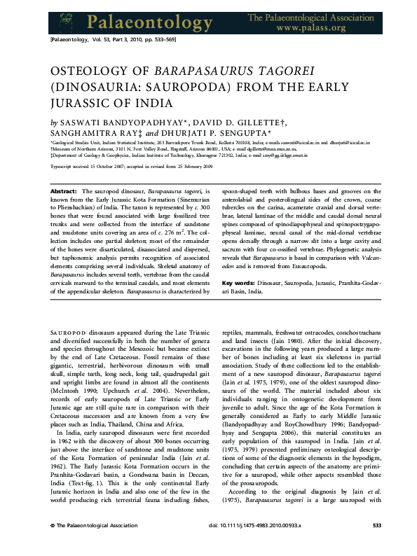
 Saswati Bandyopadhyay