NPC
2010
Vol. 5
No. 2
253 - 258
Natural Product Communications
Aristolactams, 1-(2-C-Methyl-β-D-ribofuranosyl)-uracil and
Other Bioactive Constituents of Toussaintia orientalis
Josiah O. Odaloa, Cosam C. Josepha, Mayunga H.H. Nkunyaa*, Isabel Sattlerb, Corinna Langeb,
Gollmick Friedrichb, Hans-Martin Dahseb and Ute Möllmanb
a
Department of Chemistry, University of Dar es Salaam, P.O. Box 35061, Dar es Salaam, Tanzania
b
Leibniz Institute for Natural Product Research and Infection Biology, Hans Knöll Institute,
Beutenbergstrasse 11a, 07745 Jena, Germany
nkunya@chem.udsm.ac.tz, mnkunya@tcu.go.tz
Received: September 1st, 2009; Accepted: November 23rd, 2009
The new aristolactam alkaloid toussalactam {2-hydroxy-1,6-dimethoxy-5H-dibenzo[cdf]indol-4-one} and the known ones,
namely aristolactam AII, aristolactam BII, piperolactam C and aristolactam FII; 1-(2-C-methyl-β-D-ribofuranosyl)-uracil,
3,4,5-trimethoxyphenyl-β-D-glucopyranoside, and three catechinoids were isolated from the cytotoxic Toussaintia orientalis
Verdc stem and root bark extracts, and their structures established based on analysis of spectroscopic data. The aristolactams
exhibited antimicrobial and antiinflammatory activity, aristolactam FII showing almost the same level of activity as the
standard anti-inflammatory agent Indomethacin. The compounds also exhibited either mild or no antiproliferative and
cytotoxic activities, except aristolactam FII that showed the same level of cytotoxicity as the standard drug Camptothecin. 1-(2C-Methyl-β-D-ribofuranosyl)-uracil, which is being reported for the first time as a natural product, was inactive in the
antibacterial, antifungal, antiinflammatory, antiproliferative and cytotoxicity assays.
Keywords: Toussaintia orientalis, Annonaceae, Aristolactams, 1-(2-C-methyl-β-D-ribofuranosyl)-uracil, Antimicrobials,
Antiproliferative, Cytotoxicity, Antiinflammatory.
In East Africa several Annonaceae species are used as
herbal medicines [1,2]. This has inspired us to
investigate the nearly 90 Annonaceae species occurring
in Tanzania, some of which having been taxonomically
described only recently [3,4]. Others, such as
Toussaintia oriantalis Verdc., are reported to occur
only in Tanzania where their ecological habitats are
systematically being destroyed through human
activities, thus threatening them with imminent
extinction. Therefore, our investigations are focused on
determining the bioactive or other chemical
constituents of such endangered plant species.
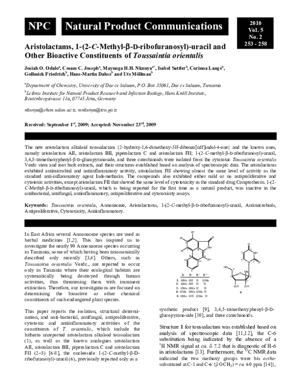
This paper reports the isolation, structural determination, and anti-bacterial, antifungal, antiproliferative,
cytotoxic and antiinflammatory activities of the
constituents of T. oriantalis, which include the
hitherto unreported aristolactam alkaloid toussalactam
(1), as well as the known analogues aristolactam
AII, aristolactam BII, piperolactam C and aristolactam
FII (2–5) [6-8], the nucleoside 1-(2-C-methyl-β-Dribofuranosyl)-uracil (6), previously reported only as a
R''
O
R'
2
O
NH
1
R
5a
10
4
5
5
NH
6
10a
6
R'''
6a
N
2
O
HO
5'
8
R
1:
2:
3:
4:
5:
OMe
OMe
OMe
OMe
OMe
R'
R''
R'''
OH
OH
OMe
OMe
OH
H OMe
H
H
H
H
OMe H
OMe H
O
Me
4'
1'
2'
OH
OH
6
synthetic product [9], 3,4,5-trimethoxyphenyl-β-Dglucopyranoside [10], and three catechinoids.
Structure 1 for toussalactam was established based on
analysis of spectroscopic data [11,12], the C-6
substitution being indicated by the absence of a
1
H NMR signal at ca. δ 7.2 that is diagnostic of H-6
in aristolactams [13]. Furthermore, the 13C NMR data
indicated the two methoxy groups were bis orthosubstituted at C-1 and C-6 {δ OCH3) = ca. 60 ppm [14]},
�254 Natural Product Communications Vol. 5 (2) 2010
Odalo et al.
O
H
HN
O
H
H
N
H
H
O
CH3
HO
H
H
OH
OH
Figure 1: Important H/C HMBC correlations for toussalactam (1).
the third substituent being a C-2 OH group. The COSY,
HMQC and HMBC interactions (Figure 1) indicated the
inter-atomic connectivity of the aristolactam skeleton
and the substitution pattern of the phenanthrenoid
carbacyclic system, thus confirming structure 1.
Aristolactams form a small group of modified
aporphinoids that exhibit antibacterial, antimalarial,
cytotoxic, and platelet aggregation inhibition activities.
These compounds are distributed in the families
Annonaceae,
Aristolochiaceae,
Menispermaceae,
Monimimaceae and Piperaceae [7,15].
Structure 6 for 1-(2-C-methyl-β-D-ribofuranosyl)-uracil
was based on analysis of the 1H and 13C NMR spectra,
as well as HMBC interactions (Figure 2), indicating the
presence of a C-2 methylated pentafuranose sugar
linked to the uracil moiety through the anomeric carbon.
In the 1H NMR spectrum the J3,4 value of 9.2 Hz was
indicative of the β-configuration at the anomeric carbon
[16-19], as further corroborated from the UV and CD
spectra [21-23]. The CD and 1H NMR spectral data, as
well as the strong H-6/H-3α NOE correlation, also
indicated both the anti and C-3α endo conformation for
the sugar unit in solution [17], and an anti orientation
for the base. NOE further indicated a cis configuration
for 2β-C-Me/H-3α.
Compound 6 is reported for the first time as a plant
natural product, which is also unprecedented among
nucleosides since so far they have been obtained only
from marine sponges [23,24]. Nucleoside analogues
have been used to treat viral infections [26-28] and are
potential leads to new antiviral agents [28-30].
The light petroleum, CH2Cl2 and MeOH stem and root
bark extracts showed activity in the brine shrimp test
(LC50 72.4, 22.3 and 19.2, and 57.6, 1.3 and 17.2 µg/mL
respectively), the stem bark extracts being the most
active. This is the source of the aristolactams that
showed the highest antibacterial, antiinflammatory,
antiproliferative and cytotoxic activities. In the
antibacterial assay the aristolactams 1 – 5 exhibited
growth inhibition effects against M. vaccae, but only
Figure 2: Important H/C HMBC correlations for 1-(2-C-methyl-β-Dribofuranosyl)-uracil (6).
mild activity against all other microbial strains, with 1
and 5 more active against the fungi and M. vaccae than
against the other organisms tested (Table 1).
Generally, the active compounds showed better efficacy
against bacterial than fungal strains, the fungus P.
notatum (P1) being more susceptible as compared with
the other fungi, S. salmonicolor and C. albicans. Most
of the compounds exhibited moderate activity against P.
notatum (P1), compound 1 being the most active against
this fungal strain (Table 1). Against all the test
organisms, compound 6 and 3,4,5-trimethoxyphenyl-βD-glucopyranoside showed no activity.
The results in Table 1 indicate that increased
oxygenation of the aristolactam skeleton generally
enhanced antibacterial activity, especially at C-6, which
also increased antifungal activity. Since inhibition of rat
liver cytosol NAD (P) linked 3α-hydroxysteroid
dehydrogenase is correlated to anti-inflammatory
activity in humans, that test was applied to compounds
3–6, 3,4,5-trimethoxy-phenyl-β-D-glucopyranoside and
epicatechin-4β,8-epicatechin (Table 2). Aristolactams
3–5 were moderately active, epicatechin-4β,8epicatechin was only mildly active, while both 6 and
were
3,4,5-trimethoxyphenyl-β-D-glucopyranoside
inactive.
Compared with the standard anticancer drugs Taxol®,
Colchicine and Camptothecin, the aristolactams also
exhibited some antiproliferative activity, compound 4
being the most potent against L-929, whereas 5 was the
most active against the K-562 cell lines (Table 3),
having the same level of cytotoxicity as Camptothecin.
Compound
6
and
3,4,5-trimethoxyphenyl-β-Dglucopyranoside were inactive in the antiproliferative
and cytotoxicity assays.
Experimental
General experimental procedures: CC: Silica gel 60
(0.063-0.200 mm, Merck) and Sephadex® LH-20
(Pharmacia); TLC: Kieselgel 60 F 254 precoated on
�Bioactive constituents of Toussaintia orientalis
Natural Product Communications Vol. 5 (2) 2010 255
Table 1: Antibacterial and antifungal activity (zones of inhibition in mm) of the isolated compounds.
Compounds/microorganisms
Toussalactam (1)
Aristolactam AII (2)
Aristolactam BII (3)
Piperolactam C (4)
Aristolactam FII (5)
1-(2-C-Methyl-β-D-ribofuranosyl)-uracil (6)
3,4,5-Trimethoxyphenyl-β-D-glucopyranoside
Ciprofloxacin (5 µg/ml)
Amphotericin (10 µg/ml)
B1
14
0
0
12
12
0
0
28
--
B3
13
0
0
12
15p
0
0
18
--
B4
0
0
0
0
0
0
0
23/32p
--
B7
--0
0
-0
0
---
B9
0
0
0
0
0
0
0
22/27
--
M4
23/29p
14
16p
13p
20p
0
0
22p
--
H4
0
0
--13p
0
0
-14
H8
13
0
0
0
11p
0
0
-20
P1
17
15
0
0
13
0
0
-18
p = Partial inhibition; 0 = inactive; -- = Test not carried out on these microorganisms because of small quantity of the compounds. Bacterial species tested: B1 =
Bacillus subtilis ATTC 6633 (IMET) NA; B3 = Staphylococcus aureus (IMET 10760) SG511; B4 = Escherichia coli SG 458; B7 = Pseudomonas aeruginosa SG 137
(IMET 10480); B9 = Pseudomonas aeruginosa K 799/61 and M4 = Mycobacterium vaccae IMET 10670. Fungal species used: H4 = Sporobolomyces salmonicolor
SBUG 549; H8 = Candida albicans BMSY 212 and P1 = Penicilium notatum.
Table 2: Antiinflammatory activity of the isolated compounds.
Compounds
% 3α-Hydroxysteroid dehydrogenase inhibition
Aristolactam BII (3)
Piperolactam C (4)
Aristolactam FII (5)
1-(2-C-Methyl-β-D-ribofuranosyl)-uracil (6)
3,4,5-Trimethoxyphenyl-β-D-glucopyranoside
Epicatechin-4β,8-epicatechin
Indomethacin (standard)
30 µg/mL
87
65
47
0
0
11
93
3 µg/mL
31
19
30
0
0
0
27
IC50 (µg/mL)
0.3 µg/mL
11
16
24
0
0
0
10
4.6
14.5
--0
0
0
4.6
Table 3: Antiproliferative and cytotoxic activity of the isolated compounds.
Compounds
Aristolactam BII (3)
Piperolactam C (4)
Aristolactam FII (5)
1-(2-C-Methyl-β-D-ribofuranosyl)-uracil (6)
3,4,5-Trimethoxyphenyl-β-D-glucopyranoside
Taxol®
Colchicine
Camptothecin
plastic plates (Merck, 0.20 mm); visualizing: UV/VIS
and Dragendorff’s or anisaldehyde spray [31]; m.p.
Electrothermal 9100 (uncorrected); UV spectra: Hitachi
200-20 spectrophotometer; IR spectra: JASCO FT/IR4100 spectrometer; 1H NMR (300 and 500 MHz) and
13
C NMR (75 MHz) in DMSO-D6: Bruker DRX-300,
internal standard TMS (1H NMR) and solvent signal for
13
C NMR; ESIMS: 70 eV on an HP 5990/5988A
spectrometer; HRESIMS: JEOL JMS-HX 110 mass
spectrometer.
Plant materials: The stem and root barks were
collected in October 2004 from Zaraninge forest reserve
at the edge of Saadani National Park, Bagamoyo
District in Tanzania. The plant species was identified at
the Herbarium of the Department of Botany, University
of Dar es Salaam, where a voucher specimen No. FMM
3330 is preserved.
Extraction and isolation: The air-dried and powdered
stem and root barks (1 Kg each) were subjected to
sequential extraction in light petroleum, CH2Cl2 and
Antiproliferative activity (µg/mL)
L-929 (GI50)
K-562 (GI50)
50
50
13.2
21.8
19.1
5.7
50
50
>50
>50
0.1
0.01
0.9
0.02
0.02
0.002
Cytotoxicity (µg/mL)
HeLa (CC50)
9.6
36.3
0.2
50
>50
0.01
0.006
0.2
MeOH at room temp, each 2 x 72 h. Vacuum liquid
chromatography (VLC) of the concd. cytotoxic (brine
shrimp test) CH2Cl2 stem bark extract (6.9 g) on silica
gel and then CC (silica gel: light petroleum/EtOAc
gradient) yielded 1 (4.2 mg), 4 (48.1 mg), 5 (26.6 mg),
3 (6.5 mg), and 2 (5.3 mg) in that sequence. VLC (silica
gel: light petroleum/EtOAc gradient) of the MeOH
extracts, then CC (silica gel: light petroleum/EtOAc
gradient), then Sephadex® LH-20, MeOH/CH3Cl, 1:1
v/v) yielded (-)-epicatechin, a mixture of (-)-epicatechin
and (+)-catechin, epicatechin-4β,8-epicatechin, 6 and
3,4,5-trimethoxyphenyl-β-D-glucopyranoside.
Toussalactam
(2-hydroxy-1,6-dimethoxy-5Hdibenzo[cdf]indol-4-one, 1)
Brownish yellow powder (yield 4.2 mg).
MP: 255-258°C.
Anisaldehyde spray – yellow.
UV, λmax (log ε) 202 (4.42), 236 (4.54), 278 (4.45), 289
(4.43) and 391 (3.82) nm.
�256 Natural Product Communications Vol. 5 (2) 2010
IR (film) νmax: 3224, 2926, 1684, 1647, 1609, 1558,
1541, 1507, 1456, 1437, 1362, 1330, 1248, 1200, 1173,
1110, 1037, 982, 960, 926, 869, 844, 802, 766, 744 and
720 cm-1.
1
H NMR: δ 4.01 (3H, s, 1-OMe), 4.05 (3H, s, 6-OMe),
7.60 (1H, ddd, J = 7.8, 7.2, 1.7 Hz, H-6), 7.61 (1H, s,
H-3), 7.65 (1H, ddd, J = 7.8, 7.2, 1.7 Hz, H-8), 8.17
(1H, dd, J = 7.5, 1.8 Hz, 9), 9.16 (1H, dd, J = 7.5, 2.2
Hz, H-10) and 10.95 (1H, br s, N-H).
13
C NMR: δ 168.1 (C=O), 151.5 (C-2), 149.0 (C-1),
133.8 (C-6), 130.2 (C-6a), 127.3 (C-10a), 127.2 (C-8),
126.9 (C-10), 126.0 (C-5a), 125.8 (C-9), 122.3 (C-7),
120.9 (C-3a), 120.6 (C-5), 117.7 (C-10b), 113.4 (C-3),
60.8 (6-OMe) and 59.3 (1-OMe).
HRESIMS, m/z 296.0967 ([MH]+, calculated for
C17H14NO4 = 296.0893), 295 ([M]+, 15), 294 (100), 280
(9), 279 (71) and 264 (18).
1-(2-C-Methyl-β-D-ribofuranosyl)-uracil (6)
White crystals (yield, 561 mg).
MP: 117-118oC (MeOH/EtOAc).
[α]D20: +67.81 (c 0.20, MeOH).
Anisaldehyde spray – pink.
UV, λMeOHmax (log ε): 213 (3.14) and 263 (3.88).
IR (film) νmax: 3595, 3097, 1697, 1662, 1631, 1473,
1418, 1398, 1379, 1274, 1184, 1123, 1094, 1079, 1051,
1033, 949, 880 and 732 cm-1.
HRESIMS, m/z 258.22804 (calc. for C10H14N2O6,
258.08519), m/z (% rel. int.) 257 (23), 214 (100) and
166 (40).
1
H NMR: δ 1.15 (3H, s, 2'-Me), 3.77 (1H, dd, J = 12.5,
2.6 Hz, H-5'a), 3.83 (1H, d, J = 9.2 Hz, H-3'), 3.91 (1H,
td, J = 9.2, 2.3, H-4'), 3.97 (1H, dd, J = 12.5, 2.6 Hz,
H-5'b), 5.67 (1H, d, J = 8.1 Hz, H-5), 5.95 (1H, s, H-1')
and 8.13 (1H, d, J = 8.1, H-6).
13
C NMR: δ 166.0 (C-4), 152.4 (C-2), 142.5 (C-6),
102.3 (C-5), 93.1 (C-1'), 83.9 (C-4'), 80.0 (C-2'), 73.4
(C-3') 60.5 (C-5) and 20.2 (2'-Me).
Brine shrimp test: This was carried out according to a
standard procedure [32], using brine shrimp (Artemia
salina Leach) larvae as the indicator organisms, which
were hatched in artificial seawater prepared from sea
salt (3.8 g) in distilled water (1000 mL), and then
filtered. The mature nauplii were collected after 48 h of
hatching. Each sample was tested at concentrations of
240, 120, 80, 40, 24 and 8 μg/mL in DMSO, in
triplicate vials, each containing 10 brine shrimp larvae.
An additional vial with only the solvent, DMSO, and 10
shrimp larvae was used as the control. The number of
surviving larvae after 24 h exposure was established
and the LC50 values (the concentration required to kill
50% of the larvae) were determined using Probit
analysis [33].
Odalo et al.
Agar diffusion assay for antimicrobial activity
(antibacterial and antifungal activity): The agar
diffusion method was used for the determination of
antibacterial and antifungal activity of the isolated
compounds against the microorganisms listed in Table
5, obtained from the Hans Knolls Institute for Natural
Product Research and Infectious Biology (HKI) in Jena,
Germany. Ca. 9 mL of Müller-Hinton agar for bacteria
and Sabouraud Dextrose agar for fungi (Oxoid, UK)
were poured into Petri dishes (9 cm diameter) and
inoculated with the respective test organisms. Wells
(4 cm) were punched out of the solid agar using
pipette tips, and 1 mL of 50 µg/mL solutions of the
test compounds and control antibiotics (Ciprofloxacin,
5 µg/mL and Amphotericin, 10 µg/mL) were placed in
each well. The Petri dishes were then incubated at 30ºC
and 35ºC for the test bacterial and fungal strains,
respectively, for 20 h and the average diameter of the
inhibition zone surrounding the wells was measured.
Microplate dilution assay for minimum inhibitory
concentrations (MIC) determination: The minimum
inhibitory concentrations (MIC) of the isolated
compounds with good antimicrobial activity in the agar
diffusion tests were determined using a serial
microplate dilution assay against each test bacterial
species. This was determined by 2-fold serial dilution of
the compounds beyond the level where no inhibition of
growth of the bacterial strains Bacillus subtilis ATTC
6633 (IMET) NA (B1), Staphylococcus aureus (IMET
10760) SG511 (B3), Mycobacterium smegmatis SG987
(M2), M. aurum SB66 (M3), M. vaccae IMET 10670
(M4) and M. fortuitum M. Fort. (M5) was observed.
The compounds were reconstituted to 100 µg/mL in
DMSO and 100 µL aliquots of the fractions were
serially diluted by 50% with water in 96-well
microplates. Muller-Hinton (MH) broth culture (1%)
was inoculated with the test bacteria and then incubated
at 37ºC overnight, and 100 µL aliquots of the resulting
culture were added to each well. Ciprofloxacin
(100 µg/mL) was used as the reference antibiotic and
two wells were used as sterility and growth controls,
respectively, with the sterility control containing only
Oxoid MH broth, while the negative growth control
contained both MH broth as well as the test organism.
The microplates were sealed and incubated at 37ºC at
100% relative humidity for 18 h. As an indicator of
bacterial growth, 40 µL aliquots of a 0.2 mg/mL
solution of p-iodonitrotetrazolium violet (INT)
dissolved in water were added to the microplate wells.
Antiinflammatory
activity
[3α-hydroxysteroid
dehydrogenase (3α-HSD) assay]: The source of 3αhydroxysteroid dehydrogenase used in the bioassay was
liver from adult male Sprague-Dawley rats (150-200 g).
The liver was excised and homogenized in 3 volumes of
�Bioactive constituents of Toussaintia orientalis
50 mM Tris-HCl of pH 8.6 containing 250 mM sucrose,
1 mM dithiothreitol and 1 mM EDTA. Homogenates
were centrifuged (100,000 x g for 30 min) and
the resulting supernatant (cytosol containing 3αhydroxysteroid dehydrogenase) was used for enzyme
assays without further processing, after being prepared
according to the method described by Penning [34], and
having attained a final specific activity of 3.58 µM of
5β-dihydrocortisone reduced/min/mg of protein).
The reduction of 5β-dihydrocortisone was monitored by
measuring the changes in the absorbance of the pyridine
nucleotide at 340 nm. Each assay (1.0 mL) contained
potassium phosphate buffer (pH 6.0, 0.840 mL of 1M),
NADPH (20 µL of 9 M), 5β-dihydrocortisone (10 µL of
5 mM), and acetonitrile (30 µL). The reactions were
initiated by the addition of enzyme (30-50 µg of either
cytosolic protein or 0.6 µg of purified enzyme), and
optical density change was followed over a period of 5
min. Control incubation experiments by addition of the
cytosol in which either 5β-dihydrocortisone or NADPH
was absent indicated that the presence of both
substances was required before the cytosol would
promote a change in absorbance at 340 nm. The %
inhibition of the isolated compounds was generated at
30, 3 and 0.3 µg/mL concentration. Increasing amounts
of the isolated compound were added to the standard
assay system, and the concentration of the compound
required to reduce the rate of 5β-dihydrocortisone
Natural Product Communications Vol. 5 (2) 2010 257
reductions by 50% (IC50) was computed from the
resulting natural logarithm dose-response curves.
Antiproliferative and cytotoxicity assay: This assay
was carried out by the Molecular Natural Product
Research group of HKI in Jena, Germany, as described
in the literature [35], using the cell lines K-562
(human chronic myeloid leukemia) and L-929
(mouse fibroblast) for antiproliferative effects
(GI50, concentration which inhibited cell growth by
50%), and against HeLa cells for cytotoxicity
(GC50 = concentration at which cells were destroyed by
50%; used partially in referring to the lysis of cells).
Taxol®, Colchicine and Camptothecin were used as the
standard antiproliferative and cytotoxic drugs.
Acknowledgements - JOO thanks the Germany
Academic Exchange Services (DAAD) and NAPRECA
for
funding
these
studies
through
the
DAAD/NAPRECA Fellowship Scheme, which also
supported the collaborative research engagement with
the Leibniz Institute for Natural Product Research and
Infection Biology, Hans Knöll-Institute in Jena,
Germany. Part of the research was funded through
Sida/SAREC support to the Faculty of Science,
University of Dar es Salaam. We thank Mr F. Mbago of
the Herbarium, Department of Botany at the University
of Dar es Salaam for the location and identification of
the investigated plant species.
References
[1]
Verdcourt B. (1971) Flora of Tropical East Africa - Annonaceae, Crown Agents, London, p. 1,9.
[2]
Kokwaro JO. (1993) Medicinal Plants of East Africa, 2nd edition, Kenya Literature Bureau, Nairobi.
[3]
Verdcourt B. (1996) Sanrafaelia, a new genus of Annonaceae from Tanzania. Garcia de Orta, Series Botanica, 13, 43-44.
[4]
Couvreur TLP, Van Der Ham RWJM, Mbele YM, Mbago FM, Johnson DM. (2009) Molecular and morphological characterization
of a new monotypic genus of Annonaceae, Mwasumbia, from Tanzania. Systematic Botany, 34, 266-276.
[5]
Deroin T, Luke Q. (2005) A new Toussaintia (Annonaceae) from Tanzania. Journal of East African Natural History, 94, 165-174.
[6]
Desai SJ, Prabhu BR, Mulchandani NB. (1988) Aristolactams and 4,5-dioxoaporphines from Piper longum. Phytochemistry, 27,
1511-1515.
[7]
Chia YC, Chang FR, Teng CM, Wu YC. (2000) Aristolactams and dioxoaporphines from Fissistigma balansae and Fissistigma
oldhamii. Journal of Natural Products, 63, 1160-1163.
[8]
Sun NJ, Antoun M, Chang CJ, Cassady JM. (1987) New cytotoxic aristolactams from Pararistolochia flos-avis. Journal of Natural
Products, 50, 843-846.
[9]
Beigelman LN, Ermolinsky BS, Gurskaya GV, Tsapkina EN, Karpeisky MY, Mikhailov SN. (1987) New synthesis of 2′-Cmethylnucleosides starting from D-glucose and D-ribose. Carbohydrate Research, 166, 219-232.
[10]
Shimomura H, Sashida Y, Oohara M, Tenma H. (1988) Phenolic glucosides from Parabenzoin praecox. Phytochemistry, 27,
644-646.
[11]
Olsen CE, Tyagi OD, Boll PM, Hussaini FA, Parmar VS, Sharma NK, Teneja P, Jain SC, (1993) An aristolactam from Piper
acutisleginum and revision of the structures of piperolactam B and D. Phytochemistry, 33, 518-520.
[12]
Chen YC, Chen JJ, Chang YL, Teng CM, Lin WY, Wu CC, Chen IS. (2004) A new aristolactam alkaloid and anti-platelet
aggregation constituents from Piper taiwanense. Planta Medica, 70, 174-177.
[13]
Likhitwitayawuid K, Wiasathien L, Jongboonprasert V, Krungkrai J, Aimi N, Takayama H, Kitajima M. (1997) Antimalarial
alkaloids from Goniothalamus tenuifolius. Pharmacology Letters, 7, 99-102.
�258 Natural Product Communications Vol. 5 (2) 2010
Odalo et al.
[14]
Makriyannis A, Knittel JJ. (1979) The conformational analysis of aromatic methoxy groups from carbon-13 chemical shifts and
spin-lattice relaxation times. Tetrahedron Letters, 20, 2753-2756.
[15]
Daneluttea AP, Costantina MB, Delgadob GE, Braz-Filho R, Kato MJ. (2005) Divergence of secondary metabolism in cell
suspension cultures and differentiated plants of Piper cernuum and P. crassinervium. Journal of Brazilian Chemical Society, 16,
1425-1430.
[16]
Davies DB. (1978) Conformations of nucleosides and nucleotides. Progress of Nuclear Magnetic Resonance Spectroscopy, 12,
135-225.
[17]
Stevens JD, Fletcher HG. (1968) The proton magnetic resonance spectra of pentofuranose derivatives. Journal of Organic
Chemistry, 33, 1799-1805.
[18]
Kam BL, Barascut JL, Imbach JL. (1979) A general method of synthesis and isolation, and an N.M.R.-spectroscopic study, of tetraO-acetyl-D-aldopentofuranoses. Carbohydrates Research, 69, 135-142.
[19]
Jenkins SR, Arison B, Walton E. (1968) Branched-chain sugar nucleoside. IV. 2’-Methyladenosine. Journal of Organic Chemistry,
33, 2490-2494.
[20]
Guo FQ, Li Ai, Huang LF, Liang YZ, Chen BM. (2006) Identification and determination of nucleosides in Cordyceps sinesis and
its substitutes by high performance liquid chromatography with mass spectrometric detection. Journal of Pharmacological and
Biomedical Analysis, 40, 623-630.
[21]
Miles DW, Inskeep WH, Robins MJ, Winkley MW, Robins RK, Eyring H. (1970) Circular dichroism of nucleoside derivatives. IX.
Vicinal effects on the circular dichroism of pyrimidine nucleosides. Journal of American Chemical Society, 92, 3872-3881.
[22]
Ingwall JS. (1972) Circular dichroism of nucleosides. I. Anomeric pairs of the D-pentofuranosides of adenine. Journal of American
Chemical Society, 94, 5487-5495.
[23]
Searle PA, Molinski TF. (1995) Trachycladines A and B: 2’-C-methyl-5’-deoxyribofuranosyl nucleosides from the marine sponge
Trachycladus laevispirulifer. Journal of Organic Chemistry, 60, 4296-4298.
[24]
Ichiba T, Nakao Y, Scheuer PJ, Sata NU, Kelly-Borges M. (1995) Kumusine, a chloroadenine riboside from a sponge, Theonella
sp. Tetrahedron Letters, 36, 3977-3980.
[25]
De Clercq E. (2005) Antiviral drug discovery and development: Where chemistry meets with biomedicine. Antiviral Research, 67,
56-75.
[26]
De Clercq E. (2003) Clinical potential of the acyclic nucleoside phosphonates cidofovir, adefovir, andtenofovir intreatment of DNA
virus and retrovirus infections. Clinical Microbiological Review, 16, 569-596.
[27]
Naesens L, De Clercq E. (2001) Recent developments in herpes virus therapy. Herpes, 8, 12-16.
[28]
Donia M, Hamann MT. (2003) Marine natural products and their potential applications as antiinfective agents. Lancet Infectious
Diseases, 3, 338-348.
[29]
Suckling CJ. (1991) Chemical approaches to the discovery of new drugs. Science Progress, 75, 323-359.
[30]
Carroll SS, Tomassini JE, Bosserman M, Getty K, Stahlhut MW, Eldrup AB, Bhat B, Hall D, Simcoe AL, LaFemina R, Rutkowski
CA, Wolanski B, Yang Z, Migliaccio G, De Francesco R, Kuo LC, MacCoss M, Olsen DB. (2003) Inhibition of hepatitis C virus
RNA replication by 2′-modified nucleoside analogs. Journal of Biological Chemistry, 278, 11979-11984.
[31]
Stahl E. (1969) Thin-layer chromatography, a laboratory handbook. Springer-Verlag, New York, p. 857.
[32]
Meyer BN, Ferrigini RN, Jacobsen LB, Nicholas DE, McLaughlin JL. (1982) Brine shrimp: A convenient general bioassay for
active plant constituents. Planta Medica, 45, 31-34.
[33]
Finney DJ. (1971) Probit analysis, 3rd ed., Cambridge University Press, Cambridge, 333 pp.
[34]
Penning TM. (1985) Inhibition of 5ß-dihydrocotisone reduction in rat liver cytosol: A rapid spectrophotometric screen for
nonsteroidal anti-inflammatory drug potency. Journal of Pharmacological Science, 74, 651-654.
[35]
Dahse HM, Schlegel B, Gräfe U. (2001) Differentiation between inducers of apoptosis and nonspecific cytotoxic drugs by means of
cell analyzer and immunoassay using the cell lines K-562 (human chronic myeloid leukemia), and L-929 (mouse fibroblast) for
antiproliferative effects and HeLa (human cervix carcinoma) for cytotoxic effects. Pharmazie, 56, 489-491.
�
 Josiah Odalo