International Journal of
Molecular Sciences
Review
Impact of Omega-3 Fatty Acids on the Gut Microbiota
Lara Costantini †
ID
, Romina Molinari †
ID
, Barbara Farinon and Nicolò Merendino *
ID
Department of Ecological and Biological Sciences (DEB), Tuscia University, Largo dell’Università snc,
01100 Viterbo, Italy; lara.cost@libero.it (L.C.); rominamolinari@libero.it (R.M.); barbara.farinon@gmail.com (B.F.)
* Correspondence: merendin@unitus.it; Tel.: +39-0761-357-133
† These authors contributed equally to this work.
Received: 31 October 2017; Accepted: 1 December 2017; Published: 7 December 2017
Abstract: Long-term dietary habits play a crucial role in creating a host-specific gut microbiota
community in humans. Despite the many publications about the effects of carbohydrates (prebiotic
fibers), the impact of dietary fats, such as omega-3 polyunsaturated fatty acids (PUFAs), on the
gut microbiota is less well defined. The few studies completed in adults showed some common
changes in the gut microbiota after omega-3 PUFA supplementation. In particular, a decrease in
Faecalibacterium, often associated with an increase in the Bacteroidetes and butyrate-producing bacteria
belonging to the Lachnospiraceae family, has been observed. Coincidentally, a dysbiosis of these
taxa is found in patients with inflammatory bowel disease. Omega-3 PUFAs can exert a positive
action by reverting the microbiota composition in these diseases, and increase the production of
anti-inflammatory compounds, like short-chain fatty acids. In addition, accumulating evidence in
animal model studies indicates that the interplay between gut microbiota, omega-3 fatty acids, and
immunity helps to maintain the intestinal wall integrity and interacts with host immune cells. Finally,
human and animal studies have highlighted the ability of omega-3 PUFAs to influence the gut–brain
axis, acting through gut microbiota composition. From these findings, the importance of the omega-3
connection to the microbiota emerges, encouraging further studies.
Keywords: omega-3 PUFAs; DHA; EPA; gut microbiota; dysbiosis; inflammation; behavioral disorders
1. Introduction
In the last few years, the emergence and growing accessibility of next-generation sequencing (NGS)
technologies have allowed advances in the understanding of the composition and functional activity
of the gut microbial community. Approximately 100 papers on gut microbiota were published in 2007,
whereas about 3000 such studies were published in 2016, and almost the same number to date in 2017
(research performed by setting the words “gut” and “microbiota” in October 2017 on PubMed and
Scopus). The importance of using NGS technology is due to the necessity of simultaneous analysis of a
large amount of genetic material. Indeed, overall, the gut microbiota is estimated to contain 150 times
more genes than the human genome. These genes have been estimated to belong to approximately
1013 –1014 microbes, with a species diversity of up to several hundred per individual [1]. However,
The Human Microbiota Project and other studies have collectively found that thousands of different
species may inhabit the human gut, pointing out the high degree of taxa variation in the microbiota
composition of different populations. Despite this variation, the human gut microbiota is characterized
by some basic similarities. Approximately 60% of the gut bacteria belong to the Bacteroidetes and
Firmicutes phyla, and, among them, Bifidobacterium, Lactobacillus, Bacteroides, Clostridium, Escherichia,
Streptococcus, and Ruminococcus are the most commonly found genera in adults [2]. However, several
factors influence the bacterial composition in taxa type and abundance, making the total gut microbiota
profile host-specific in humans. These factors include host phenotype, such as age, gender, body mass
Int. J. Mol. Sci. 2017, 18, 2645; doi:10.3390/ijms18122645
www.mdpi.com/journal/ijms
�Int. J. Mol. Sci. 2017, 18, 2645
2 of 18
index (BMI), lifestyle, and immune function; geographical belonging and environmental factors; use of
antibiotics, drugs, and probiotics; and diet.
The causal relationship between the gut microbiota and overall pathological conditions is still
unclear. Indeed, it is still unclear whether a disease-prone microbial composition exists (so-called
dysbiosis) or whether the changes in the microbial community occur after the onset of the disease [3].
Conversely, diet undoubtedly influences the composition of gut microbiota, providing nutrients for
both the host and the bacteria. This gut community has many degrading enzymes and metabolic
capabilities that are able to break down macromolecules into smaller chemical compounds, which can
then be uptaken by enterocytes [4]. Moreover, long-term dietary habits have been shown to play a
crucial role in creating an inter-individual variation in microbiota composition [5]. However, despite
the great number of publications on the effects of carbohydrates, the impacts of dietary fats and protein
on the gut microbiota are less well defined. In particular, gut microbiota changes associated with
omega-3 fatty acids are poorly understood.
Among the omega-3 polyunsaturated fatty acids (PUFAs), eicosapentaenoic acid (EPA, C20:5) and
docosahexaenoic acid (DHA, C22:6) are the two main bioactive forms in humans. These fatty acids
can be synthesized from the dietary precursor and essential fatty acid, α-linolenic acid (ALA, C18:3).
However, the synthesis pathway requires several elongation and desaturation chemical reactions,
so that the conversion of the two active forms in mammals is less efficient than dietary uptake. For
this reason, consumption of EPA- and DHA-rich foods is recommended. However, since foods rich
in these fatty acids are not widespread, EPA and DHA are widely used as nutritional supplements,
often as nutraceuticals. Several papers have demonstrated the correlation between omega-3 PUFAs
and the inflammatory response. Although the literature on this topic is discordant, omega-3 PUFAs
are generally associated with anti-inflammatory effects, in comparison with the omega-6 PUFAs that
are linked to pro-inflammatory effects, due to the different downstream lipid metabolites [6]. Also,
with regards to the link to immunity, studies have shown that the supplementation of omega-3 PUFAs
provides multiple health benefits against different chronic degenerative diseases, such as cardiovascular
diseases [7], rheumatoid arthritis [8], inflammatory bowel disease (IBD) [9], depression [10], and
cancer [11].
Considering the few insights existing in literature, in the present review, we assessed whether
omega-3 PUFAs have an impact on the composition of the human gut microbiota in adults and infants.
Moreover, a connection of this topic to inflammation and behavioral disorders was completed.
2. Omega-3 Influence on Human Gut Microbiota: State of the Art
The use of NGS technology has expanded the knowledge about the correlation between the
human gut microbiota and omega-3 PUFAs. However, the literature in this topic is still in the initial
stages. The current literature is listed below and summarized in Table 1. The first report in the
literature about the impact of omega-3 fatty acids on human gut microbiota of adults came from a
clinical study carried out in 60 overweight (BMI > 25) healthy people, between 40 and 60 years old.
In this study, a commercially available probiotic with high concentrations of Bifidobacteria, Lactobacilli,
and Streptococcus thermophilus (named VSL#3) was provided in combination with and without an
omega-3 nutraceutical supplementation of 180 mg EPA and 120 mg of DHA for six weeks. This study
failed to elucidate differences between the probiotic group and the probiotic plus omega-3 group.
However, the limitation of this analysis was that the evaluation of microbiota changes was only
completed using colony counting on anaerobic or aerobic selective media [12]. Subsequent studies
focused more on food and diet impact instead of nutraceutical use of omega-3 PUFAs, likely because
omega-3 fatty acids integrated in a food matrix can have a higher positive impact on gut microbiota.
Supporting this hypothesis, a randomized crossover trial was completed on 20 middle-aged healthy
individuals by administering a high daily dose (4 g) of a mixed DHA/EPA supplement for eight
weeks [13]. The supplementation was performed using two different formulations: as a nutraceutical
in the form of capsules, and as functional drink that was EPA- and DHA-rich. In this study, a taxonomy
�Int. J. Mol. Sci. 2017, 18, 2645
3 of 18
classification of the whole microbiota of the samples was completed with NGS technology. In this
case, no statistically significant changes were observed in the Firmicutes/Bacteroidetes phyla ratio
for both types of supplementations. On the contrary, analyzing the data at family and genus levels
revealed consistent differences associated with both omega-3 PUFA supplementations. In particular,
increases in the Clostridiaceae, Sutterellaceae, and Akkermansiaceae families were recorded, and these
changes were reverted by the washout period. A statistically increased abundance of Bifidobacterium
and Oscillospira genera, associated with a reduction of Coprococcus and Faecalibacterium, were found
after both omega-3 PUFA supplementations in comparison with before the study and after washout.
Instead, an increase in Lachnospira and Roseburia genera was prominent only after the functional
omega-3 drink feeding. So, as previously anticipated, the functional drink had a greater impact on gut
microbiota in comparison with nutraceutical supplementation. This study highlighted the increased
abundance of butyrate-producing bacterial genera after omega-3 PUFA supplementation [13]. Acetate,
propionate, and butyrate are the most abundant (>95%) short-chain fatty acids (SCFA) present in
gut lumen, as end products of the fermentation of dietary fibers by the gut microbiota. Among the
dominant butyrate-producing bacterial taxa, the following genera belonging to the Lachnospiraceae
family of the phylum Firmicutes were found: Eubacterium, Roseburia, Anaerostipes, and Coprococcus [14].
The importance of butyrate, and SCFAs in general, are linked to anti-inflammatory properties. Indeed,
they have been shown to ameliorate IBD, although their exact mechanism of action is still not
completely clear [15].
In another analysis named COMIT (Canola Oil Multicenter Intervention Trial), a double-blinded
randomized crossover clinical study, the effect of five different unsaturated oil blends on gut microbiota
were tested in 25 volunteers with a risk of metabolic syndrome [16]. These participants were recruited
based on the presence of at least one of these risk factors: wide waist circumference, high blood
pressure, high triglyceride level, low HDL-cholesterol, and high blood glucose. The dietary treatment
consisted of a daily intake of 60 g of one of the following dietary oils for 30 days: conventional canola
oil (35.17 g oleic acid/60 g oil), DHA-enriched high oleic canola oil (37.95 g oleic acid and 3.48 g
DHA/60 g oil), high oleic canola oil (42.88 g oleic acid/60 g oil), a blend of 25:75 corn/safflower oil
(41.61 g linolenic acid/60 g oil), and a blend of 60:40 flax/safflower (22.48 g linolenic acid and 19.19 g
ALA/60 g oil). After a pyrosequencing analysis, these dietary treatments revealed differences at the
genus level rather than the phylum level. The high oleic canola oil feeding resulted in the highest level
of Faecalibacterium among all other oils. Conversely, DHA-enriched high oleic canola oil resulted in the
lowest level. A comparison between canola and canola/DHA indicated that canola was associated
with Coprobacillus and Blautia, whereas canola/DHA was associated with the family Lachnospiraceae
of the phylum Firmicutes. Instead, the comparison between all the canola oils and the PUFA-rich
oils (i.e., corn/safflower and flax/safflower) revealed a correlation of the genera Parabacteroidetes,
Prevotella, Turicibacter, and Enterobacteriaceae family with the first group versus the genus Isobaculum,
associated with the second group. For the microbiota changes between canola and canola/DHA oils,
the authors speculated that this could be the result of an interaction between the gut microbiota and
DHA metabolites, potentially through the enterohepatic circulation of bile salts [16,17].
Another dietary intervention was the Pilchardus Study, a multicentre randomized trial in patients
diagnosed with type 2 diabetes (glycated haemoglobin level between 6.0% and 8.0%) and not subjected
to insulin treatment or antidiabetic drugs [18]. In this study, the participants followed a six-month
dietary intervention of either a standard diet for diabetes, control (n = 15), or a standard diet
supplemented with 100 g of sardines five days a week (n = 17), which provided approximately 3 g
of EPA and DHA. The analysis of the abundance of the target bacteria by quantitative real-time
polymerase chain reaction (qPCR) revealed a significant decrease in Firmicutes phylum in both
experimental groups, with the Firmicutes/Bacteroidetes ratio decreasing in the omega-3 group. Moreover,
E. coli concentrations increased in both groups and the proportions of Bacteroides-Prevotella increased in
the sardine-fed group [18].
�Int. J. Mol. Sci. 2017, 18, 2645
4 of 18
In another case report, Noriega and co-workers analyzed the effect of omega-3 PUFA
supplementation on human gut microbiota using NGS technology [19]. In this study, a daily
supplementation of 600 mg of omega-3 PUFAs through a fish protein diet was implemented for
two weeks in one 45-year-old man. This intervention led to an increase in the Firmicutes phylum, and
to a simultaneous decrease in Bacteroidetes and Actinobacteria. Moreover, a reduction in Faecalibacterium
genus versus an increase in Blautia, Roseburia, Coprococcus, Ruminococcus, and Subdoligranulum genera
was recorded. Some of these recorded genera are still associated with butyrate production. However,
after two washout weeks, a reversal trend was observed, indicating that gut microbiota is strongly
sensitive to diet changes [19].
The recent study of Menni and co-workers [20] correlated DHA circulating levels with DHA
dietary intake, determined by a Food Frequency Questionnaire. The association with major taxa
was determined in the largest population studied to date in this topic, with 876 participants, based
on a cohort of middle-aged and elderly women (mean age = 64.98 years old). They found that
a DHA intake of 350 mg/day resulted in a serum DHA concentration of 0.14 mmol/L, and was
significantly associated with 36 Operational Taxonomic Units (OTUs). Of these, 21 OTUs (58%)
belonged to Lachnospiraceae, 7 to Ruminococcaceae (19%), and 5 to Bacteroidetes (14%). In this study,
a correlation between serum DHA and faecal metabolites was evaluated, and a positive correlation with
N-carbamylglutamate was found. Even in this analysis, a positive correlation between omega-3 PUFAs
and SCFA-producing bacteria (Lachnospiraceae family) was highlighted. The authors hypothesized that
the levels of N-carbamylglutamate present in the gut lumen may mediate the association between the
found taxa and serum DHA [20].
These studies have highlighted some common changes in gut microbiota after omega-3
supplementation. In particular, a decrease in Faecalibacterium, often associated with an increase
in the Lachnospiraceae family, genus Roseburia, and Bacteroidetes, has been observed. In a cross-sectional
study, the gut microbiota composition of IBD-affected individuals was identified [21]. Notably, in the
IBD group, the authors found an increase in Escherichia, Faecalibacterium, Streptococcus, Sutterella, and
Veillonella genera, whereas Bacteroides, Flavobacterium, and Oscillospira genera decreased [21]. Therefore,
omega-3 PUFAs could improve IBD patients’ condition by reverting the microbiota to a healthier
composition. Moreover, omega-3 PUFAs can trigger a healthy chain reaction, increasing SCFA amounts;
their anti-inflammatory action can help improve this pathology. However, further studies are needed
to validate this hypothesis.
Other studies focused on the correlation between gut microbiota changes and omega-3 diet in
infants. Emerging evidence has shown that the acquisition of the microbiota community in infancy does
not start from delivery, as long-believed, through natural parturition and subsequent breastfeeding,
but rather begins in utero, demonstrated by the presence of a microbiota community in the placenta
and amniotic fluid [22]. Therefore, the mother’s diet can influence the correct development of the
infant’s microbiota during gestation.
The first evidence of the correlation between infant microbiota and omega-3 PUFAs came from
the randomized, non-blinded, 2 × 2 intervention study by Nielsen and colleagues [23]. In this
study, 114 nine-month-old infants were included and randomized to receive cow’s milk or infant
formula with or without 5 mL/day of fish oil until the 12th month. In 65 of the 114 infants, the gut
microbiota were analyzed in faeces by fingerprint profiles generated by the V3 and V6-8 PCR-DGGE
(Denaturing Gradient Gel Electrophoresis). The study revealed that consumption of fish oil in
cow’s milk groups created a differential fingerprint profile, and this difference was not found in
the infant formula groups. The authors explained that difference with the fact that cow’s milk contains
considerably less omega-3 PUFAs in comparison with infant formula, so omega-3 PUFAs can have
a dose-response effect in changing the gut microbiota profile [23]. Subsequently, the same research
group performed a double-blinded randomized parallel intervention in 132 nine-month-old infants, to
analyze microbiota differences after nine months of daily supplementation with 5 mL fish oil (1.6 g EPA
and DHA) or sunflower oil (3.1 g linolenic acid, C18:2 omega-6) [24]. Differences between groups were
�Int. J. Mol. Sci. 2017, 18, 2645
5 of 18
analyzed in faeces using fingerprint profiles generated analyzing Terminal Restriction Fragment Length
Polymorphism (T-RFLP). Interestingly, the authors found that fish oil caused significant changes in the
microbiota in comparison with sunflower oil, but only among children who had stopped breastfeeding
before the study. The authors determined that the cessation of breastfeeding opened the infant
microbiota to new bacteria. Therefore, breastfeeding likely causes a delay in gut microbial maturation.
Indeed, they found that the T-RFLP pattern of non-breastfed infants at the 9th month was more similar
to those at the 18th month than that of the partial breastfed nine-month-old infants [24].
However, the first deeper analysis in infants using NGS technology was a randomized controlled
trial, where 32 infants born premature with enterostomy were randomized to receive either the
usual nutritional therapy or an enteral supplementation of a fish and safflower blend oil until bowel
reanastomosis, for a maximum of 10 weeks [25]. The experimental PUFA group showed greater
bacterial diversity combined with lower abundance of some pathogenic bacteria, such as Streptococcus,
Clostridium, and some genera of the Enterobacteriaceae family, such as Escherichia, Pantoea, Serratia, and
Citrobacter [25]. In a population-based prospective human cohort study [26], 81 maternal-neonate
dyads were studied to understand whether a maternal high-fat diet can influence the neonatal and
infant gut microbiota. Stool and meconium were collected from neonates until six weeks of age, and a
dietary questionnaire was completed by the mothers to estimate fat, sugar, and fiber intakes. From
the questionnaire, two different groups were identified: a high-fat maternal diet group, with a 43.1%
fat intake, above the recommended limit of 20–35%, and a low-fat maternal diet group, with a 24.4%
fat intake. This cohort analysis revealed that a maternal high-fat diet during gestation influenced
the neonatal microbiota, resulting in a significant depletion of Bacteroides in the high-fat maternal
diet group that persists beyond delivery, in infants four to six weeks old [26]. In that study, fatty
acid types were not differentiated. However, considering that the levels for sugar and fiber intakes
were not in line with the recommended range (i.e., sugar mean 59.6%, recommended <25%; fiber
mean 24.9%, recommended >25%), the main fat intake was assumed to be from saturated fatty acids,
common in the Western American diet. Therefore, as discussed above, the omega-3 PUFAs favor the
butyrate-producing bacterial genera, whereas a diet rich in saturated fats can depauperate the gut
microbiota of these commensal bacteria.
Table 1. Summarized studies investigating the omega-3 influence on human gut microbiota.
Human
Studies
Studied
Population
Rajkumar et al.
(2014) [12]
60 overweight
healthy people
Watson et al.
(2017) [13]
Pu et al. (2016)
COMIT
study [16]
Diets
Commercial prebiotic, named
VSL#3, vs. VSL#3 + 180 mg EPA
and 120 mg of DHA for 6 weeks
20 middle-aged
healthy
individuals
4 g of mixed DHA/EPA
supplement (as capsules and
functional drink) for 8 weeks
25 volunteers
with risk of
metabolic
syndrome
60 g of five different unsaturated
oil blends for 30 days:
conventional canola oil (35.17 g
oleic acid), DHA-enriched high
oleic canola oil (37.95 g oleic acid
and 3.48 g DHA), high oleic
canola oil (42.88 g oleic acid),
a blend of 25:75 corn/safflower oil
(41.61 g linolenic acid), and a
blend of 60:40 flax/safflower
(22.48 g linolenic acid and
19.19 g ALA)
Method
Main Outcomes
Colony counting on
anaerobic or aerobic
selective media
No difference between groups.
Sequencing by NGS
(Illumina) of
16S rRNA gene,
V4 region
No difference for Firmicutes/Bacteroidetes
phyla ratio.
Increases in the Clostridiaceae, Sutterellaceae,
and Akkermansiaceae families in both
experimental groups.
Increased abundance of Bifidobacterium,
Oscillospira, associated with a reduction of
Coprococcus and Faecalibacterium genera in both
experimental groups. Increased abundance of
Lachnospira and Roseburia genera only in
functional drink group.
Sequencing by
pyrosequencing of
16S rRNA gene,
V1–V3 regions
No difference between groups at phylum level.
Highest level of Faecalibacterium genus in high
oleic canola oil, and lowest in DHA-enriched
high oleic canola oil. Conventional canola was
correlated with Coprobacillus and Blautia genera,
whereas canola/DHA was associated with the
family Lachnospiraceae of the phylum Firmicutes.
All the canola oils are correlated with
Parabacteroidetes, Prevotella, and Turicibacter
genera, and with Enterobacteriaceae family
versus the PUFA-rich oils (i.e., corn/safflower
and flax/safflower) correlated with the
genus Isobaculum.
�Int. J. Mol. Sci. 2017, 18, 2645
6 of 18
Table 1. Cont.
Human
Studies
Studied
Population
Balfego et al.
(2016)
Pilchardus
Study [18]
32 patients
diagnosed with
type 2 diabetes
Diets
Method
Main Outcomes
Standard diet for diabetes
supplemented with 100 g of
sardines 5 days a week for
6 months (n = 17) (~3 g of
EPA + DHA)
qPCR on target
bacterial indicators
Firmicutes/Bacteroidetes phyla ratio decrease,
while Prevotella genus increase in the
omega-3 group.
Increase of the phylum Firmicutes and a
decrease of Bacteroidetes and Actinobacteria
phyla. Reduction in Faecalibacterium genus
versus an increase in Blautia, Roseburia,
Coprococcus, Ruminococcus and
Subdoligranulum genera.
Noriega et al.
(2016) [19]
One healthy
45-year-old
man
Daily supplementation of 600 mg
of omega-3 PUFAs by fish protein
diet, for 2 weeks
Sequencing by NGS
(Ion Torrent) of
16S rRNA gene,
V4 region
Menni et al.
(2017) [20]
Cohort of 876
middle-aged
and elderly
women
DHA intake of 350 mg/day
with a serum concentration
of 0.14 mmol/L.
(DHA dietary intake
determined by Food
Frequency Questionnaire)
Sequencing by NGS
(Illumina) of
16S rRNA gene,
V4 region
This intake is correlated with 21 OTUs
belonging to Lachnospiraceae family, 7 OTUs to
the Ruminococcaceae family, and 5 to the
Bacteroidetes phylum.
Nielsen et al.
(2007) [23]
One hundred
and fourteen
9-month-old
infants
Cow’s milk or infant formula with
or without 5 mL/day of fish oil
until the 12th month
Fingerprint profiles
generated by
PCR-DGGE of
16S rRNA gene, V6-8
and V3 regions
Fish oil in cow’s milk groups has a differential
fingerprint profile, and this difference was not
found in infant formula groups.
Andersen et al.
(2011) [24]
One hundred
and thirty-two
9-month-old
infants
Daily supplementation of 5 mL
fish oil (1.6 g EPA + DHA) or
sunflower oil (3.1 g linolenic acid,
omega-6) for 9 months
Fingerprint profiles
generated by T-RFLP
of 16S rRNA gene,
whole gene
Fish oil gave significant changes in microbiota
in comparison with sunflower oil, but only
among children who had stopped
breast-feeding before the study.
Younge et al.
(2017) [25]
32 premature
infants with
enterostomy
Usual nutritional therapy and an
enteral supplementation of a fish
and safflower blend oil for a
maximum of 10 weeks
Sequencing by NGS
(Illumina) of 16S
rRNA gene,
V4 region
Lower abundance of some pathogenic bacteria
as Streptococcus, Clostridium, Escherichia,
Pantoea, Serratia, and Citrobacter genera.
3. Gut Microbiota; Inflammation; and Omega-3
Several studies have shown that the intestinal microbiota is important for the development
of the systemic and gut immune response [27,28]. Studies on germ-free mice have shown that the
lack of intestinal microbiota leads to the reduced development of the intestinal immune system and
oral tolerance [29]. Another role for the gut microbiota is the continuous stimulation of resident
macrophages to release large amounts of IL-10 that promote the induction of regulatory T cells (Treg)
and prevent excessive development of T helper 17 (Th17) cells [30]. Symbiotic intestinal bacteria are
essential for the development and function of specific lymphocyte subsets. Early exposure to microbes
in the intestine could be a critical factor modulating the original Th2-biased immune response, to
subsequently induce the differentiation of other Th cell lineages, such as Th1, Th17, and Treg cells [31].
The gut microbiota produces many immunogenicity endotoxins such as lipopolysaccharides (LPS).
In some cases, LPS pass through the intestinal wall, especially when the barrier is destroyed, causing
further damage. Even minute quantities of LPS in the systemic circulation, on the picogram scale, have
the potential to elicit an inflammatory response in humans. LPS is thought to enter the circulation
by transportation across the intestinal epithelium either via the para-cellular pathway through
the openings of intestinal tight-junctions between two epithelial cells, or through a trans-cellular
pathway [32].
Inflammation plays a role in the insurgence of various diseases and recent findings have suggested
that an altered gut microbiota, in particular a reduction of health-promoting gut bacteria such as
Lactobacilli and Bifidobacteria, has been linked to metabolic diseases, including obesity, diabetes,
cardiovascular diseases [33], cystic fibrosis [34], neurological diseases (Parkinson’s disease, Alzheimer’s
disease, and multiple sclerosis) [35], as well as musculoskeletal conditions such as frailty, osteoporosis,
and gout [36,37].
As mentioned above, diet is one of the strongest selective pressures for microbial communities
within the gastrointestinal tract. Table 2 summarizes the studies that have investigated the role of
PUFAs on microbiota. Several studies have demonstrated that feeding a high-fat diet (i.e., 45–60%
�Int. J. Mol. Sci. 2017, 18, 2645
7 of 18
kcal from fat) influences the types and amounts of gut microbes and adversely affects intestinal
health. In particular, a high-fat diet is implicated in dysbiosis, including a decrease in Bacteroidetes
and an increase in both Firmicutes and Proteobacteria in the murine model [38,39], a reduction of
microbiota richness in terms of the number of species per sample [40,41], as well as an increase
in LPS-producing bacteria such as Enterobactericeae and/or a decrease in LPS-suppressing bacteria
(those which can lower the numbers of LPS-producing bacteria, such as Bifidobacterium). Moreover,
a high-fat diet results in epithelial alterations, such as intestinal barrier dysfunction [42]; a higher
intestinal permeability [43,44]; and an increased LPS translocation that can diffuse from the gut
to the bloodstream, either by direct diffusion mediated by para-cellular permeability or through
absorption by enterocytes during chylomicron secretion [45]. Current evidence suggests that dietary
fat augments the circulating LPS concentrations. The resultant postprandial endotoxemia leads to
low-grade systemic inflammation, which has been implicated in the development of several metabolic
diseases, insulin resistance, adipocyte hyperplasia and reduction of pancreatic β-cell function [46], and
impaired glucose metabolism [47].
Studies have shown that different types of dietary fat, including saturated fatty acids (SFAs),
monounsaturated fatty acids (MUFAs), and PUFAs, and their abundance in the diet, could change
gut microbiota composition [48]. In particular, omega-3 PUFAs share the important immune
system activation/inhibition pathway with gut microbes modulating pro-inflammatory profiles [49].
For example, supplementation with an equal mixture of EPA and DHA decreased intestinal barrier
dysfunction and decreased PPAR-γ levels caused by ischemia and reperfusion intestinal injury in
a Sprague Dawley rat model [50]. Several types of fatty acids have an antimicrobial activity, and
this activity occurs after the complete enzymatic hydrolysis of fat by the gut microbiota in the lower
gastrointestinal tract [51]. The antimicrobial activity of fatty acids depends on the length of their carbon
chain and on the presence, number, position, and orientation of double bonds. Unsaturated fatty
acids tend to have greater activity than saturated fatty acids with the same length carbon chain [51].
The antimicrobial activity of PUFAs increases in the direction of the number of double bonds in
their carbon chain; the cis-orientation seems to have more activity than the trans-orientation. Some
studies have shown that omega-3 PUFAs can modify the intestinal microbiota composition [52] by
increasing the number of Bifidobacteria that decrease gut permeability [53], and increase the number of
Enterobacteria that increase intestinal permeability [54], allowing increased systemic concentration of
LPS and endotoxemia.
Studies on the effects of omega-3 PUFAs on microbiota have mainly focused on the major bacterial
phyla Bacteroidetes and Firmicutes in animal models. Omega-3 PUFAs from flaxseed seem to decrease the
proportion of Bacteroidetes [55], and those from fish oil appear to lower the population of Firmicutes [56].
An increase in the Firmicutes/Bacteroidetes ratio has been linked to weight gain and other metabolic
conditions, such as insulin resistance, in part by the synthesis of SCFAs.
Caesar and colleagues [57] showed that the type of dietary fat is a major driver of community
structure, affecting both the composition and diversity of the gut microbiota. The authors fed two
different groups of rats either a fish-oil diet or a lard diet. The results showed that mice fed fish
oil had higher levels of Lactobacillus and Akkermansia muciniphila than mice fed with lard, in which
Bilophila was abundant. The increase of Lactobacillus is associated with reduced inflammation in
several inflammatory bowel diseases. The increase of Akkermansia muciniphila improves the barrier
function and glucose metabolism, and also decreases macrophage infiltration in the white adipose
tissue (WAT) [58]. In a study comparing different types of high-fat diets on the profile of gut
bacteria in a mouse model, Liu and co-workers [55] observed that consumption of an SFA-rich
diet resulted in a significant decrease in the abundance of Bacteroidetes compared to either omega-3
PUFA-rich or omega-6 PUFA-rich diets. A mouse study [59] reported that a diet supplemented with
EPA and DHA significantly increased the abundance of Firmicutes and reduced the percentage of
Bacteroidetes, compared with a diet supplemented with oleic acid. As for human studies [16,17],
the changes in metabolic parameters after DHA intake in mice could be the result of interactions
�Int. J. Mol. Sci. 2017, 18, 2645
8 of 18
between gut microbiota and DHA metabolites, potentially through the enterohepatic circulation of
bile salts [17]. Myles et al. [60] indicated that omega-3 PUFA intake in pregnant mice could influence
offspring microbiota and immune response through the anti-inflammatory effects of omega-3 PUFAs.
These findings suggest that the administration of omega-3 PUFAs during embryonic development is
important for the proper development of the microbiota and immune system.
Studies on mice-transplanted faeces showed that the omega-3 PUFAs can modify the microbiota
through the production and secretion of intestinal alkaline phosphatase (IAP), leading to a reduction
in the number of LPS-producing bacteria, thus reducing metabolic endotoxemia [52]. Mujico et al. [59]
showed that, in diet-induced obese mice, supplementation with a combination of EPA and DHA
significantly increased the quantities of Firmicutes, and especially the Lactobacillus taxa. Evidence
suggests that some physiological effects of the microbiota could be associated with the interactions
between dietary PUFAs. Dietary PUFAs have been suggested to affect the attachment sites for the
gastrointestinal microbiota, possibly by modifying the fatty acid composition of the intestinal wall [61].
Data from animal models indicates that fish oil in particular has effects on shaping the microbiome.
Ghosh et al. [62] found that mice fed a diet supplemented with fish oil had a reduced abundance of
Enterobacteriaceae and Clostridia species compared with mice fed a diet rich in omega-6 fatty acids.
The role of omega-3 on microbiota composition and diversity has not yet been thoroughly
explored in human cohorts in comparison to animal models. As described above, increased intestinal
permeability is involved in several disorders associated with chronic low-grade inflammation,
including obesity, obesity-associated insulin resistance, type 2 diabetes, and IBD. The integrity of
the intestinal epithelium is created by the tight junctions. Tight junctions are composed of multiple
proteins, including cytosolic zonula occludin. Zonulin, a detectable protein in human serum [63],
has been shown to reflect intestinal permeability [64,65]. Serum zonulin has been used as a serum
marker for intestinal permeability in several studies [66–68]. Increased serum concentrations have
been detected in a range of metabolic conditions associated with chronic low-grade inflammation.
This marker was used by Mokkala and co-workers [69] to analyze intestinal permeability in pregnant
women. Numerous metabolic alterations accompany pregnancy that support foetal growth and
development. Initial results suggested that healthy pregnant women exhibited an increase in intestinal
permeability compared with non-pregnant women [70]. However, little is known about the effects of
pregnancy on intestinal permeability and whether this could lead to subsequent health consequences.
Mokkala et al. [69] showed that gut microbiota composition, including both microbiota richness
and the abundance of specific taxa, and dietary intakes of omega-3 PUFAs, fibers, and certain vitamins
and minerals, are linked to concentrations of serum zonulin. The gut microbiota richness differed
between the high and low zonulin groups, as exhibited by higher microbiota richness in the low
zonulin group. Mokkala et al. [69] found that a higher total intake of omega-3 PUFAs was associated
with lower serum zonulin concentrations. This was the first study to suggest that gut microbiota
richness is associated with intestinal permeability in humans in vivo. This study on pregnant women
showed a higher abundance of F. prausnitzii together with a lower abundance of Bacteroides in the
low zonulin group, indicating that these bacteria may play a role in intestinal epithelial integrity.
In a previous study [71], bacterial diversity was associated with intestinal barrier function in patients
with ulcerative colitis. This observation may be important for human health because a high amount
of pro-inflammatory species, such as Bacteroides, in relation to potentially anti-inflammatory species,
such as F. prausnitzii, has been associated with adverse metabolic outcomes, such as insulin resistance.
Instead, a higher abundance of the genus Blautia has been associated with glucose intolerance [72].
In maintaining intestinal epithelial integrity, PUFAs influence the inflammatory status of the gut
by serving as precursors to anti-inflammatory eicosanoid synthesis, or enhance intestinal integrity by
regulating the tight junction functions [73,74].
The pathology of IBDs, which include ulcerative colitis (UC) and Crohn’s disease (CD), is a
chronic inflammatory condition of the gastrointestinal tract. Several studies have indicated that the
intestinal microbiota is one of the critical factors influencing UC and CD [75]. Studies in patients with
�Int. J. Mol. Sci. 2017, 18, 2645
9 of 18
UC or CD showed an altered composition of gut microbiota with an increase in Actinobacteria and
Proteobacteria, and a decrease in Bacteroidetes and Firmicutes [76,77].
In CD patients, Joossens et al. [78] observed a reduced concentration of F. prausnitzii, B. adolescentis,
and D. invisus, and an increased abundance of R. gnavus. F. prausnitzii is a butyrate-producing
bacterium; its decline leads to a decrease in SCFA production in IBD, whereas an increase was noted in
sulfate-reducing bacteria that induce mucosal inflammation [79]. In IBD, the prolonged activation of
NF-κB leads to the production of pro-inflammatory cytokines [80]. Omega-3 PUFAs inhibit the NF-κB
pathway through resolvins and protectins. Based on the analyzed studies, omega-3 PUFAs may be a
useful tool in the prevention of diseases associated with dysbiosis. Future studies with clinical trials
are needed to analyze the relationship between omega-3 PUFAs and microbiota.
Table 2. Summarized studies investigating the omega-3 influence on animal and human gut microbiota.
Studies
Studied Population
Diets
Main Outcomes
Hildebrandt
et al. (2009) [38]
C57BL/6 and β
resistin-like molecule β
knockout mice
High-fat diet (45% fat) for
21 weeks
High fat diet caused changed in microbiota composition
with a decrease in Bacteroidetes phylum and an increase
in both Firmicutes and Proteobacteria phyla.
Zhang et al.
(2010) [40]
Apoa-I−/− and
wild-type
C57BL/6J mice
High-fat diet (34.9% fat) for
25 weeks
Sulphate-reducing, endotoxin-producing bacteria
populations were enhanced in all animals fed with the
high-fat diet.
Devkota et al.
(2012) [41]
C57BL/6 germ
free mice
Milk, lard fat, or PUFAs
(38% fat) for 3 weeks
Milk fat promotes expansion of sulfite-reducing
bacteria, Bilophila genus of Proteobacteria phylum.
PUFAs resulted in a higher abundance of Bacteroidetes
phylum and lower abundance of Firmicutes phylum.
Kaliannan et al.
(2015) [52]
C57BL/6 wild type,
fat-1 mice
Diet high in omega-6 PUFAs
(10% corn oil) or omega-3
PUFAs (5% corn oil,
5% fish oil) for 8 months
High tissue omega-6/omega-3 PUFAs ratio can increase
the proportions of LPS-producing and/or
pro-inflammatory bacteria, low n-6/n-3 PUFAs ratio
can increase LPS-suppressing and/or
anti-inflammatory bacteria.
Liu et al.
(2012) [55]
Wild-type mice
Saturated fatty acids, omega-6
PUFAs, or omega-3 PUFAs
diet for 14 weeks
Omega-6 PUFAs and the omega-3 PUFAs diet reduced
the proportion of Bacteroidetes phylum.
Yu et al.
(2014) [56]
Imprinting Control
Region mice
Natural saline group,
high-dose fish oil group
(10 mg/kg), and low dose fish
oil group (5 mg/kg) for
2 weeks
Fish oil treatment resulted in a decrease
in Firmicutes phylum.
Caesar et al.
(2015) [57]
C57Bl/6 Wild-type
germ free mice
High fat diet (45%) for fish oil
or lard
Fish-oil diet increases levels of Lactobacillus genera and
Akkermansia muciniphila species, lard diet increases
levels of Bilophila genus of Proteobacteria phylum.
Mujico et al.
(2013) [59]
Imprinting Control
Region mice
Control diet (4% fat), high fat
diet (43.3% fat, saturated
16.1%, MUFAs 12.7%, PUFAs
5.5%) for 19 weeks
PUFAs increases Firmicutes phylum.
Ghosh et al.
(2013) [62]
C57BL/6 mice
Corn oil diet or corn oil + fish
oil diet for 5 weeks
Omega-6 PUFAs enriched the microbiota with
Enterobacteriaceae family, omega-3 PUFA enriched
microbiota with Lactobacillus and Bifidobacteria genera
of Firmicutes phylum.
Mokkala et al.
(2016) [69]
Pregnant women
Diet with high intake of
omega-3 PUFAs
Pregnant women with high intake of omega-3 PUFAs
have shown higher abundance of F. prausnitzii species
of Firmicutes phylum and a lower abundance of
Bacteroides genera of Bacteroidetes phylum.
4. Gut Microbiota, Behavioral Disorders, and Omega-3
Inflammation and dysbiosis are conditions associated with different behavioral, mood, and
psychological disorders, including major depressive disorder (MDD), anxiety, and autism spectrum
disorder (ASD). Increasing evidence shows that gut microbiota influences mammalian behavior.
For instance, the complete absence of microbiota in germ-free mice induced depressive-like behavior
and impairments in sociability [81], whereas bacterial colonization of these mice improved social
behavior [82]. Furthermore, psychological disorders, such as MDD and ASD, are characterized
by higher intestinal permeability, chronic low-grade inflammation [83], neurotransmitter signaling
alteration, and Hypothalamic–Pituitary–Adrenal (HPA) axis dysfunction [84], leading to excessive
�Int. J. Mol. Sci. 2017, 18, 2645
10 of 18
stress-induced corticosterone release. These are all processes that are influenced by gut microbiota.
Indeed, the gut microbiota is an integral part of the microbiota–gut–brain axis, a bidirectional
crosstalk between the gut microbiota and brain, essential in the regulation of many physiological
functions, such as digestive and gastrointestinal functions, as well as inflammation, neurogenesis,
neurodevelopment [85], behavior, and stress responses. Through this axis, the gut microbiota and
central nervous system (CNS) communicate by different pathways, including endocrine, immune, and
neural pathways, using the gastrointestinal tract as a scaffold [86].
Both MDD and ASD are characterized by similar alterations in gut microbiota composition
associated with a pro-inflammatory microbial profile [81,87,88]. Since gut microbiota can modulate
neurogenesis, neurodevelopment, and mammalian behavior, and since dysbiosis is linked to
inflammation, neurodevelopmental, and behavioral disorders, correct microbiota development
appears to be fundamental to guaranteeing proper brain function and avoiding behavioral and social
impairments later in life. Various environmental factors that impair gut microbiota composition may
impact neurodevelopment and increase the risk of behavioral disorders. With respect to this, omega-3
PUFAs, and in particular EPA and DHA, are essential nutrients for brain development and health
as they play a pivotal role in the regulation of synaptic plasticity, neurogenesis [89], dopaminergic
and serotonergic neurotransmission [90], and HPA axis activity [91]. An omega-3 PUFA deficiency,
especially during intrauterine and early life, is associated with impaired psychomotor development,
and issues with attention, cognition, and visual acuity [92]. Moreover, a substantial decrease in plasma
and brain omega-3 PUFAs levels, for DHA in particular, was found in patients with ASD [93,94];
it is also correlated with mood and behavioral disorders such as anxiety and depression later in
life [64,95]. Conversely, DHA supplementation has been shown to improve the symptoms of these
conditions [96–98]. These omega-3 PUFA benefits on the brain may be due to their ability to modulate
gut microbiota composition.
To date, data are limited showing that omega-3 PUFA administration leads to benefits for
behavioral disorders by modulating gut microbiota composition; the few studies on this subject, mostly
completed in animal models, are summarized in Table 3. For instance, Pusceddu and colleagues [99]
showed that long-term EPA/DHA administration can lead to a beneficial anti-inflammatory effect
associated with a composition restoration of altered gut microbiota on maternal-separated rats.
Particularly, maternal-separated rats showed an increase in Bacteroidetes, and non-separated rats
showed a decrease in Firmicutes, in agreement with the results obtained by Jiang et al. [88] on
depressed human patients. In these early-life-stressed rats, a long-term administration of EPA/DHA
led to the restoration of the normal Firmicutes/Bacteroidetes ratio. Furthermore, long-term EPA/DHA
administration in separated mice improved the inflammatory condition typically associated with
stress by increasing the abundance of butyrate-producing bacteria and decreasing the levels of
pro-inflammatory bacterial genera, such as Akkermansia and Flexibacter, which have been reported
to be related to an inflammatory state [100,101]. These taxa changes align with those noted in a
previously-mentioned case report [19]. Since inflammation plays an important role in depression,
the gut microbiota shift observed in maternal-separated rats is likely protective of the behavioral disorders.
At a more molecular level, Kaliannan and co-workers [52] provided information about how
omega-3 PUFAs modulate gut microbiota composition by enriching it with beneficial species through
the modulation of IAP expression. Nevertheless, how omega-3 is able to modulate IAP expression
must be clarified. One hypothesis is that lipid mediators obtained by omega-3 PUFA metabolizing,
such as resolvin E1, are directly responsible for IAP expression [102].
Outcomes from another study by Davis et al. [103] on the stress-induced adult mouse model
through social isolation demonstrated that environmental stress can cause significant changes in
the adult gut microbiota, and these changes may be countered with the introduction of DHA into
the diet, providing evidence of the ability of omega-3 PUFAs to positively modulate gut microbiota
composition. In this survey, a sexual dimorphism was found in response to stress and to DHA
treatment, with adult males being more sensitive than females. Gut microbiota changes appearing
�Int. J. Mol. Sci. 2017, 18, 2645
11 of 18
in males after isolation are linked with depressive-like behavior and showed a decrease in bacteria
implicated in SCFA production, such as Allobaculum, and an increase in those involved in tryptophan
metabolism, such as Ruminococcus species. Of note, this bacterial genus was also found in high levels
in ASD children [104] and may lead to an increase in tryptophan biosynthesis that has been found
to be higher in males. Enrichment of tryptophan biosynthesis supposedly leads to an increase in the
concentration of quinolinic acid, a neuroactive compound able to cross the blood–brain barrier that
has been correlated with anxiety behavior.
Another recent study on mice by Robertson and co-workers [92] highlighted that in utero and
early life omega-3 PUFA intake, particularly EPA and DHA, regulates the gut microbiota development
influencing bacterial abundance and types in adolescence and adulthood, and affects social and
communicative behavior throughout one’s lifespan. In particular, mice born from mothers fed a diet
lacking in omega-3 PUFAs and themselves fed the same diet displayed anxiety and depressive-like
behavior, as well as a cognitive and sociability impairment, compared with those fed an omega-3
PUFA-enriched diet. These behavioral features were significantly more obvious in adulthood than in
adolescence. Furthermore, mice groups lacking omega-3 PUFAs displayed a systemic inflammation
activated by high LPS plasma levels and altered HPA axis activity, as well as an imbalance in the
normal Firmicutes/Bacteroidetes ratio. However, mice fed an omega-3-enriched diet showed significantly
enhanced cognition, and dampened HPA-axis activity and inflammation, as well as an improved
intestinal epithelial integrity due to a higher abundance of the Bifidobacteria genus.
This evidence supports the idea of a novel mechanistic hypothesis by which omega-3 PUFAs
exert their beneficial effects on health, brain functions, and behavior by influencing gut microbiota
composition and, thus, gut–brain axis functionality.
Table 3. Summarized studies investigating the omega-3 effects on microbiota composition in stressed
and depressed animal models.
Studies
Robertson et al.
(2017) [92]
Studied
Population
Diets
C57BL/6J mice
Control standard chow or omega-3
PUFA supplemented diet contained 1 g
EPA + DHA/100 g diet (O3+), or
omega-3 PUFA deficient diet (O3−)
O3+ diet leads to an increase of the abundance of
Bifidobacterium and Lactobacillus genera; enhances
cognition and dampens HPA axis activity.
Main Outcomes
Pusceddu et al.
(2015) [99]
Maternally
separated
female rats
Saline water or EPA/DHA
0.4 g/kg/day (low dose) or
EPA/DHA 1 g/kg/day (high dose)
Long-term administration of high dose of EPA/DHA
leads to restoration of the normal
Firmicutes/Bacteroidetes phyla ratio; increases level of
the butyrate-producing bacteria Butyrivibrio genus;
increases the levels of several members of
anti-inflammatory Actinobacteria phylum
(such as Aerococcus genus); decreases the
abundance of pro-inflammatory Proteobacteria
phylum (such as Undibacterium genus); and
decreases other pro-inflammatory bacteria genera
including Akkermansia and Flexibacter.
Davis et al.
(2016) [103]
Socially
isolated
C57BL/6J mice
Control diet (modified AIN-93G diet
composed by soybean, soy, and corn
oils) or modified AIN-93G diet with
the addition of 0.1% by weight DHA
or modified AIN-93G diet with the
addition of 1% by weight DHA
Addition of DHA leads to sex-specific compositional
shifts within the Firmicutes phylum, more
accentuated in male than in female, with an increase
of Allobaculum genus (SCFAs-producing bacteria)
and a decrease of Ruminococcus genus (involved in
tryptophan metabolism).
5. Conclusions
The evidence is growing for a correlation between gut microbiota dysbiosis and pathological
status. In particular, some metabolic disorders of the host are thought to be associated with an
inflammation-related composition of the gut microbiota. Different bacterial taxa modulate immune
functionality that can play pro- and anti-inflammatory roles, and, thus, the composition of the
microbiota community determines, in part, the level of resistance to infection and susceptibility
to inflammatory diseases. Omega-3 PUFAs exert significant effects on the intestinal environment;
�Int. J. Mol. Sci. 2017, 18, 2645
12 of 18
on mood and cognitive functioning, such as anxiety and depression; and modulating the gut microbiota
composition (Figure 1). In summary, based on conducted studies, the omega-3 PUFAs can be
considered prebiotics. Therefore, the consumption of an omega-3-rich diet has been thought to
be beneficial for health, but the gut microbiota changes in humans associated with omega-3 PUFAs
are poorly understood. Future research with well-conducted clinical trials is needed to analyze the
relationships between omega-3 PUFAs and the gut microbiota.
Figure 1. Omega-3 polyunsaturated fatty acid (PUFA) potential action in restoring eubiosis in gut
microbiota. Dysbiosis of the Firmicutes/Bacteroidetes ratio is associated with several conditions, such
as weight gain and obesity [56], insulin resistance [56], high-fat diet [38,39], gut permeability [54],
IBDs [21], and depression [88]. Similarly, a Bifidobacteria decrease combined with a Enterobacteria
increase leads to the establishment of endotoxemia that causes a chronic low-grade inflammation
associated with some pathological conditions, like insulin resistance [46], gut permeability [43,44],
and depression [92]. Initial evidence shows that omega-3 PUFAs are able to reverse this condition
by restoring the Firmicutes/Bacteroidetes ratio, and increasing Lachnospiraceae taxa [13,16,18–20], both
associated with an increased production of the anti-inflammatory short-chain fatty acid (SCFA)
butyrate [13,19,20]. Moreover, animal studies showed the ability of omega-3 PUFAs to increase
lipopolysaccharide (LPS)-suppressing bacteria, Bifidobacteria, and to decrease LPS-producing bacteria,
Enterobacteria, negating the endotoxemia phenomenon [52]. For all these actions, omega-3 PUFAs can
be considered as prebiotics, able to restore gut eubiosis in some pathological conditions.
Conflicts of Interest: The authors declare no conflict of interest.
References
1.
Qin, J.; Li, R.; Raes, J.; Arumugam, M.; Burgdorf, K.S.; Manichanh, C.; Nielsen, T.; Pons, N.; Levenez, F.;
Yamada, T.; et al. A human gut microbial gene catalogue established by metagenomic sequencing. Nature
2010, 464, 59–65. [CrossRef] [PubMed]
�Int. J. Mol. Sci. 2017, 18, 2645
2.
3.
4.
5.
6.
7.
8.
9.
10.
11.
12.
13.
14.
15.
16.
17.
18.
19.
20.
21.
13 of 18
Human Microbiome Project Consortium. Structure, function and diversity of the healthy human microbiome.
Nature 2012, 486, 207–214.
Power, S.E.; O’Toole, P.W.; Stanton, C.; Ross, R.P.; Fitzgerald, G.F. Intestinal microbiota, diet and health.
Br. J. Nutr. 2014, 111, 387–402. [CrossRef] [PubMed]
Flint, H.J.; Scott, K.P.; Duncan, S.H.; Louis, P.; Forano, E. Microbial degradation of complex carbohydrates in
the gut. Gut Microbes 2012, 3, 289–306. [CrossRef] [PubMed]
Flint, H.J.; Duncan, S.H.; Louis, P. The impact of nutrition on intestinal bacterial communities.
Curr. Opin. Microbiol. 2017, 38, 59–65. [CrossRef] [PubMed]
Bagga, D.; Wang, L.; Farias-Eisner, R.; Glaspy, J.A.; Reddy, S.T. Differential effects of prostaglandin derived
from omega-6 and omega-3 polyunsaturated fatty acids on COX-2 expression and IL-6 secretion. Proc. Natl.
Acad. Sci. USA 2003, 100, 1751–1756. [CrossRef] [PubMed]
Watanabe, Y.; Tatsuno, I. Omega-3 polyunsaturated fatty acids for cardiovascular diseases: Present, past and
future. Expert Rev. Clin. Pharmacol. 2017, 10, 865–873. [CrossRef] [PubMed]
Miles, E.A.; Calder, P.C. Influence of marine n-3 polyunsaturated fatty acids on immune function and
a systematic review of their effects on clinical outcomes in rheumatoid arthritis. Br. J. Nutr. 2012, 107
(Suppl. S2), S171–S184. [CrossRef] [PubMed]
Calder, P.C. Fatty acids and immune function: Relevance to inflammatory bowel diseases. Int. Rev. Immunol.
2009, 28, 506–534. [CrossRef] [PubMed]
Arnold, L.E.; Young, A.S.; Belury, M.A.; Cole, R.M.; Gracious, B.; Seidenfeld, A.M.; Wolfson, H.; Fristad, M.A.
Omega-3 fatty acids plasma levels before and after supplementation: Correlation with mood and clinical
outcomes in the omega-3 and therapy studies. J. Child Adolesc. Psychopharmacol. 2017, 27, 223–233. [CrossRef]
[PubMed]
Merendino, N.; Costantini, L.; Manzi, L.; Molinari, R.; D’Eliseo, D.; Velotti, F. Dietary omega ω-3
polyunsaturated fatty acid DHA: A potential adjuvant in the treatment of cancer. Biomed. Res. Int. 2013,
310186. [CrossRef]
Rajkumar, H.; Mahmood, N.; Kumar, M.; Varikuti, S.R.; Challa, H.R.; Myakala, S.P. Effect of probiotic (VSL#3)
and omega-3 on lipid profile, insulin sensitivity, inflammatory markers, and gut colonization in overweight
adults: A randomized, controlled trial. Mediat. Inflamm. 2014, 2014, 348959. [CrossRef]
Watson, H.; Mitra, S.; Croden, F.C.; Taylor, M.; Wood, H.M.; Perry, S.L.; Spencer, J.A.; Quirke, P.; Toogood, G.J.;
Lawton, C.L.; et al. A randomised trial of the effect of omega-3 polyunsaturated fatty acid supplements on
the human intestinal microbiota. Gut 2017. [CrossRef] [PubMed]
Louis, P.; Flint, H.J. Formation of propionate and butyrate by the human colonic microbiota. Environ. Microbiol.
2017, 19, 29–41. [CrossRef] [PubMed]
Sun, M.; Wu, W.; Liu, Z.; Cong, Y. Microbiota metabolite short chain fatty acids, GPCR, and inflammatory
bowel diseases. J. Gastroenterol. 2017, 52, 1–8. [CrossRef] [PubMed]
Pu, S.; Khazanehei, H.; Jones, P.J.; Khafipour, E. Interactions between obesity status and dietary intake of
monounsaturated and polyunsaturated oils on human gut microbiome profiles in the canol oil multicentre
intervention trial (COMIT). Front. Microbiol. 2016, 7, 1612. [CrossRef] [PubMed]
Yokota, A.; Fukiya, S.; Islam, K.B.; Ooka, T.; Ogura, Y.; Hayashi, T. Is bile acid a determination of the gut
microbiota on a high-fat diet? Gut Microbes 2012, 3, 455–459. [CrossRef] [PubMed]
Balfego, M.; Canivell, S.; Hanzu, F.A.; Sala-Vila, A.; Martinez-Medina, M.; Murillo, S.; Mur, T.; Ruano, E.G.;
Linares, F.; Porras, N.; et al. Effects of sardine-enriched diet on metabolic control, inflammation and gut
microbiota in drug-naive patients with type 2 diabetes: A pilot randomized trial. Lipids Health Dis. 2016,
15, 78. [CrossRef] [PubMed]
Noriega, B.S.; Sanchez-Gonzalez, M.A.; Salyakina, D.; Coffman, J. Understanding the Impact of Omega-3
Rich Diet on the Gut Microbiota. Case Rep. Med. 2016, 2016, 3089303. [CrossRef] [PubMed]
Menni, C.; Zierer, J.; Pallister, T.; Jackson, M.A.; Long, T.; Mohney, R.P.; Steves, C.J.; Spector, T.D.; Valdes, A.M.
Omega-3 fatty acids correlate with gut microbiome diversity and production of N-carbamylglutamate in
middle aged and elderly women. Sci. Rep. 2017, 7, 11079. [CrossRef] [PubMed]
Santoru, M.L.; Piras, C.; Murgia, A.; Palmas, V.; Camboni, T.; Liggi, S.; Ibba, I.; Lai, M.A.; Orru, S.;
Loizedda, A.L.; et al. Cross sectional evaluation of the gut-microbiome metabolome axis in an Italian
cohort of IBD patients. Sci. Rep. 2017, 7, 9523. [CrossRef] [PubMed]
�Int. J. Mol. Sci. 2017, 18, 2645
22.
23.
24.
25.
26.
27.
28.
29.
30.
31.
32.
33.
34.
35.
36.
37.
38.
39.
40.
14 of 18
Collado, M.C.; Rautava, S.; Aakko, J.; Isolauri, E.; Salminen, S. Human gut colonisation may be initiated
in utero by distinct microbial communities in the placenta and amniotic fluid. Sci. Rep. 2016, 6, 23129.
[CrossRef] [PubMed]
Nielsen, S.; Nielsen, D.S.; Lauritzen, L.; Jakobsen, M.; Michaelsen, K.F. Impact of diet on the intestinal
microbiota in 10-month-old infants. J. Pediatr. Gastroenterol. Nutr. 2007, 44, 613–618. [CrossRef] [PubMed]
Andersen, A.D.; Molbak, L.; Michaelsen, K.F.; Lauritzen, L. Molecular fingerprints of the human fecal
microbiota from 9 to 18 months old and the effect of fish oil supplementation. J. Pediatr. Gastroenterol. Nutr.
2011, 53, 303–309. [CrossRef] [PubMed]
Younge, N.; Yang, Q.; Seed, P.C. Enteral High Fat-Polyunsaturated Fatty Acid Blend Alters the Pathogen
Composition of the Intestinal Microbiome in Premature Infants with an Enterostomy. J. Pediatr. 2017, 181,
93–101.e6. [CrossRef] [PubMed]
Chu, D.M.; Antony, K.M.; Ma, J.; Prince, A.L.; Showalter, L.; Moller, M.; Aagaard, K.M. The early infant
gut microbiome varies in association with a maternal high-fat diet. Genome Med. 2016, 8, 77. [CrossRef]
[PubMed]
Honda, K.; Littman, D.R. The microbiota in adaptive immune homeostasis and disease. Nature 2016, 535,
75–84. [CrossRef] [PubMed]
Sanz, Y.; De Palma, G. Gut microbiota and probiotics in modulation of epithelium and gut-associated
lymphoid tissue function. Int. Rev. Immunol. 2009, 28, 397–413. [CrossRef] [PubMed]
Round, J.L.; Mazmanian, S.K. The gut microbiota shapes intestinal immune responses during health and
disease. Nat. Rev. Immunol. 2009, 9, 313–323. [CrossRef] [PubMed]
Rivollier, A.; He, J.; Kole, A.; Valatas, V.; Kelsall, B.L. Inflammation switches the differentiation program
of Ly6Chi monocytes from anti-inflammatory macrophages to inflammatory dendritic cells in the colon.
J. Exp. Med. 2012, 209, 139–155. [CrossRef] [PubMed]
Ohnmacht, C.; Park, J.H.; Cording, S.; Wing, J.B.; Atarashi, K.; Obata, Y.; Gaboriau-Routhiau, V.; Marques, R.;
Dulauroy, S.; Fedoseeva, M.; et al. The microbiota regulates type 2 immunity through RORγt(+) T cells.
Science 2015, 349, 989–993. [CrossRef] [PubMed]
Ghoshal, S.; Witta, J.; Zhong, J.; de Villiers, W.; Eckhardt, E. Chylomicrons promote intestinal absorption of
lipopolysaccharides. J. Lipid Res. 2009, 50, 90–97. [CrossRef] [PubMed]
Komaroff, A.L. The Microbiome and Risk for Obesity and Diabetes. JAMA 2017, 317, 355–356. [CrossRef]
[PubMed]
Bruzzese, E.; Callegari, M.L.; Raia, V.; Viscovo, S.; Scotto, R.; Ferrari, S.; Morelli, L.; Buccigrossi, V.;
Lo Vecchio, A.; Ruberto, E.; et al. Disrupted intestinal microbiota and intestinal inflammation in children
with cystic fibrosis and its restoration with Lactobacillus GG: A randomised clinical trial. PLoS ONE 2014, 9,
e87796. [CrossRef] [PubMed]
Berer, K.; Mues, M.; Koutrolos, M.; Rasbi, Z.A.; Boziki, M.; Johner, C.; Wekerle, H.; Krishnamoorthy, G.
Commensal microbiota and myelin autoantigen cooperate to trigger autoimmune demyelination. Nature
2011, 479, 538–541. [CrossRef] [PubMed]
Britton, R.A.; Irwin, R.; Quach, D.; Schaefer, L.; Zhang, J.; Lee, T.; Parameswaran, N.; McCabe, L.R. Probiotic
L. reuteri treatment prevents bone loss in a menopausal ovariectomized mouse model. J. Cell. Physiol. 2014,
229, 1822–1830. [CrossRef] [PubMed]
Vieira, A.T.; Macia, L.; Galvao, I.; Martins, F.S.; Canesso, M.C.; Amaral, F.A.; Garcia, C.C.; Maslowski, K.M.;
de Leon, E.; Shim, D.; et al. A Role for Gut Microbiota and the Metabolite-Sensing Receptor GPR43 in a
Murine Model of Gout. Arthritis Rheumatol. 2015, 67, 1646–1656. [CrossRef] [PubMed]
Hildebrandt, M.A.; Hoffmann, C.; Sherrill-Mix, S.A.; Keilbaugh, S.A.; Hamady, M.; Chen, Y.Y.; Knight, R.;
Ahima, R.S.; Bushman, F.; Wu, G.D. High-fat diet determines the composition of the murine gut microbiome
independently of obesity. Gastroenterology 2009, 137, 1716–1724. [CrossRef] [PubMed]
Graham, C.; Mullen, A.; Whelan, K. Obesity and the gastrointestinal microbiota: A review of associations
and mechanisms. Nutr. Rev. 2015, 73, 376–385. [CrossRef] [PubMed]
Zhang, C.; Zhang, M.; Wang, S.; Han, R.; Cao, Y.; Hua, W.; Mao, Y.; Zhang, X.; Pang, X.; Wei, C.; et al.
Interaction between gut microbiota, host genetics and diet relevant to development of metabolic syndrome
in mice. ISME J. 2010, 4, 232–241. [CrossRef] [PubMed]
�Int. J. Mol. Sci. 2017, 18, 2645
41.
42.
43.
44.
45.
46.
47.
48.
49.
50.
51.
52.
53.
54.
55.
56.
57.
58.
59.
60.
15 of 18
Devkota, S.; Wang, Y.; Musch, M.W.; Leone, V.; Fehlner-Peach, H.; Nadimpalli, A.; Antonopoulos, D.A.;
Jabri, B.; Chang, E.B. Dietary-fat-induced taurocholic acid promotes pathobiont expansion and colitis in
Il10−/− mice. Nature 2012, 487, 104–108. [CrossRef] [PubMed]
Moreira, A.P.; Texeira, T.F.; Ferreira, A.B.; Peluzio Mdo, C.; Alfenas Rde, C. Influence of a high-fat diet on gut
microbiota, intestinal permeability and metabolic endotoxaemia. Br. J. Nutr. 2012, 108, 801–809. [CrossRef]
[PubMed]
Ji, Y.; Sakata, Y.; Tso, P. Nutrient-induced inflammation in the intestine. Curr. Opin. Clin. Nutr. Metab. Care
2011, 14, 315–321. [CrossRef] [PubMed]
Cani, P.D.; Delzenne, N.M. The gut microbiome as therapeutic target. Pharmacol. Ther. 2011, 130, 202–212.
[CrossRef] [PubMed]
Laugerette, F.; Vors, C.; Geloen, A.; Chauvin, M.A.; Soulage, C.; Lambert-Porcheron, S.; Peretti, N.; Alligier, M.;
Burcelin, R.; Laville, M.; et al. Emulsified lipids increase endotoxemia: Possible role in early postprandial
low-grade inflammation. J. Nutr. Biochem. 2011, 22, 53–59. [CrossRef] [PubMed]
Krajmalnik-Brown, R.; Ilhan, Z.E.; Kang, D.W.; DiBaise, J.K. Effects of gut microbes on nutrient absorption
and energy regulation. Nutr. Clin. Pract. 2012, 27, 201–214. [CrossRef] [PubMed]
Wu, H.; Tremaroli, V.; Backhed, F. Linking Microbiota to Human Diseases: A Systems Biology Perspective.
Trends Endocrinol. Metab. 2015, 26, 758–770. [CrossRef] [PubMed]
Patterson, E.; O’Doherty, R.M.; Murphy, E.F.; Wall, R.; O’Sullivan, O.; Nilaweera, K.; Fitzgerald, G.F.;
Cotter, P.D.; Ross, R.P.; Stanton, C. Impact of dietary fatty acids on metabolic activity and host intestinal
microbiota composition in C57BL/6J mice. Br. J. Nutr. 2014, 111, 1905–1917. [CrossRef] [PubMed]
Candido, F.G.; Valente, F.X.; Grzeskowiak, L.M.; Moreira, A.P.B.; Rocha, D.M.U.P.; Alfenas, R.C.G. Impact of
dietary fat on gut microbiota and low-grade systemic inflammation: Mechanism and clinical implication in
obesity. Int. J. Food Sci. Nutr. 2017, 4, 1–19. [CrossRef] [PubMed]
Wang, X.; Pan, L.; Lu, J.; Li, N.; Li, J. N-3 PUFAs attenuate ischemia/reperfusion induced intestinal barrier
injury by activating I-FABP-PPARγ pathway. Clin. Nutr. 2012, 31, 951–957. [CrossRef] [PubMed]
Desbois, A.P.; Smith, V.J. Antibacterial free fatty acids: Activities, mechanisms of action and biotechnological
potential. Appl. Microbiol. Biotechnol. 2010, 85, 1629–1642. [CrossRef] [PubMed]
Kaliannan, K.; Wang, B.; Li, X.Y.; Kim, K.J.; Kang, J.X. A host-microbiome interaction mediates the opposing
effects of omega-6 and omega-3 fatty acids on metabolic endotoxemia. Sci. Rep. 2015, 5, 11276. [CrossRef]
[PubMed]
Cani, P.D.; Neyrinck, A.M.; Fava, F.; Knauf, C.; Burcelin, R.G.; Tuohy, K.M.; Gibson, G.R.; Delzenne, N.M.
Selective increases of bifidobacteria in gut microflora improve high-fat-diet-induced diabetes in mice through
a mechanism associated with endotoxaemia. Diabetologia 2007, 50, 2374–2383. [CrossRef] [PubMed]
Lam, Y.Y.; Ha, C.W.; Campbell, C.R.; Mitchell, A.J.; Dinudom, A.; Oscarsson, J.; Cook, D.I.; Hunt, N.H.;
Caterson, I.D.; Holmes, A.J.; et al. Increased gut permeability and microbiota change associate with
mesenteric fat inflammation and metabolic dysfunction in diet-induced obese mice. PLoS ONE 2012,
7, e34233. [CrossRef] [PubMed]
Liu, T.; Hougen, H.; Vollmer, A.C.; Hiebert, S.M. Gut bacteria profiles of Mus musculus at the phylum and
family levels are influenced by saturation of dietary fatty acids. Anaerobe 2012, 18, 331–337. [CrossRef]
[PubMed]
Yu, H.N.; Zhu, J.; Pan, W.S.; Shen, S.R.; Shan, W.G.; Das, U.N. Effects of fish oil with a high content of
n-3 polyunsaturated fatty acids on mouse gut microbiota. Arch. Med. Res. 2014, 45, 195–202. [CrossRef]
[PubMed]
Caesar, R.; Tremaroli, V.; Kovatcheva-Datchary, P.; Cani, P.D.; Bäckhed, F. Crosstalk between gut microbiota
and dietary lipids aggravates WAT inflammation through TLR signaling. Cell Metab. 2015, 22, 658–668.
[CrossRef] [PubMed]
David, L.A.; Maurice, C.F.; Carmody, R.N.; Gootenberg, D.B.; Button, J.E.; Wolfe, B.E.; Ling, A.V.; Devlin, A.S.;
Varma, Y.; Fischbach, M.A.; et al. Diet rapidly and reproducibly alters the human gut microbiome. Nature
2014, 505, 559–563. [CrossRef] [PubMed]
Mujico, J.R.; Baccan, G.C.; Gheorghe, A.; Díaz, L.E.; Marcos, A. Changes in gut microbiota due to
supplemented fatty acids in diet-induced obese mice. Br. J. Nutr. 2013, 110, 711–720. [CrossRef] [PubMed]
Myles, I.A.; Pincus, N.B.; Fontecilla, N.M.; Datta, S.K. Effects of parental omega-3 fatty acid intake on
offspring microbiome and immunity. PLoS ONE 2014, 9, e87181. [CrossRef] [PubMed]
�Int. J. Mol. Sci. 2017, 18, 2645
61.
62.
63.
64.
65.
66.
67.
68.
69.
70.
71.
72.
73.
74.
75.
76.
77.
16 of 18
Kankaanpaa, P.E.; Salminen, S.J.; Isolauri, E.; Lee, Y.K. The influence of polyunsaturated fatty acids on
probiotic growth and adhesion. FEMS Microbiol. Lett. 2001, 194, 149–153. [CrossRef] [PubMed]
Ghosh, S.; DeCoffe, D.; Brown, K.; Rajendiran, E.; Estaki, M.; Dai, C.; Yip, A.; Gibson, D.L. Fish oil
attenuates omega-6 polyunsaturated fatty acid-induced dysbiosis and infectious colitis but impairs LPS
dephosphorylation activity causing sepsis. PLoS ONE 2013, 8, e55468. [CrossRef] [PubMed]
Fasano, A.; Not, T.; Wang, W.; Uzzau, S.; Berti, I.; Tommasini, A.; Goldblum, S.E. Zonulin, a newly discovered
modulator of intestinal permeability, and its expression in coeliac disease. Lancet 2000, 355, 1518–1519.
[CrossRef]
Liu, J.J.; Galfalvy, H.C.; Cooper, T.B.; Oquendo, M.A.; Grunebaum, M.F.; Mann, J.J.; Sublette, M.E. Omega-3
polyunsaturated fatty acid (PUFA) status in major depressive disorder with comorbid anxiety disorders.
J. Clin. Psychiatry 2013, 74, 732–738. [CrossRef] [PubMed]
Tripathi, A.; Lammers, K.M.; Goldblum, S.; Shea-Donohue, T.; Netzel-Arnett, S.; Buzza, M.S.; Antalis, T.M.;
Vogel, S.N.; Zhao, A.; Yang, S.; et al. Identification of human zonulin, a physiological modulator of tight
junctions, as prehaptoglobin-2. Proc. Natl. Acad. Sci. USA 2009, 106, 16799–16804. [CrossRef] [PubMed]
Zak-Golab, A.; Kocelak, P.; Aptekorz, M.; Zientara, M.; Juszczyk, L.; Martirosian, G.; Chudek, J.;
Olszanecka-Glinianowicz, M. Gut microbiota, microinflammation, metabolic profile, and zonulin
concentration in obese and normal weight subjects. Int. J. Endocrinol. 2013, 2013, 674106. [CrossRef]
[PubMed]
Moreno-Navarrete, J.M.; Sabater, M.; Ortega, F.; Ricart, W.; Fernandez-Real, J.M. Circulating zonulin, a
marker of intestinal permeability, is increased in association with obesity-associated insulin resistance.
PLoS ONE 2012, 7, e37160. [CrossRef] [PubMed]
Jayashree, B.; Bibin, Y.S.; Prabhu, D.; Shanthirani, C.S.; Gokulakrishnan, K.; Lakshmi, B.S.; Mohan, V.;
Balasubramanyam, M. Increased circulatory levels of lipopolysaccharide (LPS) and zonulin signify novel
biomarkers of proinflammation in patients with type 2 diabetes. Mol. Cell. Biochem. 2014, 388, 203–210.
[CrossRef] [PubMed]
Mokkala, K.; Roytio, H.; Munukka, E.; Pietila, S.; Ekblad, U.; Ronnemaa, T.; Eerola, E.; Laiho, A.; Laitinen, K.
Gut Microbiota Richness and Composition and Dietary Intake of Overweight Pregnant Women Are Related to
Serum Zonulin Concentration, a Marker for Intestinal Permeability. J. Nutr. 2016, 146, 1694–1700. [CrossRef]
[PubMed]
Kerr, C.A.; Grice, D.M.; Tran, C.D.; Bauer, D.C.; Li, D.; Hendry, P.; Hannan, G.N. Early life events influence
whole-of-life metabolic health via gut microflora and gut permeability. Crit. Rev. Microbiol. 2015, 41, 326–340.
[CrossRef] [PubMed]
Persborn, M.; Soderholm, J.D. Commentary: The effects of probiotics on barrier function and mucosal pouch
microbiota during maintenance treatment for severe pouchitis in patients with ulcerative colitis—Authors’
reply. Aliment. Pharmacol. Ther. 2013, 38, 1406–1407. [CrossRef] [PubMed]
Egshatyan, L.; Kashtanova, D.; Popenko, A.; Tkacheva, O.; Tyakht, A.; Alexeev, D.; Karamnova, N.;
Kostryukova, E.; Babenko, V.; Vakhitova, M.; et al. Gut microbiota and diet in patients with different
glucose tolerance. Endocr. Connect. 2016, 5, 1–9. [CrossRef] [PubMed]
Li, Q.; Zhang, Q.; Wang, M.; Zhao, S.; Xu, G.; Li, J. n-3 polyunsaturated fatty acids prevent disruption
of epithelial barrier function induced by proinflammatory cytokines. Mol. Immunol. 2008, 45, 1356–1365.
[CrossRef] [PubMed]
Mani, V.; Hollis, J.H.; Gabler, N.K. Dietary oil composition differentially modulates intestinal endotoxin
transport and postprandial endotoxemia. Nutr. Metab. 2013, 10, 6. [CrossRef] [PubMed]
Nishikawa, J.; Kudo, T.; Sakata, S.; Benno, Y.; Sugiyama, T. Diversity of mucosa-associated microbiota in
active and inactive ulcerative colitis. Scand. J. Gastroenterol. 2009, 44, 180–186. [CrossRef] [PubMed]
Mondot, S.; Kang, S.; Furet, J.P.; Aguirre de Carcer, D.; McSweeney, C.; Morrison, M.; Marteau, P.; Dore, J.;
Leclerc, M. Highlighting new phylogenetic specificities of Crohn’s disease microbiota. Inflamm. Bowel Dis.
2011, 17, 185–192. [CrossRef] [PubMed]
Willing, B.P.; Dicksved, J.; Halfvarson, J.; Andersson, A.F.; Lucio, M.; Zheng, Z.; Jarnerot, G.; Tysk, C.;
Jansson, J.K.; Engstrand, L. A pyrosequencing study in twins shows that gastrointestinal microbial profiles
vary with inflammatory bowel disease phenotypes. Gastroenterology 2010, 139, 1844–1854.e1. [CrossRef]
[PubMed]
�Int. J. Mol. Sci. 2017, 18, 2645
78.
79.
80.
81.
82.
83.
84.
85.
86.
87.
88.
89.
90.
91.
92.
93.
94.
95.
96.
97.
17 of 18
Joossens, M.; Huys, G.; Cnockaert, M.; De Preter, V.; Verbeke, K.; Rutgeerts, P.; Vandamme, P.; Vermeire, S.
Dysbiosis of the faecal microbiota in patients with Crohn’s disease and their unaffected relatives. Gut 2011,
60, 631–637. [CrossRef] [PubMed]
David, R.-C.; Patricia, R.-M.; Margolles, A.; Gueimonde, M.; de Los Reyes-Gavilan, C.G.; Salazar, N. Intestinal
short chain fatty acids and their link with diet and human health. Front. Microbiol. 2016, 7, 185. [CrossRef]
Silverberg, M.S.; Satsangi, J.; Ahmad, T.; Arnott, I.D.; Bernstein, C.N.; Brant, S.R.; Caprilli, R.; Colombel, J.F.;
Gasche, C.; Geboes, K.; et al. Toward an integrated clinical, molecular and serological classification
of inflammatory bowel disease: Report of a Working Party of the 2005 Montreal World Congress of
Gastroenterology. Can. J. Gastroenterol. 2005, 19 (Suppl. A), 5A–36A. [CrossRef] [PubMed]
Zheng, P.; Zeng, B.; Zhou, C.; Liu, M.; Fang, Z.; Xu, X.; Zeng, L.; Chen, J.; Fan, S.; Du, X.; et al. Gut microbiome
remodeling induces depressive-like behaviors through a pathway mediated by the host’s metabolism.
Mol. Psychiatry 2016, 21, 786–796. [CrossRef] [PubMed]
Desbonnet, L.; Clarke, G.; Shanahan, F.; Dinan, T.G.; Cryan, J. Microbiota is essential for social development
in the mouse. Mol. Psychiatry 2014, 19, 146–148. [CrossRef] [PubMed]
Leonard, B.; Maes, M. Mechanistic explanations how cell-mediated immune activation, inflammation
and oxidative and nitrosative stress pathways and their sequels and concomitants play a role in the
pathophysiology of unipolar depression. Neurosci. Biobehav. Rev. 2012, 36, 764–785. [CrossRef] [PubMed]
O’Brien, S.M.; Scott, L.V.; Dinan, T.G. Cytokines: Abnormalities in major depression and implications for
pharmacological treatment. Hum. Psychopharmacol. 2004, 19, 397–403. [CrossRef] [PubMed]
Sampson, T.; Mazmanian, S.K. Control of Brain Development, Function, and Behavior by the Microbiome.
Cell Host Microbe 2015, 17, 565–576. [CrossRef] [PubMed]
Cryan, J.F.; Dinan, T.G. Mind-altering microorganisms: The impact of the gut microbiota on brain and
behaviour. Nat. Rev. Neurosci. 2012, 13, 701–712. [CrossRef] [PubMed]
Naseribafrouei, A.; Hestad, K.; Avershina, E.; Sekelja, M.; Linlokken, A.; Wilson, R.; Rudi, K. Correlation
between the human fecal microbiota and depression. Neurogastroenterol. Motil. 2014, 26, 1155–1162.
[CrossRef] [PubMed]
Jiang, H.; Ling, Z.; Zhang, Y.; Mao, H.; Ma, Z.; Yin, Y.; Wang, W.; Tang, W.; Tan, Z.; Shi, J.; et al. Altered fecal
microbiota composition in patients with major depressive disorder. Brain Behav. Immun. 2015, 48, 186–194.
[CrossRef] [PubMed]
Mulder, K.A.; King, D.J.; Innis, S.M. Omega-3 Fatty Acid Deficiency in Infants before Birth Identified Using a
Randomized Trial of Maternal DHA Supplementation in Pregnancy. PLoS ONE 2014, 9, e83764. [CrossRef]
[PubMed]
Chalon, S. Omega-3 fatty acids and monoamine neurotransmission. Prostaglandins Leukot. Essent. Fat. Acids
2006, 75, 259–269. [CrossRef] [PubMed]
Chen, H.F.; Su, H.M. Exposure to a maternal n-3 fatty acid-deficient diet during brain development provokes
excessive hypothalamic-pituitary-adrenal axis responses to stress and behavioral indices of depression and
anxiety in male rat offspring later in life. J. Nutr. Biochem. 2013, 24, 70–80. [CrossRef] [PubMed]
Robertson, R.C.; Oriach, C.S.; Murphy, K.; Moloney, G.M.; Cryan, J.F.; Dinan, T.G.; Ross, R.P.; Stanton, C.
Omega-3 polyunsaturated fatty acids critically regulate behaviour and gut microbiota development in
adolescence and adulthood. Brain Behav. Immun. 2017, 59, 21–37. [CrossRef] [PubMed]
Jory, J. Abnormal fatty acids in Canadian children with autism. Nutrition 2016, 32, 474–477. [CrossRef]
[PubMed]
Al-Farsi, Y.M.; Waly, M.I.; Deth, R.C.; Al-Sharbati, M.M.; Al-Shafaee, M.; Al-Farsi, O.; Al-Khaduri, M.M.;
Al-Adawi, S.; Hodgson, N.W.; Gupta, I.; et al. Impact of nutrition on serum levels of docosahexaenoic acid
among Omani children with autism. Nutrition 2013, 29, 1142–1146. [CrossRef] [PubMed]
Lin, P.Y.; Huang, S.Y.; Su, K.P. A meta-analytic review of polyunsaturated fatty acid compositions in patients
with depression. Biol. Psychiatry 2010, 68, 140–147. [CrossRef] [PubMed]
Grosso, G.; Pajak, A.; Marventano, S.; Castellano, S.; Galvano, F.; Bucolo, C.; Drago, F.; Caraci, F. Omega-3
fatty acids and depression: Scientific evidence and biological mechanisms. Oxid. Med. Cell. Longev. 2014,
2014, 313570. [CrossRef] [PubMed]
Jacka, F.N.; Pasco, J.A.; Williams, L.J.; Meyer, B.J.; Digger, R.; Berk, M. Dietary intake of fish and PUFA, and
clinical depressive and anxiety disorders in women. Br. J. Nutr. 2013, 109, 2059–2066. [CrossRef] [PubMed]
�Int. J. Mol. Sci. 2017, 18, 2645
98.
99.
100.
101.
102.
103.
104.
18 of 18
Amminger, G.P.; Berger, G.E.; Schafer, M.R.; Klier, C.; Friedrich, M.H.; Feucht, M. Omega-3 fatty acids
supplementation in children with autism: A double-blind randomized, placebo-controlled pilot study.
Biol. Psychiatry 2007, 61, 551–553. [CrossRef] [PubMed]
Pusceddu, M.M.; El Aidy, S.; Crispie, F.; O’Sullivan, O.; Cotter, P.; Stanton, C.; Kelly, P.; Cryan, J.F.; Dinan, T.G.
N-3 Polyunsaturated Fatty Acids (PUFAs) Reverse the Impact of Early-Life Stress on the Gut Microbiota.
PLoS ONE 2015, 10, e0139721. [CrossRef] [PubMed]
Ganesh, B.P.; Klopfleisch, R.; Loh, G.; Blaut, M. Commensal Akkermansia muciniphila exacerbates gut
inflammation in Salmonella Typhimurium-infected gnotobiotic mice. PLoS ONE 2013, 8, e74963. [CrossRef]
[PubMed]
Frank, D.N.; St Amand, A.L.; Feldman, R.A.; Boedeker, E.C.; Harpaz, N.; Pace, N.R. Molecular-phylogenetic
characterization of microbial community imbalances in human inflammatory bowel diseases. Proc. Natl.
Acad. Sci. USA 2007, 104, 13780–13785. [CrossRef] [PubMed]
Campbell, E.L.; MacManus, C.F.; Kominsky, D.J.; Keely, S.; Glover, L.E.; Bowers, B.E.; Scully, M.;
Bruyninckx, W.J.; Colgan, S.P. Resolvin E1-induced intestinal alkaline phosphatase promotes resolution of
inflammation through LPS detoxification. Proc. Natl. Acad. Sci. USA 2010, 107, 14298–14303. [CrossRef]
[PubMed]
Davis, D.J.; Hecht, P.M.; Jasarevic, E.; Beversdorf, D.Q.; Will, M.J.; Fritsche, K.; Gillespie, C.H. Sex-specific
effects of docosahexaenoic acid (DHA) on the microbiome and behavior of socially-isolated mice.
Brain Behav. Immun. 2017, 59, 38–48. [CrossRef] [PubMed]
Wang, L.; Christophersen, C.T.; Sorich, M.J.; Gerber, J.P.; Angley, M.T.; Conlon, M.A. Increased abundance of
Sutterella spp. and Ruminococcus torques in feces of children with autism spectrum disorder. Mol. Autism
2013, 4, 42. [CrossRef] [PubMed]
© 2017 by the authors. Licensee MDPI, Basel, Switzerland. This article is an open access
article distributed under the terms and conditions of the Creative Commons Attribution
(CC BY) license (http://creativecommons.org/licenses/by/4.0/).
�
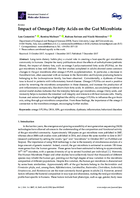
 Romina Molinari