Prenatal diagnosis and management of congenital cystic
adenomatoid malformation of the lung
Gregory J. Duncombe, FRANZCOG, DDU,a,b,c Jan E. Dickinson, FRANZCOG, DDU, CMFM,a,b,c
and Colin S. Kikiros, FRACSa,d
Perth, Australia
OBJECTIVE: Our purpose was to review the management and outcome of pregnancies with a prenatal diagnosis of fetal congenital cystic adenomatoid malformation of the lung (CCAM).
STUDY DESIGN: A retrospective review was performed of all cases since 1995 with a prenatal diagnosis of
fetal CCAM from the sole tertiary perinatal referral center in Western Australia.
RESULTS: Twenty-one pregnancies with CCAM were identified. The gestational age at diagnosis was <22
weeks in 86% of cases. Macrocysts were seen in 76% of cases during ultrasound examination. Seventeen
pregnancies continued until term. Regression of the sonographic appearances was observed in 19% of
cases. Fetal hydrops complicated two cases. One neonate died within 24 hours of delivery because of pulmonary hypoplasia. Twelve children have required pulmonary lobectomies. No adverse sequelae are evident
in surviving children.
CONCLUSION: Most cases of prenatally diagnosed CCAM have had a good outcome. This review has positively influenced the counseling of women with this diagnosis. (Am J Obstet Gynecol 2002;187:950-4.)
Key words: Congenital cystic adenomatoid malformation, prenatal diagnosis
Congenital cystic adenomatoid malformation of the
lung (CCAM) is an uncommon fetal anomaly with fewer
than 1000 cases reported in the medical literature. The
reported incidence of CCAM is approximately 1 in 25,000
pregnancies.1 This abnormality is believed to be the result of hamartomatous change in the tertiary bronchioles
or an arrest in their embryologic development between 7
and 15 weeks’ gestation.1-6
CCAM is observed as a cystic mass occupying part or all
of one fetal hemithorax, with up to 15% of cases having
bilateral involvement. The cysts within the mass may be
macrocystic (single or multiple cysts with diameters ≥5
mm) or microcystic (echodense homogeneous lung).7
The vascular supply for a CCAM arises from pulmonary
vessels. Some communication may occur between the
mass and surrounding normal lung tissue.
The ultrasound differential diagnoses are related to the
appearances of the cysts, the location of the lesion, the vas-
From the King Edward Memorial Hospital for Women,a the Women and
Infants Research Foundation,b the Department of Obstetrics and Gynaecology, University of Western Australia,c and the Department of Pediatric
Surgery, Princess Margaret Hospital.d
Presented at the Twenty-second Annual Meeting of the Society for Maternal-Fetal Medicine, New Orleans, La, January 14-19, 2002.
Reprint requests: Greg Duncombe, FRANZCOG, DDU, Department of
Obstetrics and Gynaecology, University of Western Ontario, Lawson
Health Research Institute, 268 Grosvenor St, London, Ontario N6A
4V2, Canada. E-mail: gduncomb@uwo.ca
© 2002, Mosby, Inc. All rights reserved.
0002-9378/2002 $35.00 + 0 6/6/127460
doi:10.1067/mob.2002.127460
950
cular supply as assessed by color Doppler analysis, and the
lesion’s effects on dependent tissues and structures. These
differential diagnoses include pulmonary sequestration,
bronchogenic cyst, diaphragmatic hernia, mediastinal cystic teratoma, and congenital pulmonary emphysema.2,6-8
In 1949, Chin and Tang9 first reported a case of CCAM.
The current descriptions are based on studies from
Stocker et al10 and Adzick et al.11 The Stocker classification is founded on the histologic appearances of cyst size
and the mixture of sizes within the specimen, whereas the
Adzick classification is based on sonographic appearances and cyst dimensions.
This review of the outcome of cases with a prenatal diagnosis of CCAM was stimulated by an apparent increase
in the incidence of this diagnosis within our institution.
The objectives of this study were to assess the accuracy of
the prenatal diagnosis of CCAM of the lung and the potential prognostic features and to review perinatal outcomes in relation to the current management practices at
our institution.
Material and methods
Twenty-one pregnancies with a prenatal diagnosis of
CCAM were identified between 1995 and 2001 at the
Fetal Medicine Service of King Edward Memorial Hospital for Women with follow-up at the Princess Margaret
Hospital for Children in Perth, Australia. These two institutions are the sole tertiary referral centers for perinatal
medicine and pediatric surgery within the state of Western Australia.
�Duncombe, Dickinson, Kikiros 951
Volume 187, Number 4
Am J Obstet Gynecol
A
Fig 2. Transverse ultrasound image of a 19-week fetus with a leftsided CCAM. The mass volume was >50% of the fetal thorax, and
the fetal heart was compressed and deviated to the right. Other
mass effects included tenting of the diaphragm and the production of gross ascites.
B
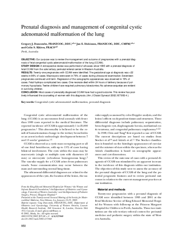
Fig 1. Examples of the ultrasound examination images of the different types of CCAM of the lung. A, Microcystic type. B, Mixed
macrocystic and microcystic type.
Table I. Obstetric and neonatal characteristics
Maternal age (y)
Parity
Gestational age at diagnosis (wk)
Period in utero after diagnosis (wk)*
Gestational age at delivery (wk)*
Birth weight (g)*
Apgar score, 5 min
30.5 (28-33)
1 (0-2)
20 (19-22)
18 (6-20)
39 (36-40)
3150 (2805-3510)
9 (9-10)
All data are displayed as median (interquartile range).
*In cases that didn’t undergo therapeutic termination.
Twelve cases had preliminary examinations at other
centers before referral to our unit for evaluation and
management. Data before 1999 was ascertained and assessed retrospectively through the Fetal Medicine Service
database.
On referral each fetus was evaluated sonographically,
and a CCAM classification was provided on the basis of
the ultrasound features evident. All ultrasound examinations were performed with an ATL 3000 or ATL 5000 machine (ATL, Philips Medical Systems, Bothell, Wash)
equipped with multihertz 5 to 2, 7 to 4 curvilinear transducers and a 7 to 4 vector transducer. Amniocentesis for
fetal karyotype was not performed routinely. In all but
one case, antenatal, neonatal, and pediatric care was performed within our tertiary institution. Complete data ascertainment was achieved.
Specific information collected included maternal age,
parity, gestational age at diagnosis, gestational age at delivery, and indication for delivery. Ultrasound examination details included referral base, gestational age,
biometry, appearance of lesion (side, laterality, size, type
[Adzick criteria])(Fig 1), cardiac axis deviation, polyhydramnios, appearance of hydrops, color Doppler analysis, and presence or absence of lesion regression.
Pediatric characteristics included birth weight, sex, Apgar
scores, postnatal imaging results, time and type of
surgery, surgical pathologic results, and general health.
In all cases involving termination of pregnancy or neonatal death, pathologic examination results were available.
Results
This series involved 21 pregnancies with a prenatal diagnosis of CCAM made or confirmed by our department
occurring over a 6-year period, 15 of which occurred in
the last 3 years, consistent with our clinical impression of
a sporadic change in the frequency of prenatal diagnosis.
The median maternal age at diagnosis was 30.5 years
(Table I). Most diagnoses were made before 22 weeks’
gestation and the fetal thoracic lesion was principally observed during the conduct of the second-trimester fetal
morphologic examination. Referrals at later gestational
ages were generated by smaller obstetric units and
prompted by abnormal pregnancy symptoms and signs
such as polyhydramnios.
By use of the Adzick descriptive classification, the majority of fetal CCAM lesions contained macrocysts (Table
II). This descriptive diagnosis was usually made on the
�952 Duncombe, Dickinson, Kikiros
October 2002
Am J Obstet Gynecol
Table II. Prenatal features of the CCAM lesions (n = 20)
Microcystic only
Macrocysts present
Right-sided lesion laterality
Cardiac deviation
Mass volume >50% of fetal thoracic volume
Incidence of spontaneous regression
Hydrops
5 (24%)
16 (76%)
13 (62%)
11 (52%)
5 (24%)
4 (19%)
2 (9%)
Data displayed as number (%).
Table III. Outcome of pregnancies and postnatal care
Live births
Termination of pregnancy
Neonatal deaths
Surgery (n = 16)*
Observation only
Pulmonary resection
Presurgery
17 (81%)
4 (19%)
1 (5%)
2 (12.5%)
12 (75%)
2 (12.5%)
Data displayed as number (%).
*Median (interquartile range) age at time of surgery was 265
days (12,280).
first ultrasound examination (primary or tertiary referral), although in a few cases a second examination was
necessary. No case had a bilateral thoracic mass, although
one large lesion initially gave that impression. The mass
filled half or more of the thorax in 29% of cases. Cardiac
axis deviation was observed in 52% of cases, virtually always at the time of initial diagnosis.
In two cases hydrops was present at the time of diagnosis. In the first case, the initial presentation was at 19
weeks’ gestation and the parents selected termination of
pregnancy after counseling (Figs 2 and 3). The second
case complicated by hydrops was a late referral at 30
weeks’ gestation, the ultrasound examination prompted
by severe polyhydramnios. Two therapeutic amnioreductions, each of 2-L volume, were performed on consecutive
days. After these procedures the fetal hydrops completely
resolved and the pregnancy progressed until term.
Four women elected therapeutic termination of pregnancy. In one case cyst aspiration was performed, but rapid
reaccumulation of fluid within the major cyst occurred and
the pregnancy was interrupted. The second case exhibited
extensive microcystic change with the impression of bilateral thoracic involvement. The third case involved earlyonset hydrops, as previously described. The fourth case
chose termination after diagnosis and counseling.
Seventeen women continued their pregnancies after
the diagnosis of fetal CCAM. Our prenatal management
strategy was based on serial sonographic assessment of the
fetus, with a median of four examinations until delivery. In
four cases there was ultrasound evidence of partial or
complete resolution of the thoracic mass before delivery.
All unterminated pregnancies reached term or near
term. Delivery timing was related to the individual obstet-
Fig 3. Fixed gross pathologic specimen of the CCAM, as shown in
Fig 2, demonstrating the macrocystic and microcystic changes.
This mass measured 5.9 ⫻ 3.8 ⫻ 2.9 cm. Both fetal lungs were severely hypoplastic for this gestational age.
ric indications. Each fetus was born in good condition
(Table I). In all cases postnatal imaging techniques concurred with the prenatal diagnosis. In two cases the neonatal condition deteriorated within hours of delivery. Severe
neonatal respiratory distress necessitating surgery on day
2 occurred in the case in which amnioreduction was required antenatally. This child has subsequently done well.
Another neonate had respiratory distress and cardiac failure and died on day 1 after delivery. Autopsy revealed a
CCAM within a pulmonary sequestration; this diagnosis
was suspected antenatally because of the presence of a
large feeding vessel from the descending thoracic aorta.
Pregnancy outcomes are described in Table III. Twelve
children have had elective pulmonary resections (because of a small risk of malignant change12,13 or infection
within the mass). The timing of surgery has been primarily related to the pediatric surgical opinion of the time.
Initially performed in the first 2 weeks of life, in the last 2
years a more conservative management strategy has been
developed with surgery delayed for months or not performed at all. In all but one case with a pathology specimen available for assessment, our diagnoses were correct.
Four cases are currently under observation, with the oldest child being 7 years old (complete regression on prenatal and postnatal imaging examinations). At the time
of writing, no long-term adverse sequelae were evident in
the surviving children.
Comment
The prenatal diagnosis of CCAM is uncommon. With
use of data obtained from the Western Australian Birth
Defects Registry, the incidence of CCAM in the state of
Western Australia during the period of our review was
around 1.2 per 10,000 births (a total of 28 cases with this
diagnosis, 2-6 per year). There were seven cases not in
�Duncombe, Dickinson, Kikiros 953
Volume 187, Number 4
Am J Obstet Gynecol
our series with a notification made to the state registry.
Two cases had terminations for multiple congenital
anomalies with a CCAM lesion found on autopsy. One
live birth at our institution (not examined in our department) and four other live births in other parts of the state
had the diagnosis made after delivery.
Before the advent of prenatal ultrasound imaging, the
diagnosis of CCAM had been made at autopsy or during
investigation of respiratory disease in childhood. The earliest recorded ultrasound imaging diagnosis of CCAM
was made in 1975.14 As with many types of congenital
anomalies, improvements in ultrasound technology have
led to an increase in the number of cases of CCAM diagnosed prenatally. In addition, the diagnoses are occurring at earlier gestational ages. The majority of cases
within our cohort were detected at the 18- to 20-week
fetal morphologic examination.
Reported prenatal prognostic features for CCAM include the size and type of the mass, laterality, progression
or regression of the mass, cardiac axis deviation, presentation with or development of hydrops, and the finding of
other anomalies.1,6,12,15 The larger the thoracic mass, the
worse the prognosis tends to be. The microcystic type
tends to have a more adverse prognosis. Lesions that
regress behave in a more benign fashion.1,16 Interestingly, these reported characteristics seemed to have had a
lesser impact on the final outcomes within our cohort.
This may be due to the small number of cases in our series and the option of termination taken in four cases.
The finding of other structural anomalies is considered
an indication for a karyotyping procedure. No fetuses in
this series had other structural anomalies detected on
ultrasound examination, and amniocentesis was not required in any case. No child was born with other structural anomalies.
The development of fetal hydrops, as in most fetal disease states, is a poor prognostic feature.1,7,11,15,17,18 Fetal
hydrops is thought to be the result of a mass effect by the
volume of the CCAM on fetal swallowing or of cardiac
compromise produced by compression of the heart or
major blood vessels. In this series there were two cases of
fetal hydrops with two opposing outcomes, as previously
described. Gestational age appeared the primary factor
dictating management.
We observed several cases of partial or complete regression of the pulmonary lesion during serial sonographic review. Resolution of CCAM is supported in the
literature.1,4,15,16,19 One difficulty with these cases was a
lack of confirmation of diagnosis as a result of there
being no pathologic specimen for analysis. In cases where
partial resolution has occurred antenatally, no further
resolution was evident in the neonatal period.4 Similar to
other recent publications, unless fetal hydrops persisted,
most pregnancies continued to term. In three pregnancies, the volume of the lesion as a proportion of the chest
volume appeared to stabilize or decrease. In these cases,
the changes resulting from mass effect such as cardiac
axis deviation tended to revert to be within normal limitations. This is suggestive of a plateau or peaking of the
mass’s growth velocity and a catch up in growth of surrounding structures.
Current neonatal and pediatric management at our institution involves stabilization and observation within the
neonatal nursery. In the presence of respiratory distress,
a computed tomographic examination is performed and
surgery is undertaken immediately. If otherwise well, the
computed tomographic examination is performed with
the patient under general anaesthesia at 4 to 6 months of
age, and surgery is arranged when the patient is between
6 and 12 months old. In some circumstances, typically in
an asymptomatic infant, surgery may be even further delayed to optimize surgical access (for example, in lesions
near the hilum). The longest delay from delivery to
surgery in our cohort has been just under 4 years. The
surgery usually consists of thoracotomy and lobectomy. If
the mass is more extensive, further limited partial resection is performed to avoid chest wall deformation. Currently, on regular pediatric review no long-term adverse
sequelae are evident in our survivors.
Summary
Fetal CCAM is a rare anomaly. An accurate diagnosis
may be made at 18 to 20 weeks’ gestation. Partial or complete regression was demonstrated on serial ultrasound
examinations in 19% of cases in our series. In the majority of cases in our series, delivery occurred at term and the
outcome was satisfactory. Current pediatric surgical practice suggests surgical excision of the lesion within the first
year of life.
We thank Karen Reid, Fetal Medicine Co-coordinator
at King Edward Memorial Hospital, Dr Adrian Charles,
Perinatal Pathologist for King Edward and Princess Margaret Hospitals, and Dr Caroline Bower, Director of the
Western Australian Birth Defects Registry, for their assistance in ascertainment of case details.
REFERENCES
1. Laberge JM, Flageole H, Pugash D, Khalife S, Blair S, Filiatraut
D, et al. Outcome of the prenatally diagnosed congenital cystic
adenomatoid lung malformation: a Canadian experience. Fetal
Diagn Ther 2001;16:178-86.
2. Sakala EP, Perrott WS, Grube GL. Sonographic characteristics of
antenatally diagnosed extralobar pulmonary sequestration and
congenital cystic adenomatoid malformation. Obstet Gynecol
Surv 1994;49:647-55.
3. Waszak P, Claris O, Lapillonne A, Picaud J, Basson E, Chappuis J,
et al. Cystic adenomatoid malformation of the lung: neonatal
management of 21 cases. Pediatr Surg Int 1999;15:326-31.
4. van Leeuwen K, Teitelbaum DD II, Hirschl RB, Austin E, Adelman S, Polley T, et al. Prenatal diagnosis of congenital cystic adenomatoid malformation and its postnatal presentation, surgical
indications, and natural history. J Pediatr Surg 1999;34:794-9.
�954 Duncombe, Dickinson, Kikiros
5. De Santis M, Masini L, Noia G, Cavaliere AF, Oliva N, Caruso A.
Congenital cystic adenomatoid malformation of the lung: antenatal ultrasound findings and fetal-neonatal outcome. Fifteen
years of experience. Fetal Diagn Ther 2000;15:246-50.
6. Adzick NS, Harrison MR, Crombleholme TM, Flake AW, Howell
LJ. Fetal lung lesions: management and outcome. Am J Obstet
Gynecol 1998;179:884-9.
7. Gembruch U, Holzgreve W. The fetus with nonimmune hydrops
fetalis. In: Harrison MR, Evans MI, Adzick NS, Holzgreve W, editors. The unborn patient. Volume 1. New York: WB Saunders;
2000. p. 525-82.
8. Kuller JA, Golbus MS. Fetal therapy. In: Brock D, Rodeck CH,
Ferguson-Smith MA, editors. Prenatal diagnosis and screening.
Hong Kong: Churchill Livingstone; 1992. p. 703-15.
9. Chin KY, Tang MY. Congenital adenomatoid malformation of
one lobe of a lung with general anasarca. Arch Pathol
1949;48:221-9.
10. Stocker JT, Madewell JE, Drake RM. Congenital cystic adenomatoid malformation of the lung: classification and morphologic
spectrum. Hum Pathol 1977;8:155-71.
11. Adzick NS, Harrison MR, Glick PL, Globus MS, Anderson RL,
Mahony BS, et al. Fetal cystic adenomatoid malformation: prenatal diagnosis and natural history. J Pediatr Surg 1985;20:483-8.
12. Pinter A, Kalman A, Karsza L, Verebely T, Szemledy F. Long-term
outcome of congenital cystic adenomatoid malformation. Pediatr Surg Int 1999;15:332-5.
October 2002
Am J Obstet Gynecol
13. Ozcan C, Celik A, Ural Z, Veral A, Kandiloglu G, Balik E. Primary
pulmonary rhabdomyosarcoma arising within cystic adenomatoid malformation: a case report and review of the literature.
J Pediatr Surg 2001;36:1062-5.
14. Garrett WJ, Kossoff G, Lawrence R. Gray scale echography in the
diagnosis of hydrops due to fetal lung tumor. J Clin Ultrasound
1975;3:45-50.
15. Bunduki V, Ruano R, da Silva MM, Migudez J, Miyadahira S,
Maksoud J, et al. Prognostic factors associated with congenital
cystic adenomatoid malformation of the lung. Prenat Diagn
2000;20:459-64.
16. Hsu KF, Wu MH, Chang CH, Yao BL, Chang FM. Complete intrauterine resolution of fetal congenital cystic adenomatoid
malformation of the lung type III. J Ultrasound Med
1995;14:871-5.
17. Adzick NS. The fetus with a cystic adenomatoid malformation.
In: Harrison MR, Golbus MJ, Filly RA, editors. The unborn patient. Volume 1. New York: WB Saunders; 1990. p. 320-7.
18. Taguchi T, Suita S, Yamanouchi T, Nagano M, Satoh S, Koyamagi
T, et al. Antenatal diagnosis and surgical management of congenital cystic adenomatoid malformation of the lung. Fetal
Diagn Ther 1995;10:400-7.
19. Monni G, Paladini D, Ibba RM, Teodoro A, Zoppi M, Lamberti A,
et al. Prenatal ultrasound diagnosis of congenital cystic adenomatoid malformation of the lung: a report of 26 cases and review of
the literature. Ultrasound Obstet Gynecol 2000;16:159-62.
�
 Greg Duncombe