Journal of Hazardous Materials 351 (2018) 29–37
Contents lists available at ScienceDirect
Journal of Hazardous Materials
journal homepage: www.elsevier.com/locate/jhazmat
Washable antimicrobial polyester/aluminum air filter with a high capture
efficiency and low pressure drop
T
Dong Yun Choia,1, Ki Joon Heob,c,1, Juhee Kangb, Eun Jeong Ana, Soo-Ho Junga, Byung Uk Leec,
⁎⁎
⁎
Hye Moon Leea,d, , Jae Hee Jungb,e,f,
a
Powder and Ceramics Division, Korea Institute of Materials and Science, Changwondaero 797, Seongsan-gu, Changwon, 51508, Republic of Korea
Center for Environment, Health, and Welfare Research, Korea Institute of Science and Technology (KIST), Hwarang-ro 14-gil 5, Seongbuk-gu, Seoul, 02792, Republic of
Korea
c
Aerosol and Bioengineering Laboratory, Department of Engineering, Konkuk University, Neungdong-ro 120, Gwangjin-gu, Seoul, 05029, Republic of Korea
d
Alink Co. Ltd., Chanwondaero 797, Seongsan-gu, Changwon, 51508, Republic of Korea
e
Green School, Korea University, Anam-ro 145, Seongbuk-gu, Seoul, 02841, Republic of Korea
f
Division of Energy & Environment Technology, KIST School, Korea University of Science and Technology, Hwarang-ro 14-gil 5, Seongbuk-gu, Seoul, 02792, Republic of
Korea
b
G RA P H I C A L AB S T R A C T
A R T I C L E I N F O
A B S T R A C T
Keywords:
Air filter
Antimicrobial filter
Bioaerosol
Conductive fiber
Particulate matter
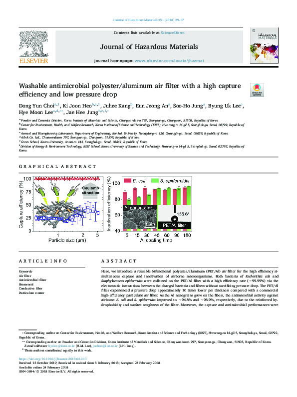
Here, we introduce a reusable bifunctional polyester/aluminum (PET/Al) air filter for the high efficiency simultaneous capture and inactivation of airborne microorganisms. Both bacteria of Escherichia coli and
Staphylococcus epidermidis were collected on the PET/Al filter with a high efficiency rate (∼99.99%) via the
electrostatic interactions between the charged bacteria and fibers without sacrificing pressure drop. The PET/Al
filter experienced a pressure drop approximately 10 times lower per thickness compared with a commercial
high-efficiency particulate air filter. As the Al nanograins grew on the fibers, the antimicrobial activity against
airborne E. coli and S. epidermidis improved to ∼94.8% and ∼96.9%, respectively, due to the reinforced hydrophobicity and surface roughness of the filter. Moreover, the capture and antimicrobial performances were
⁎
Corresponding author at: Center for Environment, Health, and Welfare Research, Korea Institute of Science and Technology (KIST), Hwarang-ro 14-gil 5, Seongbuk-gu, Seoul, 02792,
Republic of Korea.
⁎⁎
Corresponding author at: Powder and Ceramics Division, Korea Institute of Materials and Science, Changwondaero 797, Seongsan-gu, Changwon, 51508, Republic of Korea.
E-mail addresses: hyelee@kims.re.kr (H.M. Lee), jaehee@kist.re.kr (J.H. Jung).
1
These authors contributed equally to this work.
https://doi.org/10.1016/j.jhazmat.2018.02.043
Received 13 October 2017; Received in revised form 8 February 2018; Accepted 22 February 2018
Available online 24 February 2018
0304-3894/ © 2018 Elsevier B.V. All rights reserved.
�Journal of Hazardous Materials 351 (2018) 29–37
D.Y. Choi et al.
stably maintained during a cyclic washing test of the PET/Al filter, indicative of its reusability. The PET/Al filter
shows great potential for use in energy-efficient bioaerosol control systems suitable for indoor environments.
1. Introduction
antimicrobial activity, and adsorption of gaseous pollutants. However,
multiple layers of filters result in high pressure drops along with long
airflow pathways, and require more energy consumption and frequent
filter replacement [23]. Therefore, considerable attention is being devoted to the development of multifunctional air filters that integrate at
least two layers with different functions into a single layer [24,25].
To enable high-efficiency PM capture, fibrous filters should be
thicker or be composed of densely packed nanofibers. Such filter
structures inevitably increase airflow resistance, consequently leading
to greater energy consumption [26,27]. However, if electrostatic forces
between the particles and fibers are established, the capture efficiency
can be improved substantially without an increase in filter pressure
drop [28]. Recently, our group reported a conductive polyester/aluminum (PET/Al) fibrous filter showing high-efficiency electrostatic PM
removal (∼99.99% for 30–400 nm particles) with a low pressure drop
[29]. Because the PET/Al filter is electrically conductive, strong electric
fields can be created around the filter by directly introducing electric
potential, and the charged PM is effectively deposited onto the fibers
via Coulomb forces. There have been several reports of the mild antimicrobial activity of alumina (Al2O3) particles against microorganisms
[30–32]. In addition, thin oxide layers (3–10 nm) form on the Al nanostructures of the PET/Al filter, which could confer antimicrobial
activity. Improving the antimicrobial properties of the PET/Al filter via
the structural control of the Al layers would be of great significance to
support its potential as a bifunctional filter for total air quality treatment.
In this study, we demonstrate the bifunctionality of the PET/Al filter
regarding its high effective capture and inactivation of airborne bacteria. This filter showed a pressure drop 10 times lower per thickness
compared with a commercial high-efficiency particulate air (HEPA)
filter. Both Escherichia coli and Staphylococcus epidermidis were captured
on the PET/Al filter with a high efficiency of ∼99.99% via electrostatic
forces. The antimicrobial activity of the filter itself for each species was
increased to ∼94.8% and ∼96.9%, respectively, due to the enhanced
Bioaerosols are classified as airborne biological particulate matter
(PM), including microorganisms (e.g., viruses, bacteria, and fungi),
biological particulate fragments, and toxins. Individual bioaerosols
range in size from submicroscopic particles (< 0.01 μm) to particles
larger than 100 μm, which are readily transmitted by wind and can float
for a long time in the atmosphere [1–3]. Because they can be inhaled or
attach to humans in their airborne state, they are an etiological agent
for respiratory and infectious human diseases [4]. Thus, the control of
microorganisms suspended in air is currently an active research field
driven by the increasing demand for occupational and public health
safety [5–7].
Fibrous filters are used widely as a method for removing hazardous
bioaerosols due to their fascinating features, such as a light weight,
cost-effectiveness, easy of fabrication, and universal applicability
[1,8–10]. However, microorganisms that are captured in such filter
media can remain viable, and some can even grow and propagate by
absorbing air moisture and nutrients in dust. These organisms can become resuspended in the air upon the deterioration of the filters or
accidental physical impact during maintenance [11]. In addition, volatile organic compounds, an indoor air carcinogen produced by microbial metabolism, can be emitted [12]. Hence, many efforts have
been made to inactivate biological PM by depositing antimicrobial
components onto filter fibers, such as inorganic materials (e.g., Ag, Cu,
and TiO2 nanoparticles (NPs)) and organic materials (e.g., Sophora
flavescens, Euscaphis japonica, and tea tree oil NPs) [13–20]. However,
because antimicrobial NPs physically adhere to the fiber surfaces, these
filters show poor durability against washing treatments for reuse.
Moreover, the antimicrobial activity of the filters progressively diminishes due to the accumulation of dust covering the functional particles [21,22].
Modern air-cleaning devices are composed of multiple filters with
different functions that support high-efficiency PM capture,
Fig. 1. (a) Diagram of the chemical solution process for conductive polyester/aluminum (PET/Al) filter fabrication. (b) Configuration of the electrostatic filtration device composed of a
carbon fiber ionizer and two PET/Al filters. Electric fields are formed between the front filter and the ionizer as well as the back filter and the front filter. Inflowing particles are negatively
charged by the ionizer, and are captured by Coulomb forces towards the front PET/Al filter. (c) Schematic diagram of the experimental setup used in the filtration tests. (For interpretation
of the references to colour in this figure legend, the reader is referred to the web version of this article).
30
�Journal of Hazardous Materials 351 (2018) 29–37
D.Y. Choi et al.
(particles/cm3) of the bacterial bioaerosol at the inlet and outlet of the
filter, respectively. The size and number concentration of bacterial
bioaerosols were measured with an aerodynamic particle sizer (Model
3321, TSI Inc.) at both the inlet and outlet of the electrostatic filtration
device marked with a dashed blue box in Fig. 1c. The airflow face velocity was fixed to 3.4 cm/s for all filtration tests unless noted otherwise. To eliminate any effects on time-dependent particle generation,
efficiency measurements were conducted more than four times for each
experimental condition.
surface roughness and wettability of the chemically grown Al layer.
More importantly, the capture and antimicrobial performances remained high during repeated reuse of the PET/Al filter after washing
with water. This work presents a new approach for the simultaneous
removal and inactivation of biological PM, which is attractive for lowcost, energy-efficient air quality applications.
2. Materials and methods
2.1. Al-coated conductive fibrous filter fabrication
2.5. Antimicrobial test
A PET nonwoven filter (fiber diameter = 30 μm; porosity = 78.3%)
was used as a backbone membrane to fabricate the PET/Al filter. Fig. 1a
illustrates the chemical solution (CS) process, which included two steps:
(i) catalytic treatment of the PET filter by titanium isopropoxide (Ti(Oi-Pr)4) and (ii) dip-coating of the catalytically treated filter into an Al
precursor ink, AlH3{O(C4H9)2}, which was synthesized via an ethereal
reaction of aluminum chloride (AlCl3) with lithium aluminum hydride
(LiAlH4) in dibutyl ether (O(C4H9)2). More detailed explanations of the
Al precursor ink preparation and CS process are described in our previous work [29,33–36].
After the bacteria were deposited onto the PET/Al filter for about
10 min, they were left for an additional contact time of 5 min. Then, the
filters were placed in 15 mL (Vextraction) of phosphate-buffered saline
(PBS) containing 0.01% Tween 80 and vortexed for 5 min to transfer
the bacteria from the filters to the PBS solution. The resulting bacterial
suspension was serially diluted onto plates containing nutrient agar
(catalog no. 213000, Becton Dickinson) and incubated at 37 °C for 24 h.
The colonies that grew on the plates were counted. The bacterial inactivation efficiency (ε) was calculated as follows:
2.2. Electrostatic filtration device design
λPET =
Fig. 1b presents a sketch of the electrostatic filtration device used in
this study. The device was composed of a carbon fiber ionizer to charge
bioaerosols and two conductive PET/Al filters to capture them. The
ionizer was positioned 5 cm ahead of the front of the PET/Al filter. A
constant voltage of −10 kV was applied to the ionizer when negative
ion generation was required. Thus, the bacterial aerosols gained negative charges before passing through the PET/Al filters. PET/Al filters
were installed on both sides of a 5-mm-thick plastic separator, and each
filter was connected to a metal electrode to introduce electric potential.
The front PET/Al filter was connected to a positive high-voltage source
as required; the back PET/Al filter was grounded for all filter tests.
λPET/Al =
⎜
(3)
Cinlet⋅Qsampling⋅η⋅ζ extraction
Vextraction
λPET/Al ⎞
,
ε = ⎛1 −
λPET ⎠
⎝
,
(4)
⎟
(5)
where λPET and λPET/Al are the active proportions of bacteria from
the PET filter (control) and PET/Al filter, respectively. N is the total
concentration of bacteria (particles/mL) in the extraction suspension
plated onto the agar. Cinlet is the total concentration of airborne bacteria
from the nebulizer. Qsampling is the total airflow sampling volume.
ζextraction is the physical extraction of bacteria at each filter, defined as
the ratio of the number of particles transferred from the filter to the
extraction liquid to the number of particles removed from airflow using
the filter. The values of ζextraction for all filter samples were as high as
∼96%, comparable to those obtained using the method proposed by
Wang et al. [37].
To evaluate the capture and inactivation efficiency of the PET/Al
filter, we selected two species of bacteria with different cell wall
structures, S. epidermidis (Gram-positive bacteria) and E. coli (Gramnegative bacteria). These bacteria are commonly found in indoor environments and on human skin, and are used widely in bioaerosol research. The S. epidermidis and E. coli cultures were incubated in a nutrient broth medium (catalog no. 234000, Becton Dickinson) at 37 °C
for 24 h and 18 h, respectively. When the bacterial suspension reached
an optical density at 600 nm of ∼0.8, the bacteria were harvested by
centrifugation (5000 × g, 10 min) and washed three times with distilled
water. Once the concentration of the resulting suspension reached
∼108 colony forming units (CFU)/mL, 1 mL of bacterial suspension was
mixed with 19 mL of distilled water and loaded into a one-jet Collison
Nebulizer (CN 241, BGI Inc.).
2.6. Characterization
Field-emission scanning electron microscopy (FE-SEM) images and
energy dispersive spectroscopy (EDS) mapping results were acquired
using a scanning electron microscope (SU8230, Hitachi). Sheet resistance measurements of the PET/Al filters were carried out using the
four-probe van der Pauw method (FPP-HS8, DASOLENG). The pressure
drop between the upstream and downstream sides of the PET/Al filter
was measured with an electronic manometer (FCO332, Furness
Controls) with a detection range of 100 Pa. The porosity, average pore
diameter, and pore size distribution of the filters were determined using
the mercury intrusion porosimetry (MIP) analysis (Micromeritics
AutoPore V 9600, Micromeritics Corp.). The air permeability of the
filters was evaluated with an air permeability tester (FX 3300,
TEXTEST) at a constant pressure drop of 125 Pa. Ozone emissions at the
outlet of the electrostatic filtration device were analyzed for various
electric field conditions using an ultraviolet (UV) photometric ozone
monitor (Model 49C, Thermo Environment Instruments Inc.) under a
sampling flow rate of 1.0 L/min. The static water contact angle (WCA)
of the filters was determined following the sessile drop method using a
contact angle analyzer (Phoenix 300 Plus, SEO Co., Ltd.). 2010).
2.4. Filtration test
Fig. 1c presents a schematic diagram of the apparatus used to
measure the capture efficiency of PET/Al filters. Droplets containing
test bacteria were atomized using the nebulizer supplied with 1.0 L/min
airflow under 1.0 psig. Before the bacterial bioaerosols were introduced
to the filter medium, moisture was thoroughly removed by passing
through a diffusion dryer. The capture efficiencies (η) of the PET/Al
filters were calculated using the following equation:
Coutlet
,
Cinlet
(2)
CFUPET/Al
,
NPET/Al
NPET orNPET/Al =
2.3. Bacterial aerosol preparation
η=1−
CFUPET
,
NPET
(1)
where Cinlet and Coutlet represent the particle concentrations
31
�Journal of Hazardous Materials 351 (2018) 29–37
D.Y. Choi et al.
3. Results and discussion
that affect filter performance. We evaluated the characteristics of the
pore structure and the air permeability of the PET/Al filter, and the
corresponding results were compared with those of the raw PET filter.
Fig. S1 shows pore size distribution from MIP analysis for the PET filter
and the PET/Al filter. Both filters showed a pore size distribution
consisting of air pores without mesopores and capillary pores. The
pores in each filter are mainly distributed between 30 and 300 μm. The
PET filter has a porosity of 75.9% and an average pore size of 105.1 μm,
quite similar to those of the PET/Al filter; the porosity and average pore
size of the PET/Al filter are 75.0% and 119.8 μm, respectively. The air
permeability of PET filter was 303 cm3/cm2/s, and that of PET/Al filter
was 296 cm3/cm2/s. Above results imply that our CS process for the
creation of Al thin layers can retain the integrity of the original porous
structure of the PET filters.
3.1. Conductive PET/Al fibrous filter preparation
The PET/Al filters were produced following the CS process developed in our previous work [29]. Fig. 2a describes the formation mechanism of the Al nanostructures on each unit fiber during the CS
process. The PET filter treated catalytically with Ti(O-i-Pr)4 was immersed in the Al precursor ink composed of AlH3{O(C4H9)2}. AlH3
solvated in dibutyl ether begins to decompose directly into Al and 1.5
H2 at the fiber surface with the assistance of the catalyst, which enables
the creation of conductive Al layers at room temperature. Al nuclei are
produced ubiquitously over the fiber surface and grow gradually until
they completely cover the fiber surface.
The existence of Al features coating the fiber surface can be easily
distinguished by the naked eye, as the color of the raw PET filter
changes from white to metallic gray after the chemical reaction of the
Al precursor ink (left images, Fig. 2b and c). No severe damage to the
fibers was observed and the network structure of the fibers remained
(Fig. 2b-ii and c-ii). The magnified SEM images in Fig. 2b-iii and c-iii
show the fiber surfaces of the PET and PET/Al filters, respectively. The
bumpy surface of the PET/Al fibers was clearly differentiated from the
smooth surface of the PET fibers.
Fig. 2d presents the EDS mapping images of the PET/Al filter.
Carbon (cyan dots) and aluminum (yellow dots) were distributed
evenly over the fiber surface, confirming the successful formation of Al
nanostructures on the fibers via the CS process.
Porosity and distribution of pore sizes are important characteristics
3.2. Electrical and airflow resistance properties
To demonstrate the tunable electrical properties of the PET/Al filter,
we investigated the variation in electrical resistance according to the
immersion time in the Al precursor ink. Fig. 3a displays the sheet resistance values of the PET/Al filters and the corresponding areal loading
mass of Al as a function of coating time. The quantity of Al deposited on
the fibers increased linearly with increasing time, exhibiting a constant
formation rate of Al features on the fiber surface. The growth rate of the
Al thin film decelerated as the surface increased. Therefore, the sheet
resistance decreased rapidly during a reaction time of < 45 min, but the
resistance change slowed thereafter. The PET/Al filter had a somewhat
high resistance of ∼770 Ω/sq at an immersion time of 15 min (PET/
Fig. 2. (a) Schematic illustration of the formation mechanism of Al thin films on the filter fibers. (b) Photograph of a 15 cm × 15 cm raw PET filter (left); scanning electron microscopy
(SEM) image showing its fiber structure (middle) and magnified SEM image of the fiber surface (right). (c) Photograph of a 15 cm × 15 cm PET/Al filter with size of (left); SEM image
showing its fiber structure (middle) and magnified SEM image of the fiber surface (right). (d) SEM image of the PET/Al fibers (top), and the corresponding energy dispersive spectroscopy
mapping images of carbon (middle) and aluminum (bottom). Scale bars represent 20 μm. (For interpretation of the references to colour in this figure legend, the reader is referred to the
web version of this article).
32
�Journal of Hazardous Materials 351 (2018) 29–37
D.Y. Choi et al.
Fig. 3. (a) Variations in the electrical sheet resistance (red circles) and Al loading mass (blue
squares) of the PET/Al filters according to the immersion time in the Al precursor ink. (b) Pressure
drop versus flow rate characteristics of the PET/Al
filters prepared at different coating times (0, 15, 30,
and 45 min; Raw PET, PET/Al–15, PET/AL–30, and
PET/Al–45, respectively). (For interpretation of the
references to colour in this figure legend, the reader
is referred to the web version of this article).
3.3. Bacterial capture efficiency
Al–15); however, its resistance dropped to 1.1 Ω/sq after 180 min (PET/
Al–180).
Zhang et al. [38] reported that 50% of energy costs are related to
heating, ventilation, and air conditioning systems and that 30% of these
costs are associated with air filtration in commercial buildings. To save
the energy used for protecting air quality, it is important to minimize
the pressure drop across a filter while ensuring high capture efficiency,
given that the flow resistance is related to the amount of energy consumed by a fan. Fig. 3b shows the pressure drop curves of the raw PET,
PET/Al, and commercial HEPA filters as a function of the airflow rate.
The face velocity was set in the range of 1.5–7.5 cm/s to include the
standard airflow velocity of 5.33 cm/s suggested by the US Department
of Energy (DOE) [39,40]. The PET/Al filters were prepared at coating
times of 15, 30, and 45 min. Because the pressure drop of a filter is
directly proportional to its thickness, the measured pressure drop data
were normalized to the thickness of each sample. The PET and PET/Al
filters were about 0.25 mm thick, while the HEPA filter was about
0.7 mm thick. The airflow resistance characteristics of the PET/Al filters
did not differ greatly from that of the PET filter even after the formation
of the Al thin layer. Moreover, the pressure drop per thickness of the
PET/Al filters was roughly 10 times lower than that of the HEPA filter,
showing superior air permeability.
The capture performance of the PET/Al filters was evaluated using
E. coli and S. epidermidis bioaerosols. Fig. S2 depicts the size distribution
curves of the two species, which showed similar trends. Fig. 4a shows
the capture efficiency of E. coli as a function of size. When uncharged E.
coli was introduced to the electrically grounded PET/Al–180 filter, E.
coli was removed with an efficiency of ∼31%, which was mainly driven
by the mechanical filtration mechanism. Because the PET/Al filter was
electrically conductive, the filter gained a high electric potential via the
external high-voltage device, and strong electric fields were generated
around the filter fibers. Thus, charged bioaerosols were effectively
captured by the fibers via by electrostatic attraction. To examine the
electrostatic capture performance, we applied voltages of −10 kV
and + 10 kV to the ionizer and the front PET/Al filter, respectively. The
capture efficiency of the charged E. coli was significantly augmented by
Coulomb forces, and the average efficiency increased to about 99.99%.
The overall capture performance for S. epidermidis did not differ greatly
from that of E. coli since they had similar size properties (Fig. S3). The
application of a high electric field was beneficial for augmenting the
electrostatic capture efficiency, but could cause a harmful level of
ozone generation from the ionizer. The ozone concentration was about
1.8 ppb when voltages of −10 kV and +10 kV were applied to the ionizer and the PET/Al filter, respectively (Fig. S4), far below 50 ppb,
which is the standard for electrostatic air cleaners (UL 867) [41].
Fig. 4. (a) Capture efficiency of the PET/Al–180 filter
depending on particle size of E. coli cells. (b) Bar
graph showing a comparison of the E. coli capture
efficiencies for the PET/Al filters fabricated at various
coating times. SEM images of the deposition morphology of E. coli collected by (c) mechanical filtration and (d) electrostatic filtration. Each inset shows
a magnified image of the region in the orange rectangular box.
33
�Journal of Hazardous Materials 351 (2018) 29–37
D.Y. Choi et al.
As demonstrated above, the quantity of Al on the fibers increased
proportionally with coating time, thereby improving electrical conductivity. Therefore, we investigated the filtration performance of the
conductive PET/Al filters depending on the degree of Al loading. Fig. 4b
shows the E. coli capture efficiency of the PET/Al filters prepared at
various immersion times in Al precursor ink. The efficiencies of the
PET/Al filters driven by the mechanical filtration mechanism were similar regardless of filter type, indicating that an increase in Al layer
thickness had a minimal effect on the filtration characteristics. However, the PET/Al filters conferred excellent electrostatic capture performance, independent of the electrical properties, once the electrical
networks were formed due to the formation of Al nanostructures on all
fiber surfaces. Regardless, no relationship between the capture performance for the two bacteria species and immersion time was identified,
and the difference was not significant (p-value > 0.05).
Fig. 4c and d show the difference in the deposition density of E. coli
bacteria captured on the PET/Al filters by mechanical and electrostatic
forces during the same filtration time, respectively. Bacteria were deposited sparsely onto the fibers when driven predominantly by mechanical forces. However, substantially more bacteria accumulated on
the fiber surfaces when driven by electrostatic attraction, and dendrites
of bacteria formed (inset, Fig. 4d). SEM analysis results visually convey
the outstanding electrostatic capture ability of the PET/Al filter.
from 89.3% to 94% when the Al coating time increased from 5 min to
15 min. Beyond 15 min, the performance showed no significant correlation (p-value > 0.05) with the degree of Al deposition, since it was
already saturated (> 94%). However, the efficiency of S. epidermidis
gradually improved from 79.7% to 96.9% with increasing Al coating
time. The images in Fig. 5b correspond to the cultures of both bacteria
sampled from each of the raw PET (control), PET/Al–15, and PET/
Al–180 filters; the results showed that the PET/Al filter has strong antimicrobial activity. Because Gram-positive S. epidermidis has a greater
resistance to air exposure than Gram-negative E. coli, the natural decay
of E. coli is more dominant than S. epidermidis [42,43]. Thus, the
number of S. epidermidis colonies cultured from the control filter was
always higher than that of E. coli colonies, resulting in a smaller λPET
value for E. coli. Although the inactivation performance of E. coli was
somewhat overestimated when considering the above points, the PET/
Al–180 filter showed better antimicrobial properties against S. epidermidis (96.9 ± 0.59%) than E. coli (94.8 ± 1.18%). The difference
in the antimicrobial resistance to E. coli and S. epidermidis may be due to
their dissimilar cell structures and physiologies; Gram-negative bacteria
have a cell membrane structure capable of resisting antimicrobial
agents [44,45], while the cell walls of Gram-positive bacteria can bind
larger quantities of several metals than the cell envelopes of Gram-negative bacteria [46].
3.4. Antimicrobial performance of the PET/Al filter
3.4.2. Antimicrobial mechanism of the PET/Al filter
One of the most important mechanisms by which cells adapt to
surrounding environments is their adhesive interaction with solid surfaces. The initial bacterial adhesion process is regulated by electrostatic
interactions, which is enhanced when the cell wall of a bacterium lays
flush against a filter surface. A filter surface with nanoscale roughness
3.4.1. Evaluation of the inactivation efficiency of the PET/Al filter
We investigated the antimicrobial activities of the PET/Al filter
based on the quantity of Al generated on the PET fibers, as shown in
Fig. 5a. The inactivation efficiency against E. coli increased slightly
Fig. 5. (a) Bacterial inactivation efficiencies for E.
coli (red bars) and S. epidermidis (green bars) of the
PET/Al filters prepared at different Al coating times.
(b) Digital images of recultivated E. coli colonies and
S. epidermidis colonies on agar culture plates. Changes
in the surface morphology of the PET/Al filter with Al
deposition times of (c) 15 min, (d) 45 min, (e) 90 min,
and (f) 180 min. Each inset indicates the magnified
SEM image of a fiber surface and the scale bars represents 2 μm. (g) Water contact angle (WCA) of the
PET/Al filters according to Al coating time. (h)
Images of the static WCA measurements performed
on the raw PET filter (left) and PET/Al–180 filter
(right). (i) Images describing the dynamic water adhesion behavior on the surface of the PET/Al–180
filter. (For interpretation of the references to colour
in this figure legend, the reader is referred to the web
version of this article).
34
�Journal of Hazardous Materials 351 (2018) 29–37
D.Y. Choi et al.
antimicrobial efficiencies for E. coli and S. epidermidis were improved to
∼75% and ∼85%, respectively, for an ion exposure time of 10 min. In
our study, however, the inactivation performance of the PET/Al filter
had already been saturated (> 98%) by the antimicrobial activity of the
Al nanostructures and electric filed before introducing a negative ion
treatment. Thus, these factors seem to conceal the effect of negative
ions on the antimicrobial performance. Also, it is possible that antimicrobial activity by the negative ions could be worked during transportation of bacteria to the filter medium. However, the short exposure
(less than 2 s) of bacteria to negative ions would be insignificant to
antimicrobial performance, as supported by the results of Lee et al.
[57].
could prevent close contact with the cell wall due to the relative rigidity
of the cell wall [47]. Thus, the nanorough surface could hinder the
preliminary adhesion step of bacteria, and consequently lead to apoptosis [48]. Fig. 5c–f show SEM images in sequence for the PET/Al filters
prepared at coating times of 15, 45, 90, and 180 min. The filter surface
became increasingly bumpy with longer coating times, and the size of
the Al nanograins increased. Based on these results, the enhanced
roughness of the PET/Al filter could augment its antimicrobial performance.
In addition, the morphological changes in the fiber surface could
influence the hydrophobicity of the filter, resulting in the interference
of cell-substratum interactions during proliferation and differentiation
processes [49]. To examine the wettability of the PET/Al filters depending on their surface morphology, the WCA was measured (Fig. 5g).
The contact angle of water droplets on the PET/Al filter monotonically
increased with Al loading content. For example, the WCA of the raw
PET filter was 108.4°, whereas the PET/Al–180 filter had a higher WCA
of 135.6°, indicative of enhancement of the hydrophobicity (Fig. 5h).
Fig. 5i displays an image of a 3–μL water droplet during dynamic
contact with the surface of the PET/Al–180 filter. When detaching from
the filter, the droplet was not transferred to the filter and maintained its
original shape, revealing extremely low water adhesion of the PET/
Al–180 filter surface [50]. This reinforced hydrophobicity may be attributed to the increased grain size and surface roughness of the Al
nanostructures on the PET fibers.
Together with physical antimicrobial activities, another important
mechanism driving cell death is the disruption of cell walls by reactive
oxygen species (ROS) generated from metal oxides, such as ZnO2 and
TiO2 [30,51,52]. Several studies have demonstrated that Al2O3 NPs
show mild antimicrobial properties. For example, Jiang et al. [31] observed antimicrobial activities of Al2O3 NPs against both Gram-positive
and Gram-negative bacteria under dark conditions, indicating the
possible production of free radicals under dark conditions [53]. Sadiq
et al. [32] found that Al2O3 NPs had a minor antimicrobial effect on E.
coli at high concentrations of Al2O3 NP up to 1000 μg/mL. These results
support the hypothesis that the high antimicrobial activity of our PET/
Al filter may be ascribed to both the physical (surface roughness and
hydrophobicity) and chemical (ROS and free radicals) characteristics of
the Al nanostructures formed via the CS process.
3.6. Filter reusability
To support economical maintenance, filters should be washable to
enable the recovery of their functions. We examined the performance
changes of the PET/Al filters under a cyclic washing test. Each cycle
consisted of the filtration and antimicrobial experiments, cleaning of
the test filter in an ultrasonic water bath for 10 min, UV irradiation for
10 min, and complete drying in an electric oven at 50 °C for 2 h. Fig. 7
shows the variations in the capture and inactivation performances for
both E. coli and S. epidermidis during five cycles of the washing test. The
capture efficiency was stably maintained regardless of the bacteria
species species (E. coli, 99.4 ± 0.80%; S. epidermidis, 99.6 ± 0.77%)
(Fig. 7a). Though the results are not shown here, the pressure drop
characteristic did not change during a cyclic washing test. Furthermore,
during the reusability test, the reused filter still showed high antimicrobial activities against both bacteria with no apparent degradation
of function; E. coli and S. epidermidis were inactivated with efficiencies
of 99.2 ± 0.28% and 98.8 ± 1.3%, respectively (Fig. 7b). The PET/Al
filter retained its bifunctionality even after the cleaning process owing
to the robust durability of the chemically grown Al structures on the
PET fibers. These results demonstrate the reusability of our PET/Al
filter after washing with water.
4. Conclusions
We present a bifunctional PET/Al fibrous filter showing good performance regarding the electrostatic capture and inactivation of airborne microorganisms. The PET/Al filter captured bacterial bioaerosols
of E. coli and S. epidermidis with an extremely high efficiency due to the
electrostatic attraction between the filter and bacteria, with a pressure
drop much lower than that of a commercial HEPA filter. The capture
performance of the PET/Al filter was independent of the physical
properties of the Al thin layers, such as its electrical resistance, surface
roughness, and wettability. However, the antimicrobial activity increased with increasing surface roughness and hydrophobicity. The
effects of the application of an electric field and negative ions on the
3.5. Effects of electric field and charge on antimicrobial performance
We examined the changes in the bacteria inactivation performance
of the PET/Al filter when electric fields and charges were introduced.
Fig. 6 shows the inactivation efficiencies for E. coli and S. epidermidis
according to the onset of an electric field around the filter and the
electrical charging of bacteria. The PET/Al–180 filter was used as the
test sample. For comparison, its intrinsic inactivation performance is
included in Fig. 6 for the case that electric field and charges were not
applied (left two bars). When a high electric potential of 10 kV was
applied to the filter (middle two bars), the inactivation efficiency for E.
coli increased from 94.8% to 98.7%, while that for S. epidermidis increased slightly from 96.9% to 98.2% (p-value < 0.001). The application of the electric field was more effective in E. coli because Grampositive bacteria are less sensitive to electric field effects than Gramnegative bacteria [54]. The survival rate of exposed bacteria depends
on a combination of the electric field strength and treatment time [55];
however, it is currently unknown whether the antimicrobial mechanism
on electric field effects is derived from structural damage or metabolic
dysfunction of bacteria.
When negative charges were additionally applied to bacteria (right
two bars), the inactivation performance was very slightly increased.
However, the results were not statistically significant (t-test pvalue > 0.05). This outcome is somewhat different with previous
studies of the antimicrobial effect of negative ions against bacteria
deposited on a normal air filter. Kim et al. [56] reported that the
Fig. 6. Comparison of the inactivation efficiency of the PET/Al–180 filter according to the
filtration method.
35
�Journal of Hazardous Materials 351 (2018) 29–37
D.Y. Choi et al.
[3] G.J. Smith, D. Vijaykrishna, J. Bahl, S.J. Lycett, M. Worobey, O.G. Pybus, S.K. Ma,
C.L. Cheung, J. Raghwani, S. Bhatt, Origins and evolutionary genomics of the 2009
swine-origin H1N1 influenza A epidemic, Nature 459 (2009) 1122–1125.
[4] C.E. Main, Aerobiological, ecological, and health linkages, Environ. Sci. Technol. 29
(2003) 347–349.
[5] W.W. Nazaroff, Indoor bioaerosol dynamics, Indoor Air 26 (2016) 61–78.
[6] J. Douwes, P. Thorne, N. Pearce, D. Heederik, Bioaerosol health effects and exposure assessment: progress and prospects, Ann. Occup. Hyg. 47 (2003) 187–200.
[7] J.E. Lee, B.U. Lee, G.N. Bae, J.H. Jung, Evaluation of aerosolization characteristics
of biocontaminated particles from flood-damaged housing materials, J. Aerosol Sci.
106 (2017) 93–99.
[8] W.J. Fisk, Health benefits of particle filtration, Indoor Air 23 (2013) 357–368.
[9] B. Stephens, J. Siegel, Ultrafine particle removal by residential heating, ventilating,
and air‐conditioning filters, Indoor Air 23 (2013) 488–497.
[10] J.S. Kang, H. Kim, J. Choi, H. Yi, S.C. Seo, G.N. Bae, J.H. Jung, Antimicrobial air
filter fabrication using a continuous high-throughput aerosol-based process, Aerosol
Air Qual. Res. 16 (2016) 2059–2066.
[11] B.U. Lee, Life comes from the air: a short review on bioaerosol control, Aerosol Air
Qual. Res. 11 (2011) 921–927.
[12] R. Simmons, S. Crow, Fungal colonization of air filters for use in heating, ventilating, and air conditioning (HVAC) systems, J. Ind. Microbiol. Biotechnol. 14
(1995) 41–45.
[13] J.P. Ruparelia, A.K. Chatterjee, S.P. Duttagupta, S. Mukherji, Strain specificity in
antimicrobial activity of silver and copper nanoparticles, Acta Biomater. 4 (2008)
707–716.
[14] J.H. Jung, G.B. Hwang, J.E. Lee, G.N. Bae, Preparation of airborne Ag/CNT hybrid
nanoparticles using an aerosol process and their application to antimicrobial air
filtration, Langmuir 27 (2011) 10256–10264.
[15] K. Donaldson, R. Aitken, L. Tran, V. Stone, R. Duffin, G. Forrest, A. Alexander,
Carbon nanotubes: a review of their properties in relation to pulmonary toxicology
and workplace safety, Toxicol. Sci. 92 (2006) 5–22.
[16] G.N. Bae, J.H. Jung, Aerosol-processed nanomaterials for antimicrobial air filtration, J. Nanosci. Nanotechnol. 5 (2016) 4487–4492.
[17] J.H. Jung, G.B. Hwang, S.Y. Park, J.E. Lee, C.W. Nho, B.U. Lee, G.-N. Bae,
Antimicrobial air filtration using airborne Sophora flavescens natural-product nanoparticles, Aerosol Sci. Technol. 45 (2011) 1510–1518.
[18] G.B. Hwang, K.J. Heo, J.H. Yun, J.E. Lee, H.J. Lee, C.W. Nho, G.N. Bae, J.H. Jung,
Antimicrobial air filters using natural euscaphis japonica nanoparticles, Plos One 10
(2015) e0126481.
[19] G.B. Hwang, K.M. Sim, G.N. Bae, J.H. Jung, Synthesis of hybrid carbon nanotube
structures coated with Sophora flavescens nanoparticles and their application to
antimicrobial air filtration, J. Aerosol Sci. 86 (2015) 44–54.
[20] J. Choi, B.J. Yang, G.N. Bae, J.H. Jung, Herbal extract incorporated nanofiber
fabricated by an electrospinning technique and its application to antimicrobial air
filtration, ACS Appl. Mater. Interfaces 45 (2015) 25313–25320.
[21] Y.H. Joe, W. Ju, J.H. Park, Y.H. Yoon, J. Hwang, Correlation between the antibacterial ability of silver nanoparticle coated air filters and the dust loading,
Aerosol Air Qual. Res. 13 (2013) 1009–1018.
[22] K.K. Foarde, J.T. Hanley, Determine the efficacy of antimicrobial treatments of fibrous air filters, ASHRAE Trans. 107 (2001) 156.
[23] A. Luengas, A. Barona, C. Hort, G. Gallastegui, V. Platel, A. Elias, A review of indoor
air treatment technologies, Rev. Environ. Sci. Biotechnol. 14 (2015) 499–522.
[24] Y. Zhao, Z.-X. Low, S. Feng, Z. Zhong, Y. Wang, Z. Yao, Multifunctional hybrid
porous filters with hierarchical structures for simultaneous removal of indoor vocs,
dusts and microorganisms, Nanoscale 9 (2017) 5433–5444.
[25] H. Souzandeh, K.S. Johnson, Y. Wang, K. Bhamidipaty, W.-H. Zhong, Soy-proteinbased nanofabrics for highly efficient and multifunctional air filtration, ACS App.
Mater. Interfaces 8 (2016) 20023–20031.
[26] D. Thomas, P. Contal, V. Renaudin, P. Penicot, D. Leclerc, J. Vendel, Modelling
pressure drop in HEPA filters during dynamic filtration, J. Aerosol Sci. 30 (1999)
235–246.
[27] W.J. Fisk, D. Faulkner, J. Palonen, O. Seppanen, Performance and costs of particle
air filtration technologies, Indoor Air 12 (2002) 223–234.
[28] K.M. Sim, H.S. Park, G.N. Bae, J.H. Jumg, Antimicrobial nanoparticle-coated electrostatic air filter with high filtration efficiency and low pressure drop, Sci. Total.
Environ. 533 (2015) 266–274.
[29] D.Y. Choi, S.-H. Jung, D.-K. Song, E.J. An, D. Park, T.-O. Kim, J.H. Jung, H.M. Lee,
Al-coated conductive fibrous filters with low pressure drop for efficient electrostatic
capture of ultrafine particulate pollutants, ACS Appl. Mater. Interfaces 9 (2017)
16495–16504.
[30] X. Zhu, L. Zhu, Z. Duan, R. Qi, Y. Li, Y. Lang, Comparative toxicity of several metal
oxide nanoparticle aqueous suspensions to zebrafish (Danio rerio) early developmental stage, J. Environ. Sci. Health A. Tox. Hazard. Subst. Envrion. Eng. 43 (2008)
278–284.
[31] W. Jiang, H. Mashayekhi, B. Xing, Bacterial toxicity comparison between nano-and
micro-scaled oxide particles, Environ. Pollut. 157 (2009) 1619–1625.
[32] I.M. Sadiq, B. Chowdhury, N. Chandrasekaran, A. Mukherjee, Antimicrobial sensitivity of Escherichia coli to alumina nanoparticles, Nanomedicine 5 (2009)
282–286.
[33] H.M. Lee, S.Y. Choi, K.T. Kim, J.Y. Yun, D.S. Jung, S.B. Park, J. Park, A novel
solution‐stamping process for preparation of a highly conductive aluminum thin
film, Adv. Mater. 23 (2011) 5524–5528.
[34] H.M. Lee, H.B. Lee, D.S. Jung, J.-Y. Yun, S.H. Ko, S.B. Park, Solution processed
aluminum paper for flexible electronics, Langmuir 28 (2012) 13127–13135.
[35] H.M. Lee, S.Y. Choi, A. Jung, S.H. Ko, Highly conductive aluminum textile and
paper for flexible and wearable electronics, Angew. Chem. 125 (2013) 7872–7877.
Fig. 7. Reusability of the conductive PET/Al filter. Monitoring of the (a) capture efficiency and (b) inactivation efficiency over five cleaning cycles.
antimicrobial performance were not significant, as most of the bacteria
for both bioaerosols were inactivated by the PET/Al filter alone. The
bifunctionality of the PET/Al filter was stably maintained after multiple
washings due to the robust durability of the chemically grown Al layers,
indicative of its reusability. In biological settings, a variety of environmental factors must be considered, such as humidity and temperature. Thus, field experiments are required to confirm the applicability of the PET/Al filter in real environments. This study provides
valuable information on the development of an energy-efficient
bioaerosol control system suitable for use in indoor environments.
Notes
The authors declare no competing financial interest.
Acknowledgments
This work was supported by a grant from the Railway Technology
Research Project of the Ministry of Land, Infrastructure and Transport
(18RTRP-B082486-05) and was partially supported by the Korea
Institute of Science & Technology (KIST) Institutional Program, by the
Ministry of Environment (2016000160008, Public Technology Program
based on Environmental Policy), and by the Basic Science Research
Program through the National Research Foundation of Korea (NRF)
funded by the Ministry of Science (2015R1D1A1A09056879). We thank
Alink Co., Ltd. for their help in fabricating the filters and Al precursor
ink.
Appendix A. Supplementary data
Supplementary material related to this article can be found, in the
online version, at doi:https://doi.org/10.1016/j.jhazmat.2018.02.043.
References
[1] W.C. Hinds, Aerosol Technology: Properties, Behavior, and Measurement of
Airborne Particles, John Wiley & Sons, 2012.
[2] C. Beggs, The airborne transmission of infection in hospital buildings: fact or fiction? Indoor Built Environ. 12 (2003) 9–18.
36
�Journal of Hazardous Materials 351 (2018) 29–37
D.Y. Choi et al.
(1985) 1893–1898.
[47] J.T. Seil, T.J. Webster, Antimicrobial applications of nanotechnology: methods and
literature, Int. J. Nanomed. 7 (2012) 2767.
[48] K. Shellenberger, B.E. Logan, Effect of molecular scale roughness of glass beads on
colloidal and bacterial deposition, Environ. Sci. Technol. 36 (2002) 184–189.
[49] S. Kim, U.T. Jung, S.-K. Kim, J.-H. Lee, H.S. Choi, C.-S. Kim, M.Y. Jeong,
Nanostructured multifunctional surface with antireflective and antimicrobial
characteristics, ACS Appl. Mater. Interfaces 7 (2015) 326–331.
[50] S. Zhang, N. Tang, L. Cao, X. Yin, J. Yu, B. Ding, Highly integrated polysulfone/
polyacrylonitrile/polyamide-6 air filter for multilevel physical sieving airborne
particles, ACS Appl. Mater. Interfaces 8 (2016) 29062–29072.
[51] R. Brayner, R. Ferrari-Iliou, N. Brivois, S. Djediat, M.F. Benedetti, F. Fiévet,
Toxicological impact studies based on Escherichia coli bacteria in ultrafine ZnO
nanoparticles colloidal medium, Nano Lett. 6 (2006) 866–870.
[52] S.M. Dizaj, F. Lotfipour, M. Barzegar-Jalali, M.H. Zarrintan, K. Adibkia,
Antimicrobial activity of the metals and metal oxide nanoparticles, Mater. Sci. Eng.
C Mater. Biol. Appl. 44 (2014) 278–284.
[53] M. Green, E. Howman, Semiconductor quantum dots and free radical induced DNA
nicking, Chem. Commun. (Camb.) (2005) 121–123.
[54] H. Hülsheger, J. Potel, E.-G. Niemann, Electric field effects on bacteria and yeast
cells, Radiat. Environ. Biophys. 22 (1983) 149–162.
[55] M. Yao, G. Mainelis, H.R. An, Inactivation of microorganisms using electrostatic
fields, Environ. Sci. Technol. 39 (2005) 3338–3344.
[56] Y.S. Kim, K.Y. Yoon, J.H. Park, J. Hwang, Application of air ions for bacterial decolonization in air filters contaminated by aerosolized bacteria, Sci. Total Environ.
409 (2011) 748–755.
[57] S.G. Lee, J. Hyun, S.H. Lee, J. Hwang, One-pass antibacterial efficacy of bipolar air
ions against aerosolized Staphylococcus epidermidis in a duct flow, J. Aerosol Sci.
69 (2014) 71–81.
[36] H.M. Lee, S.-Y. Choi, A. Jung, Direct deposition of highly conductive aluminum thin
film on substrate by solution-dipping process, ACS Appl. Mater. Interfaces 5 (2013)
4581–4585.
[37] Z. Wang, T. Reponen, S.A. Grinshpun, R.L. Górny, K. Willeke, Effect of sampling
time and air humidity on the bioefficiency of filter samplers for bioaerosol collection, J. Aerosol Sci. 32 (2001) 661–674.
[38] R. Zhang, C. Liu, P.-C. Hsu, C. Zhang, N. Liu, J. Zhang, H.R. Lee, Y. Lu, Y. Qiu,
S. Chu, Nanofiber air filters with high temperature stability for efficient PM2. 5
removal from the pollution sources, Nano Lett. 16 (2016) 3642–3649.
[39] D.O.E. Standard, Specification for HEPA Filters Used by DOE Contractors, in, DOESTD-3020-97, (1997).
[40] S. Zhang, H. Liu, F. Zuo, X. Yin, J. Yu, B. Ding, A controlled design of ripple‐like
polyamide‐6 nanofiber/nets membrane for high‐efficiency air filter, Small 13
(2017).
[41] Q. Zhang, P.L. Jenkins, Evaluation of ozone emissions and exposures from consumer
products and home appliances, Indoor Air 27 (2017) 386–397.
[42] B.U. Lee, S.H. Yun, J.H. Jung, G.-N. Bae, Effect of relative humidity and variation of
particle number size distribution on the inactivation effectiveness of airborne silver
nanoparticles against bacteria bioaerosols deposited on a filter, J. Aerosol Sci. 41
(2010) 447–456.
[43] G.B. Hwang, B.M. Kwon, S.J. Lee, B.U. Lee, K.M. Sim, G.N. Bae, J.H. Jung, Effects of
antimicrobial air filters on the viability and culturability of airborne bacteria, Clean
(Weinh) 10 (2016) 1264–1427.
[44] X. Liang, M. Sun, L. Li, R. Qiao, K. Chen, Q. Xiao, F. Xu, Preparation and antibacterial activities of polyaniline/Cu 0.05 Zn 0.95 O nanocomposites, Dalton Trans.
41 (2012) 2804–2811.
[45] A. Azam, A.S. Ahmed, M. Oves, M. Khan, A. Memic, Size-dependent antimicrobial
properties of CuO nanoparticles against Gram-positive and-negative bacterial
strains, Int. J. Nanomed. 7 (2012) 3527–3535.
[46] T. Beveridge, W. Fyfe, Metal fixation by bacterial cell walls, Can. J. Earth Sci. 22
37
�
 rick naranjo lopez