zy
zy
SYNAF'SE 24:l-11 (1996)
Low Calcium-InducedDisruption of
Active Zone Structure and Function at
the Frog Neuromuscular Junction
zyxwv
zyxwv
zyx
zyxwv
STEPHEN D. MERINEY, BIRGIT WOLOWSKE, ELHAM EZZATI, AND ALAN D. GRINNELL
Department of Neuroscience, University of Pittsburgh, Pittsburgh, Pennsylvania 15260 (S.D.M.);
Jerry Lewis Neuromuscular Research Center, UCLA School of Medicine,
Los Angeles, California 90024 (B.W, E.E., A.D.G.)
KEY WORDS
Freeze fracture, Transmitter release, Tetanic potentiation, Pairedpulse facilitation
ABSTRACT
Transmitter release from frog motor nerve terminals occurs at specialized sites on the nerve terminal called active zones (AZs). We have used a low calcium
(0.1 nM) saline treatment to disrupt AZ structure and correlated these changes with
alterations in transmitter release from the nerve terminal. Exposure to 0.1 nM free
calcium saline for 3 h caused many individual AZs to break into two or three pieces,
apparently unorganized particles drifted free of the AZ array, and the normally ordered
alignment of AZ particles was loosened. Despite these forms of disruption in AZ organization, physiological function remained remarkably normal. Although the size of the
endplate potential recorded in response to a single nerve stimulus was little affected,
paired-pulse facilitation and tetanic potentiation were significantly increased. Synaptic
depression was not apparent during the tetanus, but was revealed following the cessation
of the stimulation. The results are consistent with the hypothesis that 0.1 nM calcium
treatment detached AZ segments from the anchoring molecules that normally hold these
proteins in alignment with other synapse-specific molecules. We propose that the ordered
AZ organization serves to bring the calcium channels that regulate transmitter release
in close proximity to other proteins that are critical to the modulation of release, especially
during periods of high frequency stimulation. We hypothesize that the drifting AZ segments, although capable of apparently normal transmitter release, may not be tightly
coupled with the intracellular calcium handling proteins that normally restrict the time
that calcium ions have to act on the transmitter release apparatus following each action
potential. 0 1996 Wiley-Liss, Inc.
zyxwvuts
INTRODUCTION
The tightly regulated release of neurotransmitter is
known to occur at active zones (AZs).Active zones are
particularly conspicuous in freeze-fractured presynaptic membranes of frog motor nerve terminals, where
there are regularly spaced double rows of large (approximately 10 nm) intramembranous proteins running
across the width of the terminal on either side of a n
elevated membrane ridge (Heuser et al., 1974, 1979;
Peper et al., 1974; see Fig. 1). Comparison of these
observations with those made using transmission electron microscopy reveals that each such AZ unit is associated with several "docked" synaptic vesicles and a localized cluster of additional vesicles in the terminal
cytoplasm (Couteaux, 1974). AZs are also precisely
aligned opposite postsynaptic junctional folds, where
acetylcholine receptors are densely packed (Heuser and
0 1996 WILEY-LISS, INC.
Reese, 1981). Direct evidence for physical intercellular
connections of some type, particularly strong a t AZ
sites, have been demonstrated in mammalian junctions
where hypertonic saline is used to shrink the terminal.
The pre- and postsynaptic membranes tend to pull away
from each other except at AZ sites, where something
continues to hold them together (Robbins and Polak,
1989). Whatever constitutes this connection very likely
also contributes to the localization and anchoring of the
AZ structures in the presynaptic membrane. Candidate
filamentous strands in the extracellular matrix (ECM)
that appear to attach selectively at the AZ in the presynaptic membrane have been visualized by Hirokawa and
Heuser (1982) in deep-etch freeze-fracture cross sections of frog neuromuscular junctions.
zyxw
Received J u n e 25, 1995; accepted in revised form August 27, 1995.
�2
S.D. MERINEY ET AL.
zyxwvutsrqp
zyxwvutsrqp
zyxwv
zyxwvut
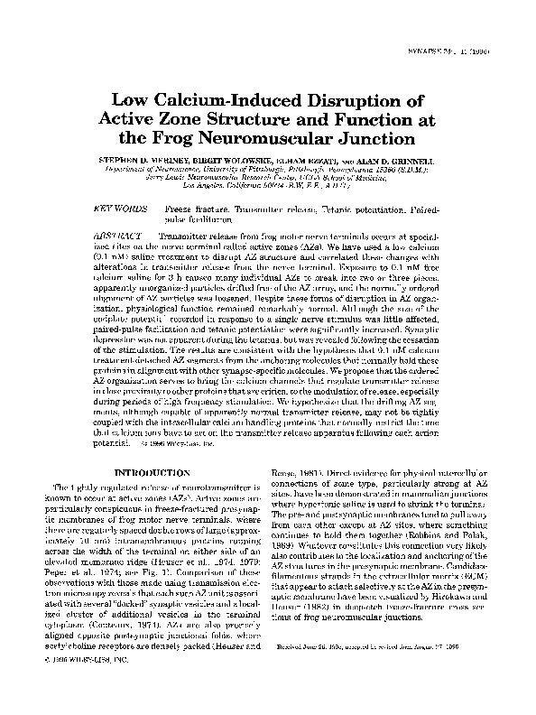
Fig. 1. Freeze-fracture through the “p” face of a control frog nerve terminal. High magnification
(printed a t 90,OOOX for analysis) montage of a representative series of AZ segments. The active zones are
defined as the two double rows of large (10nm) particles. Most of the parameters used for quantification of
AZ integrity are labeled. Brackets identifying “AZ length” indicate a single AZ segment, DP = dispersed
particles, calibration = 200 nM.
It is widely accepted that quantal release occurs near
the large intramembranous particles (Heuser et al.,
1979), which are thought to represent both voltage sensitive calcium (Ca++)channels (Cohen et al., 1991; Pumplin e t al., 1981; Robitaille et al., 1990) and calciumactivated K+ channels (Robitaille and Charlton, 1992;
Robitaille et al., 1993). The details of events inside the
terminal that couple Ca+ influx to vesicle fusion and
release are poorly understood, but are likely to involve
interactions between immediately available Cat +-semitive molecules and one or more of the complex of membrane and cytoplasmic proteins implicated in vesicle
docking and release, and depend critically on their proximity and organization (Jahn and Sudhof, 1994; Mastrogiacomo e t al., 1994; Sollner et al., 1993). I n addition,
it may be essential that Cat+channels be in close proximity to other proteins in the active zone (Cat ’ activated
potassium channels, local Ca++buffers, etc.) to account
for cooperativity in release and the normal properties
of paired-pulse facilitation, tetanic potentiation, tetanic
and post-tetanic depression, and post-tetanic potentiation. Thus one might postulate that the high degree of
organization of AZ structures in the presynaptic membrane is reflective of a similar organization inside the
terminal, and that both are of importance. In fact, the
importance of AZ integrity has been demonstrated in
the study of the disease Lambert-Eaton Syndrome
(LES). This autoantibody-mediated disease is characterized by a disruption of active zone organization a t
the motor nerve terminal that results in both a breakdown in the alignment of AZ particles and a decrease
in the total number of particles (Fukunaga et al., 1982,
1983). Associated with this disruption is a decrement
in transmitter release characterized by a reduction in
quantal content (Elmqvist and Lambert, 1968; see Vincent e t al., 1989 for review).
Despite extensive study of the structure of the frog
AZ, the functional significance of the high degree of
organization is still poorly understood. In a n effort to
better understand the relationship between AZ organization and physiological function, experimental protocols that cause a disruption of this organization have
been developed. It has been reported that a 2-3-h treatment of neuromuscular preparations with a zero Ca++
Ringer causes severe disruption of active zones and
dispersion of the 10 nm intramembranous particles
(Ceccarelli e t al., 1979; however, see Pumplin, 1983).
We confirm that 3 h of exposure to a n EGTA-buffered
saline containing 0.1 nM free Ca++causes significant
disruption of active zones, and have used such preparations to ask whether changes in AZ structure result in
alterations in the physiology of synaptic transmission.
Some of this work has been presented in preliminary
form (Meriney et al., 1989).
zyxwv
zyxwvu
zyxwvut
MATERIALS AND METHODS
Freeze-fracture electron microscopy
Adult Rana pipiens frogs were anesthetized with
0.1% tricaine methanesulfonate (Sigma Chemical Co.,
St. Louis, MO) and pithed. Cutaneous pectoris nervemuscle preparations were dissected out and pinned to
a thin layer of Sylgard in a bath of normal frog Ringer
�ACTIVE ZONE STRUCTURE AND FUNCTION
zyxwv
3
zyxwvutsrq
(NFR; 116 mM NaCl, 1mM NaHC03, 2 mM KC1, 1.8
CaC12,1mM MgCl2,5 mM Hepes, pH 7.4). Experimental
preparations were either fixed immediately, incubated
in control saline for 3 h, incubated in 0.1 nM free Ca++
saline for 3 h at room temperature, or incubated in 0.1
nM free Ca++for 3 h and then returned to control saline
for 2 h before fixation. The 0.1 nM free C a t +saline had
the following composition (in mM): 94 NaCl, 1NaHC03,
2 KC1, 10 Hepes, 0.02 CaC12,2.2 MgC12, 20 EGTA, 2.5
glucose, pH 7.2. Control saline was buffered to 1.8 mM
free Ca++using the following composition (in mM): 64
NaC1, 1 NaHC03, 2 KC1, 10 Hepes, 21.8 CaC12, 1.0
MgC12,ZO EGTA, 2.5 glucose, pH 7.2. Free Ca++concentrations were calculated by a custom software package.
All salines had a free Mg++ concentration of 1 mM.
Preparations were fixed for 1h with 2% glutaraldehyde
in NFR and washed in NFR. Small (0.5-1 mm) nerve
terminal-containing pieces were processed by standard
freeze-fracture methodology (Heuser et al., 1974; KO,
1981), and viewed in a Zeiss EM 109 transmission electron microscope at 80 kV.
To analyze the organization of this structure, the cytoplasmic halves (“p”faces) of nerve terminal membrane
leaflets were photographed and printed at low (9,000~)
and high (90,000 x ) magnification for construction of
montages. Low magnification montages were used to
determine nerve terminal position relative to the nerve
trunk and innervating axon and to verify that fractured
nerve terminal segments were all on a single muscle
fiber. High magnification montages were used to quantify details of AZ structure. For terminals in which a
significant portion (greater than 50 Fm) of the total
length had been fractured, we characterized the disruption of this structure by the parameters shown in Figures 1and 3. Based on a n average nerve terminal length
in this preparation of about 675 pm (Propst and KO,
19871, 50 pm is roughly 8% of the terminal length.
We studied eight terminals from six control untreated
muscles with fractured terminal lengths that ranged
between 58-102 p,m (mean i SEM = 75 i 15 pm);
eight terminals from three muscles treated with a 3-h
exposure to 0.1 nM free calcium with lengths ranging
between 55-199 pm (mean = 87 ? 48); four terminals
from two muscles treated with 0.1 nM free calcium
followed by control saline with lengths ranging between
56-269 pm (mean = 127 i 86); three terminals from
one muscle treated for 3 h with control saline with
lengths ranging between 73-209 pm (mean = 155 i
59). AZs were defined as the complex of double rows
of large (about 10 nm) intramembranous particles on
either side of a membrane ridge remaining in the cytoplasmic half (“p” face) of the presynaptic lipid bilayer
after it has been fractured. Distinct AZ pieces were
quantified if they contained a continuous double row of
10 nm particles at least 200 nm in length, and were
positioned at least 50 nm from any other AZ piece. AZ
pieces of smaller length were grouped into a category
of short AZ segments and their length was not quantified. The number of single rows ofAZ particles per ridge,
and the number of ridges that contained apparently
unorganized, or free, 10 nm intramembranous particles
were also quantified.
Electrophysiology
Intracellular recordings of endplate potentials were
performed a s previously described (Meriney and Grinnel, 1991). Briefly, cutaneous pectoris nerve-muscle
preparations were incubated for 10 min in 2 mg/ml
FITC-labeled peanut agglutinin (Sigma Chemical Co.,
St. Louis, MO) to stain the extracellular matrix covering
nerve terminals (KO,1987). The preparation was then
rinsed several times in NFR and pinned at 1.1times
resting in situ length to the bottom of a Sylgard coated
recording chamber (5 ml capacity). The muscle nerve
was sucked into a stimulating electrode and stimulated
with 100-200 psec pulses at 2-3 times the level necessary to evoke a maximal contraction. To prevent nerveevoked muscle contractions during experiments, the
preparation was superfused continually ( 1ml/min) with
3-5 FM d-tubocurarine chloride (dTC) in NFR. Intracellular recordings of endplate potentials (EPPs) were
made a t 15 2 1°C with glass microelectrodes (20-40
m a ) filled either with 3 M KCl or with 0.6 potassium
acetate, 5 mM KC1, and 50 mM EGTA. Muscle cells
were penetrated near endplates under visual control,
alternatively using Hoffman amplitude modulation
contrast optics and fluorescence microscopy at 200 x
with a n Olympus 20x long-working distance waterimmersion objective. Endplate potentials were recorded
on magnetic tape (RACAL) and analyzed off-line using
a custom modification of the pClamp suite of programs
(Axon Instruments, Foster City, CA). Recordings were
made in NFR before, and 0.5-1 h following a 3-h treatment with 0.1 nM free Ca+’ saline.
zyxw
zyxwvuts
zyxwvut
zyxwvutsrq
zyxwvutsrqpo
RESULTS
The normal structure of the AZ
Control frog nerve terminals typically have well organized AZ structure a s visualized by freeze-fracture techniques. Figure 1 shows a freeze-fracture replica of a
control nerve terminal a t high magnification (90,OOOX).
Normally, the 10 nm intramembranous AZ particles are
arranged in neatly organized rows that usually span
the width of the nerve terminal (see “AZ l e n g t h in Fig.
1).In control terminals, a double row of AZ particles is
conspicuous on either side of the crests of AZ ridges
in p-face replicas. These particle arrays are arranged
roughly perpendicular to the long axis of the terminal
(see “angle” in Fig. 1) and spaced roughly 1pm apart
(see “interzone distance” in Fig. 1).
Structural disruption of active zone integrity
Although control nerve terminals occasionally show
some disruption in organization, treatment for 3 h with
�4
zyxwvutsrqpon
S.D. MERINEY ET AL
zyxwvu
zyxw
zyxw
zyxwvuts
Fig. 2. Freeze-fracture through the "p" face of a 3 h 0.1 nM Ca"-treated frog nerve terminal. Most
AZs show many of the characteristics of disrupted structure outlined in the text and Table I (SR = single
rows; DP = dispersed particles; AZ segments are bracketed for one sample AZ ridge). Calibration = 200 nM.
0.1 nM free Ca++resulted in a clear-cut increase in
the frequency with which a disruption of the normally
ordered structure could be observed. Figure 2 shows a
freeze-fracture replica taken from a 0.1 nM free Ca++treated nerve terminal. To quantify the disruption in
AZ structure observed, we measured the number of AZ
segments (>200 nm in length) per ridge, the percentage
of ridges that contained very short ((200 nm) AZ pieces,
the percentage of ridges that contained single rows of
10 nm particles (defined as either an unopposed double
row of particles on one side of the AZ ridge, or a true
single row of particles), the percentage of ridges that
contained clusters of apparently unorganized particles,
and the total amount of AZ per unit length. Representative examples of the characteristics of disruption that
were quantified are indicated in Figures 1 and 2.
In control nerve terminals, 91% of AZ ridges con-
zyxw
zyxwvu
TABLE I. Disruption of AZ integrity is revealed as treatment-induced
changes in the number of active zone pieces or segments per presynaptic
ridee exuressed as a uercent of total'
zyxwvuts
zyxwvu
zyxwvu
~~
Number ofAZ uieces uer ridge (% of total)
No. AZ Dieces
Control
3 h control
3 h 0.1 nM Ca-3 h 0.1 nM Ca' '; 2 h NFR
1
2
3
4
90.9
92.9
76.9
88.2
9.1
6.1
18.4
11.4
0
0.5
0
0
0.4
0
4
0.4
'For each of t h e four experimental conditions, t h e percentage of active zone ridges that
contained one or more active zone pieces is shown.
tained a single, long AZ segment (see Table I). Occasionally (9% of the time), control ridges had two AZ segments. In contrast, a 3-h treatment in 0.1 nM free Ca++
resulted in a significant increase in the percentage of
ridges that showed more than one AZ segment (about
�zy
zyxwvu
zyx
zyxw
5
ACTIVE ZONE STRUCTURE AND FUNCTION
A
AZ segments/ridge
*
T
B
Ridges with A2 fragments
20
16
4
9
12
0
k
a
8
4
0
c
Ridges with single rows
n
m
*
zyxw
zyxwv
zy
D E dges with
T
free particles
Control
3 h r . NFR
3 hr. 0.1 nM Calcium
3 hr. 0.1 nM Calcium then 2 hr. NFR
Fig. 3. Quantification of the disruption of active zone integrity. A
Control active zones have a n average of 1.08 active zone segments per
ridge (open bar). A 3-h exposure to control saline (hatched bar) does
not affect this structural arrangement, however, a 3-h treatment with
0.1 nM free Ca" significantly increases that number to 1.27 segments
per ridge (dense hatched bar). This effect can be reversed with a
recovery exposure to control saline for a n additional 2 h (solid bar).
B Only 4.2% of control active zone ridges (open bar) have very small
active zone fragments (less than 200 nm in length), and this number
does not change with a 3-h exposure to control saline (hatched bar).
However, a 3-h exposure to 0.1 nM free Ca-- significantly increases
this percentage to 16 (dense hatched bar), and this disruption shows
only slight recovery following a subsequent 2-h exposure to control
saline (solid bar). C: Only 5% of control active zone ridges have single
rows of particles, but this number can be significantly increased by a
3-h incubation in control saline (hatched bar) or 0.1 nM free Ca"
(densely hatched bar), and this form of disruption is not significantly
different following a further 2-h exposure to control saline (solid bar).
D. Seventeen percent of control active zone ridges have clusters of
free 10 nm particles that do not appear to be organized (open bar).
Although this number has a tendency to increase following incubation
in control saline (hatched bar), the disruption is not significantly different from control unless the incubation is performed in 0.1 nM free
Ca" (dense hatched bar). This form of disruption does not recover
following a subsequent exposure to control saline for 2 h (solid bar).
* = significantly different from control ( P < 0.05) using a one-way
analysis of variance.
23%; see Table I). This disruption in 0.1 nM free Cat+
showed some reversal following a 2-h return to control
saline (see Table I). When the mean numbers of AZ
segments per ridge were compared, 0.1 nM free Ca+'
treatment significantly increased the number from 1.08
in control terminals, to 1.27 (see Fig. 3A). Furthermore,
the percentage of AZ ridges that contained very small
( ~ 2 0 nm)
0 AZ segments also increased from about 4% to
greater than 16%,and this effect only partially reversed
following a 2-h return to control saline (Fig. 3B). Therefore, it is clear that the normally continuous array of
10 nm intramembranous particles spanning most of the
width of the nerve terminal along the crest of the AZ
ridge, was fragmented into smaller pieces by exposure
to a very low free Ca++saline.
Control AZ ridges occasionally (about 5% of the time)
had single rows of 10 nm particles that did not appear
to have parallel counterparts. The percentage of AZ
ridges with single rows increased to about 25% in nerve
terminals that had been incubated for 3 h in either
control saline or 0.1 nM free Ca++saline, and this form
of disruption persisted following a further 2-h exposure
to control saline (see Fig. 3C). The appearance of clusters of apparently unorganized particles also had a tendency to increase in terminals bathed in control saline
for 3 h (Fig. 3D), but this increase was not statistically
significant. This form of disruption did increase significantly when terminals were exposed to 0.1 nM free
Ca++,and became even more prevalent when these
treated terminals were returned to control saline for
a n additional 2 h (Fig. 3D).
Even though the individual significant differences
zyxw
zyxwvutsrqpo
�6
zyxwvutsrqp
zyxwvutsrqp
zyxwvu
S.D. MERINEY ET AL
described above are modest when examined in isolation
(see Fig. 3), taken together, these disruptions of AZ
structure are a n obvious departure from normal. Furthermore, the data presented in Figure 3 represent only
the quantification of specific AZ structural characteristics and do not convey every aspect of the disruption in
organization. The clear disruption of AZ structure is
prominent when one compares Figures 1 and 2. For
example, although this parameter was not quantified,
nerve terminals treated with 0.1 nM free Cat+ showed
a n apparent decrease in the precision with which 10 nm
particles were aligned in their ordered arrays. Control
nerve terminal AZ segments were constructed of ordered arrays of particles that appeared precisely
aligned with one another. A 3-h treatment in low Ca++
appeared to loosen that alignment, so that the AZ segment often appeared wavy and disordered, but with 10
nm particles still within 50 nm of each other, and as
such, not classified as disrupted by our definitions outlined above. Although this type of disruption did not
appear in our quantification, it is probable that this
loosening is reflective of a significant disruption of organization.
Despite these forms of disorganization, measurements of the total amount of AZ/km of terminal length,
i.e., the summed length of the long and short fragments
showing a t least one row of 10 nm particles on either
side of a n intramembranous ridge, was not statistically
different in 0.1 nM Ca++-treated(1.27 t- 0.22pm AZ/
pm terminal length) vs. control (1.09 5 0.22) muscles.
Thus the low Ca++treatment does not appear to cause
a n absolute loss or internalization ofAZ structures, only
their dispersion or disorganization in the membrane.
1.0 I
0
zyxwz
I
I
I
I
I
I
I
5
10
15
20
25
30
35
1
1
1
40
45
50
Interval (msec)
Fig. 4. Paired-pulse facilitation measured i n control (filled circles)
and experimental neuromuscular junctions treated for 3 h in 0.1 nM
free Ca", and then returned for 1 h to NFR with 5 pM dTC (open
circles). A t all intervals tested (5-50 msec.) a 3-h treatment with
0.1 nM free Ca+- significantly increased the magnitude of pairedpulse facilitation.
zyxwvu
zyxwvut
zyxwvut
zyxwvutsrq
zyxwv
Physiological consequences of active
zone disruption
Despite significant disruption of AZ structure, alterations in physiological function were surprisingly limited. In fact, EPPs recorded in curarized preparations
and compared before and after the 0.1 nM free Ca++
treatment did not differ in latency or time course, and
were quite similar in amplitude, although the mean
was slightly reduced (4.95 2 3.5 mV after 0.1 nM C a t +
vs. 5.2 ?I 3.3 mV in controls; n = 27 neuromuscular
junctions in four muscles). Although transmission did
not appear affected when examined following a single
stimulus, we used paired pulses and trains of stimuli
to examine more complex features of release. The response of endplate potentials to paired-pulse stimuli
of varying inter-pulse intervals is plotted in Figure 4.
Control endplates (filled circles, Fig. 4)showed significant paired-pulse facilitation at intervals between 5 and
50 msec. Preparations treated with 0.1 nM free Ca++
saline displayed significantly larger paired-pulse facilitation than controls at all intervals tested (open circles,
Fig. 4).
This difference in potentiation was even more con-
spicuous in recordings of EPPs during trains of stimuli.
Figure 5 shows EPPs recorded in response to 20 Hz
nerve stimulation from a representative endplate before
(Fig. 5A) and following a 3-h exposure to 0.1 nM free
Ca++saline (Fig. 5B). In control nerve terminals, EPP
size usually increased during the first few stimuli (tetanic potentiation), and subsequently decreased in size
(tetanic depression). Strikingly, following a 3-h exposure to 0.1 nM free Ca++saline and return to control
saline for 0.5 to 1h, the tetanic potentiation that developed in the first few stimuli persisted throughout the
stimulation period with little or no apparent depression.
The mean response measured in 10 endplates (before
and after treatment) from one experiment is plotted in
Figure 6. In addition, Figure 6 plots recovery after the
20 Hz tetanus tested with stimulation a t 0.5 Hz. Control
endplates showed no significant change in the depressed EPP in the first post-tetanic test stimulus (5
seconds after the tetanic stimulus was terminated), but
a rapid recovery of EPP size that, within 5-10 seconds,
was within control ranges. I n contrast, following treatment with 0.1 nM free Ca++saline, the first post-tetanic
stimulus pulse resulted in a n EPP that was significantly depressed to near control depression levels (see
open circles in Fig. 6). This depressed EPP rapidly potentiated over the next 5-10 seconds to a amplitude
that was significantly larger than the control EPP size.
Therefore, despite apparently normal release measured
following a single nerve stimulus, endplates treated
with 0.1 nM free Ca++saline showed increased paired-
�ACTIVE ZONE STRUCTURE AND FUNCTION
zyxwv
zy
zyx
zy
7
5 0 0 ms
‘
7
I
3mV
zyxwvut
Fig. 5. Endplate potentials recorded from a muscle fiber during 20 Hz nerve stimulation in NFR
containing 5 )LMdTC. A: A control neuromuscular junction displays a characteristic facilitation following
the first few stimuli, followed by a depression of release. B Following a 3-h treatment in 0.1 nM free
Ca*’, and 1 h after return to NFR plus 5 )LM dTC, 20 Hz stimulation results in facilitation that is
maintained over the course of the stimulation.
o
0
zyxwv
Low calcium treatment
Control
zy
zyxwvu
zy
1
3
’
’
6
8
10
12
14
1 2 3
5 6 7 8 91011
91-100
I
/
4
20 Hz Stimulus Number
/
I
2
4
I
I
f
I
Recovery at 0.5 Hz (sec)
Fig. 6. Plot of the mean response of 10 nerve terminals to 20 Hz
nerve stimulation. Although endplate potential (EPP) size (expressed
as a percentage of control EPP size) of control nerve terminals (filled
circles) shows facilitation over the first 11 stimuli, pretreatment with
0.1 nM free Ca++shows significantly greater facilitation. Furthermore,
this facilitation is maintained only in the terminals pretreated for 3
h with 0.1 nM free Ca*’ (see average EPP size for the last 10 stimuli
in the train: stimuli #91-100). Interestingly, although treated terminals do not show significant depression of EPP size during the 20 Hz
5 sec train, there is a significant depression of transmitter release
during the first few seconds of recovery immediately following the
train. This post-tetanic depression is not significant in control terminals, and is quickly overcome by a post-tetanic potentiation that is
significantly greater in terminals treated with 0.1 nM free Ca“ for 3 h.
pulse facilitation, tetanic potentiation, and post-tetanic
potentiation. The presence of normal tetanic depression
was revealed in low Ca++-treatedpreparations only following the removal of tetanic nerve stimulation.
control preparations, there are instances in which this
organization has been disrupted, especially near the
ends of terminal branches (see Pumplin, 1983; Pawson
and Grinnell, unpublished observations). Presumably
this disruption in AZ organization in control preparations, particularly at the ends of nerve terminal
branches, reflects remodeling associated with ongoing
extension and retraction of terminal branches. This
type of remodeling has been described during alterations in AZ morphology associated with changes in
activity of animals in “summer”vs. “winter” frogs (Dor-
DISCUSSION
Frog motor nerve terminal active zones are normally
highly organized linear arrays of intramembranous proteins precisely aligned both with respect to acetylcholine receptor accumulations in the muscle membrane
and with intraterminal release-associated proteins. In
�8
zyxwvu
zyxwvu
zyxwvutsrq
S.D. MERINEY ET AL
lochter et al., 1993). Similar disorganizations has been
described a t regenerating frog neuromuscularjunctions
(KO,1984).
Surprisingly, it is possible to disrupt AZ organization
without grossly affecting transmitter release. Ceccarelli
and his colleagues (1979) reported that when a frog
neuromuscular preparation was bathed in zero Ca++
saline for 2-3 h, the AZs became highly disorganized,
breaking into fragments that drifted apart in the presynaptic membrane and lost their normal orientation.
This disruption persisted following a 1h return to Ca++containing saline a t which time recordings of EPPs
showed normal quantal contents and mEPP frequency
(Haimann e t al., 1980). On the other hand, they reported a small drop in mean quantal size, which was
interpreted as reflecting the fact that some release sites
were further from acetylcholine receptor concentrations
(Haimann e t al., 1980). Pumplin (1983) has disputed
the morphological finding, reporting no greater disruption of AZs in zero Ca++saline than in control saline.
Here, we quantify clear-cut disruption of AZ organization following treatment with a saline that buffers Ca++
to 0.1 nM. The number of AZ segments per ridge increased as AZs broke into smaller pieces (Fig. 3A,B).
The fragmentation of large pieces showed essentially
complete reversal following a 2-h return to control saline (Fig. 3A). In contrast, when small AZ pieces (<200
nm) broke free, they were much less likely to re-connect
during 2-h return to control saline (Fig. 3B). The presence of single rows of AZ particles cannot be attributed
to the low Ca++treatment, since prolonged exposure to
control saline was almost equally effective at creating
this form of disruption (Fig. 3C). The presence of dispersed AZ particles, apparently free-floating in the
membrane, was significantly increased by exposure to
low-Ca++saline, although partial disorganization of this
type increased with time in control saline, and a return
to control saline after 0.1 nM Ca++treatment did not
prevent further dispersion (Fig. 3D). Presumably these
AZ particles were completely dissociated from their
original organized arrangement, and despite a 2-h return to control saline, they were unable to be reorganized. The low Ca++(0.1 nM) treatment may be able to
disrupt connections that extend between AZ proteins
and the postsynaptic membrane near the tops of the
junctional folds (Hirokawa and Heuser, 1982; Robbins
and Polak, 1989), or, more likely, from the corresponding
part of the basal lamina in both directions. I t seems
reasonable to suggest that this connection is composed
of one or more Ca++-sensitiveadhesion molecules t h a t
serve to direct the alignment of pre- and postsynaptic elements.
Despite significant disruption of AZ organization,
physiological function was remarkably robust. The
principal physiological differences between 0.1 nM
Ca++-treatedand control preparations were a slightly
reduced mean EPP amplitude, increased paired pulse
facilitation, and sustained tetanic potentiation. The
slight reduction in mean EPP amplitude might be a
reflection of the smaller mean mEPP size reported in
similarly treated preparations by Haimann et al.
(1980). There is no evidence for significantly reduced
quantal content following treatment with low Ca++salines. If the quantal contents were sharply decreased,
one would have expected enhanced facilitation and reduced depression. However, Haimann et al. (1980)
found normal quantal contents in such preparations,
and we observed enhanced facilitation in identified
junctions studied both before and following treatment
in which the EPP size did not change. Additionally,
0.1 nM Ca++-treatedjunctions revealed close to control
levels of depression following the cessation of tetanic
stimulation (see Fig. 5). These data show that the increased tetanic potentiation was not associated with
reduced depression. I t seems probable, therefore, that
these physiological abnormalities represent true departures from normal since they dissociate increases in
paired-pulse facilitation and tetanic potentiation from
changes in tetanic and post-tetanic depression. Disruption of these features of the release process might be
expected if components of the release apparatus are not
lost, but simply drift apart sufficiently that properties of
release that depend on cooperativity would be affected.
The disease LES is characterized by both disruption
of AZ organization and a loss of AZ particles (Fukunaga
et al., 1982,1983).Under these conditions, quantal content drops significantly (Vincent e t al., 1989). Similarly,
comparisons of AZ structure in fast vs. slow frog neuromuscular junctions (Verma and Reese, 1984) or twitch
vs. tonic lizard neuromuscular junctions (Walrond and
Reese, 1985)have identified differences in AZ structure
that correlate with physiological properties in these
junctions. However, as with LES antibody-treated preparations, physiological differences here are most likely
to be due to the large differences in the total number
of AZ particles per terminal length rather than differences in the organization of those particles. Furthermore, comparisons made between nerve terminals in
different species, or types of neuromuscular junctions
within a single species, are complicated by potentially
large differences in organization and function of the
intraterminal proteins that buffer Cat+ and regulate
exocytosis. In fact, single types of neuromuscular junctions can show significant differences in structure-function relationships when examined a t different developmental ages, or following regeneration. At newly
regenerated neuromuscular junctions, AZs are small
and poorly organized, and evoked release of transmitter
recovers before reinnervation is complete such that release per unit length of the nerve terminal is initially
higher than in control junctions and is occurring from
AZ structures t h a t are smaller (KO,1981, 1984; Ding,
1982b). At endplates that reinnervate ectopic sites on
muscle, nerve terminals are very small, yet quantal
zyxwvutsr
�zyxwvut
ACTIVE ZONE STRUCTURE AND FUNCTION
content is often five times larger than in control reinnervated junctions (Ding, 1982a). Thus, AZ structure-function relationships can be complicated by intraterminal
differences in biochemical organization and, as such,
are difficult to interpret when comparisons are made
between different types of nerve terminals or even between similar terminals of different developmental age.
In contrast, we have studied acute changes in AZ
structure and function within a single population of
nerve terminals. We see only a disruption of AZ organization, and this is not associated with a significant drop
in quanta1 content (see Ceccarelli et al., 1979; Haimann
et al., 1980), but rather a relatively subtle alteration
in physiological function-increases in paired-pulse facilitation and tetanic potentiation. Our observations, in
comparison with acute effects in LES antibody-treated
preparations, effectively dissociate physiological effects
of AZ disruption with loss of AZ particles. Since disruption in the proximity of some AZ particles to one another
does not significantly affect the magnitude of release
following a single stimulus, it is possible that the high
degree of AZ organization a t the frog neuromuscular
junction is important not so much for bringing Ca++
channels in close proximity to one another, as in bringing individual transmitter release-associated Ca++
channels in close proximity to other important synaptic
proteins. In fact, AZ regions of frog nerve terminals
have been shown to contain calcium-activated K' channels, as well as Cat t channels (Cohen, 1991; Robitaille
and Charlton, 1992; Robitaille et al., 1990; Robitaille
et al., 1993; see also Roberts et al., 19901, and certainly
include a variety of other proteins including those involved in vesicle docking and fusion (see Sollner et al.,
1993).If AZs are organized primarily to bring a heterogenous group of proteins together for interaction, and
not to bring multiple C a t t channels in close proximity
to one another, transmitter release must be capable of
being evoked by the flow of Ca++through a single Ca++
channel. Stanley (1993) has presented evidence that
transmitter release is capable of being evoked by the
opening of single Ca++channels. At a single frog neuromuscular junction, there are about 500 AZs that collectively release several hundred vesicles following each
action potential (Katz and Miledi, 1979). Since release
appears to be well distributed along the length of nerve
terminals (DAlonzo and Grinnell, 1985), this implies
that each AZ releases no more than one or two vesicles,
and often may not release any transmitter following
each action potential. Since there are about 300 10-nm
particles within each AZ,perhaps one half or fewer of
which may represent Ca++channels, several scenarios
are possible. At one extreme, most of these Ca++channels open during each action potential and there is a
low probability that Ca++ion entry leads to transmitter
release. At the other extreme, only one or two percent
of the Cat+channels in a n AZ open following each action
potential, and following Cat+ entry, the probability that
9
a vesicle is released is high. Some combination of these
two extreme hypotheses is probable. At the frog neuromuscular junction, N-type C a t + channels are thought
to regulate evoked transmitter release (Kerr and Yoshikami, 1984). N-type Ca++channels have been shown to
display several modes of gating, some with low open
probability (Delcour and Tsien, 1993; Rittenhouse and
Hess, 1994). I n frog motoneuron-muscle co-cultures, we
have observed that Cat+ channels expressed in transmitter-releasing regions of neurites have a very low
probability of opening during short voltage depolarizations (Meriney and Grinnell, unpublished observations). Taken together, the above information leads us
to favor the following hypothesis: an action potential
induces, on average, the opening of only a few of the
Ca+ channels that exist at a n AZ and the flow of Cat+
through these individual Ca+' channels can, a significant fraction of the time, induce transmitter release.
Other proteins, normally clustered near this releaseassociated Cat+ channel in the AZ, are positioned both
to initiate vesicle fusion and to remove free Ca++,terminating the transmitter release process.
It is possible that the low Ca+' treatment significantly
reduces the intraterminal Ca++concentration such that
i t does not recover in NFR before physiological recordings are made 0.5-1 h later. However, we feel that
this is not a likely explanation for the physiological
effects observed since a reduced intraterminal Ca++concentration would be expected to decrease, not increase,
paired-pulse facilitation. On the other hand, our data
are consistent with the hypothesis that the disruption
in the anchoring of AZ pieces following exposure to low
Ca+' saline results in a decrease in the normally tight
association of AZ proteins with presumptive intraterminal Ca++ binding proteins. This physical disruption
would be expected to alter the handling of C a t + ions
following each action potential. A reduction in C a t +
handling would result in the increase in paired-pulse
facilitation and tetanic potentiation observed. Both
paired-pulse facilitation and tetanic potentiation are
short-term forms of synaptic plasticity that are thought
to be dependent on residual Ca+t (see Zucker, 1993,
for review). The relationship between these forms of
plasticity and residual Ca++suggests that these effects
are due to Cat+ acting at a different site than the lowaffinity site(s ) that are thought to trigger transmitter
release (Zucker, 1993). Recently, several Cat+ binding
proteins have been identified in neurons, and at synaptic sites, that may modulate the transmitter release
process by acting a s local Ca+' buffers (Michaelis et aI.,
1983; Roberts, 1993; Roberts, 1994; Tolosa de Talamoni
et al., 1993). When appropriately positioned, these proteins may both restrict the region in which the internal
Cat+ ion concentration exceeds 1 pM, and shorten the
time that intracellular Cat+has to act on the transmitter release apparatus. Support for this interpretation
appears strongest when one compares the size of the
+
zyxwv
zyxwvutsrqp
�10
S.D. MERINEY ET AL.
zyxwv
Ding, R. (198213) Lack of correlation between physiological and morphological features of regenerating frog neuromuscular junctions.
Brain Res., 253:47-55.
and Wernig, A. (1993) Acetylcholine recepDorlochter, M., Meurer, S.,
tor bars and transmitter release in frog neuromuscular junctions.
Neuroscience, 52:987-999.
Elmqvist, D., and Lambert, E.H. (1968)Detailed analysis ofneuromuscular transmission in a patient with the myasthenic syndrome sometimes associated with bronchogenic carcinoma. Mayo Clin. Proc.,
43:689-713.
Fukunaga, H., Engel, A,, Osame, M., and Lambert, H. (1982) Paucity
and disorganization of presynaptic active zones in the LambertEaton myasthenic syndrome. Muscle Nerve, 5:686-697.
Fukunaga, H., Engel, A,, Lang, B., Newsom-Davis, J., and Vincent,
A. (1983) Passive transfer of Lambert-Eaton myasthenic syndrome
with IgG from man to mouse depletes the presynaptic active zones.
Proc. Natl. Acad. Sci. U.S.A., 80:7636-7640.
Haimann, C., Grohovaz, F., Hurlbut, P., andceccarelli, B. (1980)Possible significance of changes in the structure of the active zones induced by Ca2+-freesolutions. In: Ontogenesis and Functional Mechanisms of Peripheral Synapses. J. Taxi, ed. Elsevier/North-Holland
Biomedical Press, New York, pp. 157-170.
Heuser, J.E., and Reese, T.S. (1981)Structural changes after transmitter release at the frog neuromuscular junction. J . Cell Biol., 88:
564-580.
Heuser, J.E., Reese, T.S., and Landis, D.M.D. (1974) Functional
changes in frog neuromuscular junctions studies with freeze-fracture. J . Neurocytol., 3:109-131.
Heuser, J.E., Reese, T.S., Dennis, M.J., Jan, Y., Jan, L., and Evans,
L. (1979) Synaptic vesicle exocvtosis captured bv auick freezinp.and
correlated -with quantal transmitter' release: i. Cell Biol, 81:
275-300.
Hirokawa, N., and Heuser, J.E. (1982)Internal and external differentiations of the postsynaptic membrane at the neuromuscular junction.
J. Neurocytol., 11:487-510.
Jahn, R., and Sudhof, T.C. (1994) Synaptic
vesicles and exocytosis.
.
.
Ann. Rev. Neurosci., 17:219-246.
Katz, B., and Miledi, R. (1979) Estimates of quantal content during
'chemical potentiation' of transmitter release. Proc. R. SOC.Lond.
[Biol.] 205:369-378.
Kerr, L.M., and Yoshikami, D. (1984) A venom peptide with a novel
presynaptic blocking action. Nature, 308:282-284.
KO, C.-P. (1981) Electrophysiological and freeze fracture studies of
changes following denervation a t the frog neuromuscular junctions.
J. Physiol. (Lond.), 321:627-639.
KO,C.-P. (1984) Regeneration of the active zone a t the frog neuromuscular junction. J. Cell Biol., 98:1685-1695.
KO,C.-P. (1987)A lectin, peanut agglutinin, as a probe for the extracelACKNOWLEDGMENTS
lular matrix in living neuromuscular junctions. J. Neurocytol.,
16567-576.
We thank Mike Kreman for technical assistance, and Mastrogiacomo,
A., Parsons, S.M., Zampighi, G., Jenden, D.J., UmDrs. S. Sesack and G. Barrioneuvo for critical evaluabach, J.A., and Gundersen, C.B. (1994) Cysteine string proteins: a
potential link between synaptic vesicles and presynaptic calcium
tion of the manuscript. This work was supported by a
channels. Science, 263:98 1-982.
National Science Foundation Grant BNS 8719613 Meriney, S.D., and Grinnell, A.D. (1991)Endogenous adenosine modulates stimulation-induced depression at the frog neuromuscular
(A.D.G.) and National Institutes of Health Grants
junction. J . Physiol., 443:441-455.
NS06232 (A.D.G.) and NS32345 (S.D.M.).
Meriney, S.D., Pawson, P.A., and Grinnell, A.D. (1989) Presynaptic
active zone integrity and ACh release from frog motor nerve terminals. SOC.Neurosci. Abst., 15258.
REFERENCES
Michaelis, E.K., Michaelis, M.L., Chang, H.H., and Kitos, T.E. (1983)
Ceccarelli, B., Grohovaz, F., and Hurlbut, W.P. (1979) Freeze-fracture
High affinity Ca2'-stimulated Mg2+-dependentATPase in rat brain
studies of frog neuromuscular junctions during intense release of
synaptosomes, synaptic membranes, and microsomes. J . Biol.
neurotransmitter. I. Effects of black widow spider venom and Ca2+
Chem., 258:6101-6108.
free solutions on the structure of the active zone. J . Cell Biol., Peper, K., Dreyer, F., Sandri, C., Akert, K., and Moor, H. (1974) Struc81:163-177.
ture and ultrastructure of frog motor endplate. Cell Tissue Res.,
Cohen, M.W., Jones, O.T., and Angelides, K.J. (1991) Distribution of
149:437-455.
Ca+ channels on frog motor nerve terminals revealed by fluorescent Propst, J.W., and KO, C.-P. (1987) Correlations between active zone
w-conotoxin. J. Neurosci., 11:1032-1039.
ultrastructure and synaptic function studied with freeze-fracture of
Couteaux, R. (1974) Remarks on the organization of axon terminals
physiologically identified neuromuscular junctions. J. Neurosci.,
in relation to secretory processes a t synapses. In: Advances in Cyto7:3654-3664.
pharmacology, vol. 2. B. Ceccarelli, F. Clementi, and J . Meldolesi, Pumplin, D.W. (1983) Normal variations in presynaptic active zones
eds. Raven Press, New York, pp. 369-379.
of frog neuromuscular junctions. J . Neurocytol., 12:317-323.
DAlonzo, A.J., and Grinnell, A.D. (1985) Profiles of evoked release Pumplin, D.W., Reese, T.S., and Llinas, R. (1981) Are the presynaptic
along the length of frog motor nerve terminals. J. Physiol.
membrane particles the calcium channels? Proc. Natl. Acad. Sci.
(Lond.), 359:235-258.
U.S.A., 78:7210-7213.
Delcour, A.H., and Tsien, R.W. (1993) Multiple gating modes ofN-type Rittenhouse, A.R., and Hess, P. (1994) Microscopic heterogeneity in
Ca2+channel activity distinguished by differences in gating kinetics.
unitary N-type calcium currents in rat sympathetic neurons. J.
J. Neurosci., 13:181-194.
Physiol. (Lond.), 474:87-99.
Ding, R. (1982a) Comparison of morphology and physiology ofsynapses Robbins, N., and Polak, J. (1989) Focal adhesions of nerve terminal
formed a t ectopic and original endplates sites in frog muscle. Brain
to synaptic matrix and schwann cell a t mouse neuromuscular juncRes., 253:57-63.
tions. SOC.
Neurosci. Abst., 15:258.
EPPs at the end of, and immediately following, the
cessation of a 20 Hz tetanic stimulus train in control
and low Ca"-treated preparations (Fig. 6). Low (0.1
nM) Ca"-treated preparations showed maintained potentiation throughout the tetanus that rapidly gave way
to depression immediately following the cessation of the
tetanus. This observation appears most consistent with
a decrease in the local Ca++buffering capacity of low
Ca++-treatednerve terminals. Ifthis were to occur, Cat+
entering the nerve terminal during each action potential would be buffered at a slower rate than in control
terminals. In this case, a n increase in facilitation and
tetanic potentiation would be expected, while at the
cessation of the tetanus, as the residual Ca++became
buffered, depression would be unmasked.
These data might also be expected if the nerve terminal calcium handling mechanisms are down-regulated
in response to the 3-h exposure to very low extracellular
calcium, independent of the relationship of AZ pieces
to intracellular nerve terminal components. If this is the
case, this compensatory response must require longer
than 1h to recover following a return to normal calcium
levels. In any event, the AZ disruption described here
does not significantly alter release triggered by single
action potentials a t low frequency. Departures from normal physiology observed with paired pulses and during
high frequency trains are either a compensatory response of the nerve terminal to the low calcium exposure, or a consequence of the disruption in AZ structure.
Both of these hypotheses explain physiological effects
by alterations in the mechanisms of handling of calcium
within the nerve terminal.
zyxwvu
zy
zyxwvut
~
zyxwvutsrqpon
�ACTIVE ZONE STRUCTURE AND FUNCTION
Roberts, W.M. (1993) Spatial calcium buffering in saccular hair cells.
Nature, 363:74-76.
Roberts, W.M. (1994) Localization of calcium signals by a mobile calcium buffer in frog saccular hair cells. J . Neurosci., 14:3246-3262.
Roberts, W.M., Jacobs, R.A., and Hudspeth, A.J. (1990) Colocalization
of ion channels involved in frequency selectivity and synaptic transmission at presynaptic active zones of hair cells. J . Neurosci.,
10:366&3684.
Robitaille, R., and Charlton, M.P. (1992) Presynaptic calcium signals
and transmitter release are modulated by Caz'-activated potassium
channels. J. Neurosci., 12:297-305.
Robitaille, R., Adler, E.M., and Charlton, M.P. (1990)Strategic localization of calcium channels a t transmitter release sites of frog neuromuscular synapses. Neuron, 5:773-779.
Robitaille, R., Garcia, M.L., Kaczorowski, G.J., and Charlton, M.P.
(1993) Functional colocalization of calcium and calcium-gated potassium channels in control of transmitter release. Neuron,
11:645-655.
Sollner, T., Bennett, M.K., Whiteheart, S.W., Scheller, R.H., and Rothman, J.E. (1993) A protein assembly-disassembly pathway in vitro
that may correspond to sequential steps of synaptic vesicle docking,
activation, and fusion. Cell, 75409418.
11
Stanley, E.F. (1993) Single calcium channels and acetylcholine release
at a presynaptic nerve terminal. Neuron, 11:1007-1011.
Tolosa de Talamoni, N., Smith, C.A., Wasserman, R.H., Beltramino,
C., Fullmer, C.S., and Penniston, J.T. (1993) Immunocytochemical
localization of the plasma membrane calcium pump, calbindin-Dz8K,
and parvalbumin in purkinje cells of avian and mammalian cerebellum. Proc. Natl. Acad. Sci. U.S.A., 90:11949-11953.
Verma, V., and Reese, T.S. (1984) Structure and distribution ofneuromuscular junctions on slow muscle fibers in the frog. Neurosci.,
12:647-662.
Vincent, A,, Lang, B., and Newsom-Davis, J . (1989) Autoimmunity to
the voltage-gated calcium channels underlies the Lambert-Eaton
myasthenic syndrome, a paraneoplastic disorder. Trends Neurosci., 12-496-502.
Walrond, J.P., and Reese, T.S. (1985) Structure of axon terminals and
active zones at synapses on lizard twitch and tonic muscle fibers.
J.Neurosci., 5:1118-1131.
Zampighi, G., and Kreman, M. (1985) Intercellular fibrillar skeleton
in the basal interdigitations of kidney tubular cells. J. Membr.
Biol., 88:33-43.
Zucker, R.S. (1993) Calcium and transmitter release. J. Physiol.
(Paris), 87:25-36.
�
 Stephen Meriney