Neurophannacology,Vol. 36, No. 4[5, pp. 637447, 1997
@ 1997Elsevier Science Ltd. AUrights reserved
Printed in Great Britain
0028-3908/97$17.00 + 0.00
P
PII: S0028-3908(97)00044-0
Analysis of the Ligand Binding Site of the 5-HT3
Receptor Using Site Directed Mutagenesis: Importance
of Glutamate 106
F. G. BOESS,]T L. J. STEWARD,l J. A. STEELE,l D. LIU,l J. REID,* T. A. GLENCORSE’~ and
I. L. MARTIN’*
IDepartment of Pharmacology,Faculty of Medicine, 9-70 Medical Sciences Building, University of Alberta,
Edmonton, T6G 2H7, Canadaand 2GlaxoInstitutefor Molecular Biology S.A., 14 Chemin des Aulx, Case
Postale 674, CH-1228Plan-les-Ouates, Geneva, Switzerland
(Accepted 15 January 1997)
Summary—The 5-HT3 receptor is a ligand-gated ion channel with significant structural similarity to the
nicotinic acetylcholinereceptor. Severalregions that form the ligand binding site in the nicotinic acetylcholine
receptor are partially conservedin the 5-HT3receptor, presumablyreflecting the conservedsignal transduction
mechanism. Specific amino acid differences in these regions may account for their distinct ligand recognition
properties. Using site-directed mutagenesis, we have replaced one of these residues, glutamate 106 (EI06),
with aspartate (D), asparagine (N), alanine (A) or glutamine (Q) and characterized the ligand-binding and
electrophysiologicalpropertiesof the mutantreceptors after transientexpressionin HEK-293cells. The affinity
for the selective 5-HT3 receptor antagonist [3H]GR65630 was decreased 14-fold in the mutant E106D
(K~= 3.69 + 0.32 nM) when compared to wildtype (WT, E106) 5-HT3receptor (0.27 + 0.03 nM), while the
affinity for E106N was unchanged (0.42 + 0.07 nM, means~ SEM,n = 3–10).Decreasedaffinitiesforboth
E106D and E106N were observed for the antagonists granisetron, ondansetron and renzapride and for the
agonists 5-HT (130- and 30-fold) and 2-methyl-5-HT (250- and 20-fold), respectively. Both mutants still
formed 5-HT-activatable ion channels, but the high Hill coefficient of the concentration effect curves in
wildtype (2.0) was decreased to unity in both cases. The EC=joof 5-HT was increased seven-fold in E106N
(8.7 vM) when compared to wildtype (1.2 PM), but unchanged in E106D, and the potency of the antagonist
ondansetron for both mutants was decreased. E106A and E106Q expressed poorly preventing a detailed
characterization. These data suggestthat E106 contributes to the ligand-bindingsite of the 5-HT3receptor and
may form an ionic or hydrogenbond interaction with the primary ammoniumgroup of 5-HT. @ 1997Elsevier
Science Ltd.
Keywords—5-HT3receptor, mutagenesis, ligand recognition.
for maximum receptor activation (Jackson and Yakel,
1995).
In 1991, Maricq and colleagues isolated a cDNA
encoding a 5-HT3 receptor subunit from a mouse
neuroblastomax chinese hamster brain cell hybrid
(NCB20) cDNA library (Maricq et al.). The sequence
of the cloned subunit is 20--3O%identical to various yaminobutyric acid A (GABAA), glycine, and nicotinic
acetylcholine(nACh) receptor subunits.The members of
this superfamily share a common hydropathy profile
including a large hydrophilic N-terminal domain that is
*To whomcorrespondence
shouldbe addressed.Tel: Canada thought to be extracellular and four characteristically
(403)-492-0511; Fax: Canada (403)-492-4325; E-mail: spaced hydrophobic regions that have been proposed to
Ian.Martin@UAlberta.Ca.
span the cell membrane. Biochemical and electron
tPresent address: F. Hoffman-LaRoche AG, PharruaDivision,
Preclinical Research, P.O. Box, CH-4070 Basel, Switzer- microscopical studies confirm that the quaternary structure of the 5-HT3receptor is similar to that of the nACh
land.
$Present address: Institute of Biomedical and Life Sciences, receptor (McKernanet aZ.,1990a,b;Lummis and Martin,
Division of Molecular Genetics, Robertson Building, 1992; Unwin, 1993; Green et al., 1995; Boess et al.,
1992a, 1995). The receptor complex is a pentamer
University of Glasgow, Glasgow, G11 6NU, U.K.
The biological actions of serotonin (5-hydroxytryptamine, 5-HT) are mediated by at least 14 different
receptors, all of which are G-protein coupled, with the
exception of the 5-HT3receptor, which is a ligand-gated
ion channel (Boess and Martin, 1994).Agonistactivation
of the 5-HT3 receptor produces a depolarizing response
which desensitizes in the continued presence of agonist.
The response exhibits positive cooperativity suggesting
that occupation of two agonist binding sites is necessary
637
�638
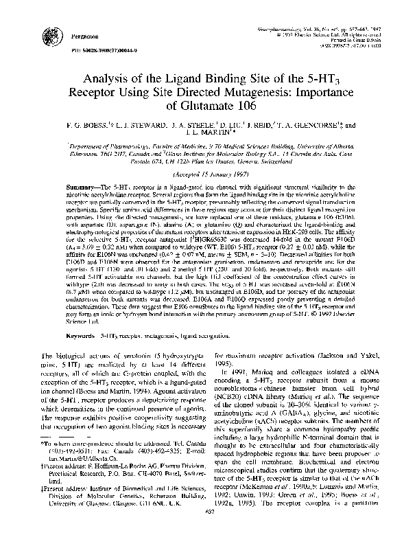
F. G. Boess et al.
N
1
‘A~c
Fig. 1. Schematic representation of a ligand-gated ion channel subunit. Dark boxed areas represent the
hydrophobic (putative transmembrane) regions, C and N represent the carboxy- and amino-ten-ninusregions,
respectively. Loops 1, 2 and 3 are the N-terminal regions contributing to the ligand binding site, as identified in
biochemical and site-directedmutagenesisstudies.A comparisonof the amino acid sequencesof the firstproposed
recognitionloop, in the mouse nACh ct7and 5-HT3ALreceptor subunitsis shown.Boxed areas indicate conserved
amino acids. Tyrosine 93 (Y93) in the nicotinic u subunitshas been identifiedas part of the ligand recognition site
of the nACh receptor. Glutamate 106 (E106) in the 5-HT3ALreceptor has been mutated in the present study to
investigate its involvement in ligand recognition in the 5-HT3receptor.
formed by five identical or related subunits arranged
symmetricallyaround a central pore (Boess et al., 1995).
In contrast to the other ligand-gated ion channels, the
native 5-HT3 receptor may be a homo-oligomer, since
only a single subunithas been identifiedin mouse,rat and
human (Hope et al., 1993;Isenberg et al., 1993; Uetz et
al., 1994; Werner et al., 1994; Belelli et al., 1995;
Miyake et al., 1995). However, in the mouse and rat,
there are two splice variants, 5-HT3-A~and 5-HT3A~,
which differ by the absence or presence of five or six
amino acids in the large intracellular loop region,
between hydrophobic regions three and four (Hope et
al., 1993; Werner et al., 1994; Miyake et al., 1995;
Miquel et al., 1995).
Evidence obtained from biochemical and mutagenesis
studies with the nACh, GABAA and glycine receptors,
suggeststhat the binding site for agonistsand competitive
antagonistsis located in the N-terminal domain (Kuhseet
al., 1995; Dunn et al., 1994; Changeux et al., 1992;
Karlin and Akabas, 1995). A chimeric protein, comprising the N-terminal domain of the U7 neuronal nAChR
with the remaining sequence derived from the 5-HT3
receptor, showed ligand-gated cation channel activity
with ct7-likepharmacology, proving that the N-terminal
domain is essential for ligand-bindingspecificity(Eise16
et al., 1993). Labelling studies with irreversible ligands
identified three regions (“loops”) in the N-terminal
domain of the nAChR a subunits that interact directly
with ligands; additional residues in the d and y subunits
were labelled by some ligands (reviewed in Changeux et
al., 1992). A role for several of these residues in ligand
binding has been confirmedby site-directedmutagenesis
(reviewed in Changeux et al., 1992; Karlin and Akabas,
1995).
Acetylcholine mustard aziridinium is an agonist in
which a reactive crosslinking group is located in the
position of the positivelycharged quaternary ammonium.
In To~edo electric organ nAChR, this irreversibleligand
labels tyrosine 93 in the LXsubunit (Cohen et al., 1991).
The same residue is also labelled by the photoactivatable
antagonist p-dimethylamino-benzenediazonium fluoroborate (DDF; Galzi et al., 1990).Mutationsof this Tyr to
Phe, Trp, Ser, Thr and several unnatural phenykdanine
derivativessuggestedthat the aromatichydroxylgroup of
Tyr forms a salt bridge or acts as hydrogen bond donor
(Grdziet al., 1991a;Aylwin and White, 1994;Sine et al.,
1994; Nowak et al., 1995). All 5-HT3 ligands possess a
positively charged nitrogen, frequently embedded in an
aliphatic or aromatic heterocycle, that may bind to the
5-HT3 receptor in a position homologous to the site of
interaction of the quaternary ammonium group of
acetylcholine with the nACh receptor. Amino acid
sequence alignment of the 5-HT3 receptor with the U7
nAChR in the region containing tyrosine 93 is shown in
Fig. 1. While significantsequence homology is apparent,
there are marked differences in the 5-HT3 receptor
sequencein the immediatevicinity of a7 nAChR tyrosine
93. Antagonist modelling studies predict that the
positively charged nitrogen in high affinity 5-HT3
receptor ligands will form an ion pair with a negatively
charged aspartate or glutamate residue on the receptor
(Gozlan and Langlois, 1992).In order to examine the role
of glutamate 106 in the recognition of agonists and
antagonists, we have modified a 5-HT3AL receptor
sequence isolated from NG108-15 cells (Werner et al.,
1994) using site-directed mutagenesis. Whole-cell patch
clamp and radioligand binding have been used to
characterizewildtypeand mutantreceptors after transient
expression in human embryonic kidney (HEK 293)
cells. A preliminary report of this work was presented
at the British Pharmacological Society (Steward et al.,
1996).
MATERIALS AND METHODS
Site-directed mutagenesisof the 5-HT3-ALcDNA
A cDNA isolated from NG108-15 cells encoding the
mouse 5-HT3-AL receptor (Werner et al., 1994) was
�Mutagenesis of the 5-HT3receptor
generously provided by Dr Eric Kawashima (Glaxo
Molecular Biology Institute, Geneva). The cDNA
sequence was subcloned into the phagemid pAlter
(Promega, Madison, WI, U.S.A.) and the eukaryotic
expression vector pRC/CMV (Promega). Mutagenesis
was performed using the Altered Sites Mutagenesis Kit
(Promega). Glutamate (E) 106 was mutated to aspartate
639
50 mg/1 and phenylmethyl sulphonyl fluoride (PMSF)
100 pM) and homogenized using an ultra-turax
(20000 rpm, 10 see). The homogenate was centrifuged
(27000g for 20 tin at 4“C), the resulting membrane
pellet gently resuspended in ice-cold 10 mhl HEPES (4(2-hydroxyethyl) -l-piperazineethanesulphonic acid) buffer (pH 7.5) and frozen at –20°C until further use. For the
(D), asparagine (N), glutamine (Q), or alanine (A) using assay, the whole cell membranes were recentrifuged
the following mutagenic oligonucleotides(WT is shown (27OOOgfor 20 tin at 4°C), and resuspended in ice-cold
HEPES.
for comparison);
WT
5’ AGA-CTT-CCC-CAC-GTC-CAC-AAA-CTC-A'lT-GAT-GAG-AAT-GTC
3’
E106D
5’ AGA-CTT-CCC-CAC-GTC-GAC-AAA-GTC-ATI'-GAT-GAG-AAT-GTC
3’
E106N
5’ AGA-CH'-CCC-CAC-GTC-GAC-AAA-ATT-ATT-GAT-GAG-AAT-GTC
3’
E106Q
5’ AGA-CTT-CCC-CAC-GTC-GAC-AAA-CTG-ATT-GAT-GAG-AAT-GTC
3’
EI06A
5’ AGA-CR-CCC-CAC-GTC-GAC-AAA-CGC-A~-GAT-GAG-AAT-GTC 3’
A silent mutation, present in each oligonucleotide,
introduced a new Sal 1 restriction site to facilitate mutant
screening. The sequence between two Bsu 361restriction
sites in the 5-HT3-AL sequence encodes the aminoterminus and the firsttwo transmembranesegmentsof the
5-HT3 receptor. Mutant pAlter-5-HT3-ALvectors were
digested with Bsu 361and the resulting 891 bp fragments
were ligated into cut pRC/CMV-5-HT3-ALvector, to
replace the wildtype Bsu 361 fragment. Mutations were
confirmed by sequence analysis using an automated
fluorescent sequencing system (Applied Biosystems,
Foster City, CA, U.S.A.) for the coding region between
and including the two Bsu 361restriction sites.
Transientexpression and preparation of membranes
Human embryonic kidney (HEK 293) cells were
cultured in Dulbecco’s modified Eagles medium
(DMEM) containing 10% fetal bovine serum, 100U/ml
penicillin, 100 pghnl streptomycinand 2 mM glutamine.
The cells were incubated in a humidified 7% COZ
atmosphereat 37°C, in 150 mm diameterplates or 35 mm
diameter plates (for electrophysiologicalstudies).
HEK 293 cells were transiently transected with 1 pg
of pRC/CMV-5-HT3-ALor mutant cDNAs for electrophysiologicalstudies using a modificationof the calcium
phosphate coprecipitation method (Chen and Okayama,
1988)and incubated under 3% C02. Eighteen hours after
transection, the cells were washed with Hanks buffered
salt solution, fresh media (DMEM) added and the cells
subsequentlyincubated under 770 C02. Recordingswere
made 1–2days after transection. For radioligandbinding
studies, HEK 293 cells were transiently transected with
35-45 pg of pRC/CMV-5-HT3-ALor mutant cDNAs.
Forty-eight hours after transection the cells were
harvested in ice-cold protease inhibitor buffer (Tris
10 mM pH 7.5, ethylenediaminetetra-aceticacid (EDTA)
1 rdvl, bacitracin 50 mg/1, soybean trypsin inhibitor
[3H]GR65630radioligandbinding assay
For [3H]GR65630 binding, assay tubes contained
800 PI of competing drug (non-specific binding was
defined in the presence of 300PM metoclopramide), or
vehicle (HEPES 10 mM buffer, pH 7.5) and 100 pl
[3H]GR65630 (0.04-12 nM for saturation studies and
0.41–1.85nM for competition studies). The assay tubes
were preincubatedfor 2 min at O“Cbefore the additionof
100pl cell membranes (equivalent to approximately
100pg protein) to initiate binding which was allowed to
proceed at O°C for 2 hr. It was terminated by rapid
filtration under vacuum through Whatman GF/B filters
(pretreated with 0.3% v/v polyethyleneimine in HEPES
buffer), followedby washingwith ice-cold HEPES buffer
for 8 sec. Bound radioactivity remaining on the filters
was determined in 4.5 ml Ecoscint A (National Diagnostics) by liquid scintillation spectroscopy, at an
efficiency of approximately6090.
Electrophysiology
Membrane currents from single cells were recorded
under voltage clamp with the whole-cell configurationof
the patch clamp technique using an Axopatch-lD
amplifier (Axon Instruments, Foster City, CA, U.S.A.).
All currents were subject to initial run-down within the
first few minutes of recording and were allowed to
stabilize before data collection began. Data acquisition,
storage and analysis were performed using pClamp 6
(Axon). Cells expressing receptors could be identified
visuallyusing phase contrastmicroscopy.These cells had
clusters (up to six) of small (approximately 2 pm in
diameter) phase bright “inclusion bodies”; 90’%0
of these
cells generated measurable currents. Borosilicate glass
pipettes were heat-polished and had resistances of 2–
5 M!2 The membrane potential was held at –60 mV and
inward currents are displayed downwards. Ligands were
applied to the cell using a gravity-feed rapid perfusion
�F. G. Boess et al,
640
Materials
HEK 293 cells were from the American Type Culture
Collection (ATCC, Rockville, MD, U.S.A.), DMEM,
Hank’s buffered salt solution (HBSS) and penicillin/
streptomycin from BioWhittaker (Marysville, MD,
U.S.A.), and fetal bovine serum (Life Technologies,
Burlington,Ontario, Canada). 5-Hydroxytryptaminewas
obtained from Sigma, meta-chlorophenylbiguanide, 2methyl-5-HT,metoclopramidefrom Research Biochemical Inc. (Natick, MA, U.S.A.), [3H]GR65630 (61.464.4 Ci/mmol) from NEN Dupont, ondansetron was
donated by Glaxo (Ware, U.K.) and renzapride and
granisetron were donated by SmithKline Beecham
Pharmaceuticals(Harlow,U.K.). All drugs were prepared
in HEPES 10 mM (pH 7.5). Other chemicals and reagents
were purchased from Sigma Chemical Company, BDH
(Toronto, Ont., Canada) and FischerBiotech (Nepean,
Ont., Canada).
(A)
[3H]GR65630 Free [nM]
-600
z
= 500
% 400
g.- 300
~ 200
r% 100
-#
ob
o
‘B)
20
40
‘/0
60
60
100
Specific
Fig. 2. [3H]GR65630saturation binding in HEK 293 cells
transiently transected with 5-HT3-ALwildtype (WT = E106
(~)) or mutant (E106D (0)) cDNA. (A) Results shown are
from a single experiment. K~ values were determined with an
iterative curve fitting program. Data is from a single
representative experiment which was repeated 3–10 times.
Mean K~values are found in Table 1. For E106D, an additional
point was obtained at 11.76nM representing over 90%
saturation. Specific binding was defined by 300 PM metoclopramide. (B) Scatchard transformation with normalized
specific binding (I&u = 100%),to compensate for differences
in transection efficiencies for WT and E106D.
system based on that described by Carbone and Lux
(1987). The cell was continuouslyperfused and solution
changes were effected by a manually-operated valve
which housed a manifold connected to solution reservoirs. The bath solution contained (in mM): NaCl, 130;
KC1,5; CaC12,1.8; MgC12,1.2; HEPES, 10; glucose, 5;
pH 7.4. The pipette solution contained (in mM): CSC1,
135; MgC12, 0.5, l,2-bis(2-aminophenoxy)ethaneN,N,N’,N’-tetraaceticacid tetrapotassium salt (BAPTA),
10; HEPES, 10; pH 7.2 with CSOH.All recordings were
made at room temperature (19–21°C).
Data analysis
Radioligandbinding data were analyzed by computerassisted iterative curve fitting (Kaleidagraph, Synergy
Software, Reading, PA, U.S.A.) according to the
equation:B = (Bm,X[L]”)/([L]n+ (K)”), where B is bound
radioligand, B~~Xis maximum binding at equilibrium, K
is the equilibrium dissociation constant for saturation
studies or the molar concentration of competing compound to reduce the specific binding by 50% for
competition studies (IC50),[L] is the free concentration
of radioligand for saturation studies or molar concentration of competing compound for competition studies and
n is the Hill coefficient.
The Cheng–Prusoff equation was used to calculate
both the Ki values of the competing drugs in radioligand
binding studies and the Kb values for antagonists of 5HT-induced currents in whole-cell patch clamp studies
(Craig, 1993).For radioligand binding studies Ki = IC5~
(1+ ([L]/K~)),where IC~ois the inhibitory concentration
of the competing compound to reduce the specific
binding by 50%, [L] is the free concentration of
radioligand and Kd the equilibrium dissociation constant
of the radioligand. For whole-cell patch clamp studies,
Kb = IC5~(1+ ([A]/EC50)),where IC50is the concentration of antagcinistrequired to inhibit the agonist response
by 50%, [A] is the agonist concentration used to induce
Table 1. Affinities of mutant and wildtype 5-HT3receptors in transiently transected HEK 293 cells
Ligand
[3H]GR65630(Kd)
5-HT
2-Methyl-5-HT
m-CPBG
Granisetron
Ondansetron
Renzapride
WT
0.27 +
15.56 ~
173 +
5.01 +
0.28 +
0.58 +
1.29 ~
0.03 (10)
3.22 (8)
18 (9)
1.09 (6)
0.03 (6)
0.10 (5)
0.17 (4)
E106D
3.69 i
2056 ~
42711 +
8.37 ~
232 ~
15.98 ~
28.87 +
0.32 (3)
564 (6)
9117 (3)
3.08 (3)
69 (3)
1.5 (3)
9.97 (3)
E106N
0.42 +
469 +
2947 t
2.89 t
154 *
5.56 ~
16.97 t
0.07 (3)
25 (3)
456 (3)
0.37 (3)
86 (3)
1.14 (5)
3.82 (3)
Ki values of the ligands were obtained by determination of IC50values obtained in competition with
[’H] GR65630,together with the Kd values obtainedby saturationwith [3H]GR65630.All values
are mean (nM)~ SEM (rI determinations).
�641
Mutagenesis of the 5-HT3receptor
u
~
1( I m
■
:
5
*
$
0zyxwvutsrqponmlkjihgfedcbaZYXWVUTSRQPONMLKJIHGFEDCBA
,
‘
Fig. 3. 5-HT competition for [3H]GR65630binding (l.592.80 nM) in HEK 293 cells transiently transected with 5-HT3ALwildtype (WT) or mutated (E106N)cDNA. Data are from a
single representative experiment which was repeated three to
eight times. Mean Ki values are found in Table 1.
Fig. 4. Granisetroncompetingfor [3H]GR65630binding (0.5&
0.57 nM) in HEK 293 cells transiently transected with 5-HT3ALwildtype (WT) or mutated (E106D)cDNA. Data are from a
single representative experiment which was repeated three to
six times. Mean Ki values are found in Table 1.
the response and
the concentration of agonist
required to produce a half-maximal response.
Concentration-effect curves were fitted to the equaby computer-assisted iteration: Z= 1~,.J(l + (
tive curve fitting as described previously, where Z and
z~= are the currents at a given agonist concentration and
the maximal value, respectively.
is the agonist
concentration required to obtain half-maximal current
and n is the Hill coefficient.
showed no significant change in affinity for the mutant
receptors when compared to WT (Table 1). Hill
coefficients for all agonists were approximately 1,
indicating that in the present study there was no evidence
of agonist cooperativity in radioligand binding for WT or
mutant receptors.
The affinities of several 5-HT3 receptor antagonists
were also altered. The largest change was observed for
granisetron, with an 800-fold and 550-fold decrease in
R
1 nA
Radioligand binding characterization of mutant 5-HT3
receptors E106D and E106N
In saturation studies with membranes obtained from
HEK 293 cells transiently expressing wildtype (WT),
E106D or E106N mutant 5-HT3 receptors, [3H]GR65630
labelled a homogeneous population of binding sites (Fig.
2). The binding site density varied with the efficiency of
both transection and expression. Therefore, for ease of
comparison, the saturation data are normalized to 11~,, in
the Scatchard transformations (Fig. 2B). The affinity of
[3H]GR65630 was decreased by 14-fold for the mutant
E106D (Kd = 3.69 f 0.32 uM, mean ~ SEM; n =3)
when compared to WT (0.27 i 0.03 nM, n = 10; Table
1, Fig. 2.); however, there was no appreciable difference
in the affinity of [3H]GR65630 for the mutant E106N
(Table 1).
Competition for [3H]GR65630 binding by a variety of
5-HT3 receptor agonist and antagonist ligands demonstrated changes in affinity for the mutant receptors, when
compared to WT. 5-HT showed a 130-fold and a 30-fold
decrease in affinity for the mutant receptors E106D
(m= 2056 f 564 uM, n = and E106N (469 f 25 nM,
n = 3), respectively,
when
compared
to WT
(15.56 ~ 3.22 nM, n =8; Fig. 3., Table 1). Similarly,
the affinity of the 5-HT3 receptor agonist 2-methyl-5-HT
was decreased 250-fold for E106D and 17-fold for
E106N (Table 1). However, the high affinity 5-HT3
receptor agonist meta-chlorophenylbiguanide (mCPBG)
“0’”
~
300 PA
“ ~
“ ~
“0”~
100 pA
150 pA
50 PA
2 sec
Fig. 5. A comparisonof 5-HT-inducedinward currents in HEK
293 cells transiently transected with 5-HT3-AL wildtype
(E106) or mutated (E106D, E106N, E106Q, E106A) cDNAs.
The horizontal bar indicates the duration of the 5-HT
application (WT, 50 ,uM; E106D, 100,uM; E106N, 100pM;
E106Q, 100YM; E106A, 10 mM). Currents were recorded
from cells voltage clam~ed at a holding Dotentialof –60 mV.
�F. G. Boess et al.
642
on application of 100 VM 5-HT, while both E106D and
1-
E106N mutant receptors exhibited maximal current
levels some 10-fold lower (Fig. 5). This may be due to
low expression levels as a similar reduction was observed
in radioligand binding studies. There was no reduction in
the respective currents for WT and mutant receptors with
application of up to 10 rnM 5-HT at a holding potential of
–60 mV indicating that agonist dependent block did not
occur.
In concentration-effect curve studies, the EC511for 5HT was increased seven-fold (8.7 PM, n =6) for the
0.8$ 0.6E
s o.40.20
w
1
1O-s 1o-7 10-s
10-5
10-4
,.-3
5-HT (M)
Fig. 6. Concentration-effect curves for 5-HT3-ALwildtype and
mutant E106N receptors in transiently transected HEK 293
cells. Data are means (n = 5–10cells). Data were fittedwith the
Hill equation Z= Z~J(l + (EC5~[5-HT])”)where Zand Z~u are
the currents at a given 5-HT concentration [5-HT] and the
maximal value respectively, and n is the Hill coefficient.
affinity for E106D (232*69 nM, n = 3) and for E106N
(154*86 uM, n =3) when compared to WT (0.28 f
0.03 nM, n =6; Fig. 4, Table 1). Renzapride and
ondansetron showed smaller decreases in affinity of
approximately 10-30-fold (Table 1).
Radioligand binding characterization of mutant 5-HT3
receptorsE106A and E106Q
The expression of E106A and E106Q was poor
compared to WT. Specific binding of [3H]GR65630(l–
2 nM) was too low to permit acceptable radioligand
binding characterization of E106A and E106Q precluding accurate Ki or Kd determinations.A direct comparison of membranes from untransfected and transected
HEK 293 cells yielded “specific” [3H]GR65630
(1.8 nM) binding of 603 (wildtype), 75 (E106A), 69
(E106Q) and 408 (NG108-15 cells; fmol/mg protein;
single determination). A structurallydistinct radioligand
[3H]-(S)-zacopride (data not shown) also yielded low
specificbinding for E106A and E106Q.
Electrophysiologicalcharacterizationof wildtype 5-HT3
receptor and mutants E106D and EI06N
The whole-cell patch clamp technique was used to
examine the functional properties of WT and mutant
receptors. WT 5-HT3 receptors transiently expressed in
HEK 293 cells produced maximal currents up to 2.5 nA
asparagine mutant E106N when compared to WT
(1.2 PM; n =9; Fig. 6, Table 2). There was no change
in EC50for 5-HT for the aspartate mutant (E106D; Table
2). The Hill coefficient for WT was 2.0 indicating
positive cooperativity of agonist activation (Table 2),
whereas for both mutant receptors E106D and E106N,
the Hill coefficientwas significantlydifferent to wildtype
and close to unity, suggesting a loss in positive
cooperativity (Fig. 6, Table 2).
meta-Chlorophenylbiguanide(mCPBG) is a full agonist in NIE-115 cells (Septilvedaet al., 1991) and has a
lower EC50 than the natural agonist 5-HT. In our
experiments, mCPBG acted as a full agonist at WT and
E106D receptors. There was no significantchange in the
EC50values of mCPBG for E106D (Table 2) which was
consistentwith the radioligandbinding studies.However,
for mCPBG, the Hill coefficient observed in concentration-effect curve with E106D was significantlyreduced
compared to WT (Table 2).
In order to determine antagonist potencies, relatively
high concentrationsof 5-HT were utilized because of the
poor expression of mutant receptors. Therefore, 10 and
20 pM 5-HT were used for E106D and E106N,
respectively,to generate an appropriateagonist response,
while 10PM 5-HT was used for similar studies with WT.
The antagonist potency of ondansetron was reduced for
both E106D and E106N mutant receptors when compared to WT receptors (Table 2). Blockade by ondansetron was readily reversed by washing (Fig. 7). The
antagonist potency of renzapride was decreased for
E106N (IC50, 121nM; Kb 36.7 nM; Table 2) when
compared to WT (IC50,11.5nM; Kb 1.23nM), while no
change was found for E106D (Table 2). These decreases
in antagonistpotencies were similar to those produced in
radioligand binding studies with the exception of the
Table 2. Whole-cell patch clamp data for mutant and wildtype 5-HT3receptors in transiently transected HEK 293 cells
m-CPBG
5-HT
Receptor
WT
E106D
E106N
Renzapride
Ondansetron
EC50(MM)
Hill
EC~o(pM)
Hill
IC50 (nM)
Kb (nM)
IC50 (nM)
Kb (nM)
1.2 (1.1–1.3)
1.2 (1.0-1.6)
8.7 (6.8-11.2)
2.0 (1.62.3)
1.1 (0.9-1.3)
1.0 (0.7–1.2)
0.3 (0.3-0.4)
0.4 (0.3-0.6)
ND
1.6 (1.3-1.9)
0.8 (0.6-1.0)
ND
6.0 (4.2-8.3)
104 (64-169)
74 (52-104)
0.64
11.1
22.4
11.5 (8.1-16.6)
15.4 (12.4-19.1)
121 (99-148)
1.23
1.65
36.5
EC511
and Hill coefficient values are given for the agonists 5-HT and mCPBG. IC~IIand Kb values are given for the antagonists renzapride and
ondansetron.Data for the antagonists were generated in the presence of 10AM 5-HT for E106 and E106D, and 20 pM 5-HI for E106N.
Values are given as means with 95% confidence intervals in parentheses. Data were collected from 6-13 cells for determination of each
value. ND= not determined.
�Mutagenesis of the 5-H’T3receptor
A
5-HT
bidansetron
5-HT
643
5-HT
300PA
1
““DL
100PA
~Q’75pA
4SSC
B
5-HT
9rsnisetron
5-HT
~
5-HT
-L.-.
--l’”pA
zyxwvutsrqponmlk
““’”ti
4sec
Fig. 7. Ondansetron(A) and granisetron (B) blockade of 5-HT-inducedinward currents recorded from HEK 293
cells transiently transected with wildtype or mutantE106Dor E106NcDNAs. (A) Cells were prepulsedfor 30 sec
with ondansetron (WT, 10 nM; E106D, 1 PM; E106N, 100nM), or (B) prepulsed with granisetron (WT, 50 nM;
E106D, 100nM) and then either 10pM (WT, E106D)or 20 PM (E106N)5-HTwas coappliedwith the antagonist.
After approximately 1 hr of washing subsequentto granisetronapplication,the maximal current was still reduced,
compared to initiat pre-granisetron5-HT current. This precluded a detailed study of this antagonist.
renzapride interaction with E106D. The 5-HT3 receptor of up to 2PM failed to antagonize 10 mM mCPBGantagonist, granisetron, blocked the current response to induced responses for E106A receptors. The EC50of 55-HT (10 PM; Fig. 7) for E106D and WT, but recovery HT for E106Q mutant receptors was approximately
from the antagonism was very slow precluding its ICSCI 50 pM (n= 7). However, mCPBG behaved as an
determination.Similar experimentshave not been carried antagonist with this mutant, blocking 100 pM 5-HTinduced inward current with an IC50of approximately
out with E106N.
50 nM (n= 5) but producing no response when applied
Electrophysiologicalcharacterizationof mutants E106A alone.
and E106Q
DISCUSSION
As observed in the radioligand binding studies, it
appeared that E106A and E106Q mutant receptors
In the nicotinic acetylcholine receptor, amino acids
expressed poorly. Currents produced with 5-HT for involved in ligand recognition are concentrated in three
E106Q and E106A were greatly reduced (<200 pA) when “loops” of the a subunitsand additional sites are present
compared to WT (Fig. 5) preventing their detailed study. on the y and d subunits (reviewed in Changeux et al.,
However, estimated values for 5-HT and mCPBG for 1992; Karlin and Akabas, 1995). A comparison of the
E106A indicated that their EC50values were greater than amino acid sequence of the 5-HT3 receptor and nAChR
1 mM (n= 3), an approximate 1000-foldincrease for 5- a subunits shows that there is significant sequence
HT and 30000-fo~d-increase for mCPBG, when com- conservation in the first of these recognition loops (Fig.
pared to WT. In addition. ondansetron at concentrations 1). However, there are also major differences, for
�644
F. G. Boess et al.
example, the triplet YNS of the nAChR IX7sequence
aligns with NEF of the 5-HT3 receptor sequence. The
tyrosine residue is conserved in all nAChR ci subunit
sequencesknown to date, with the exception of &5which
is functionally atypical. Irreversible labelling and mutagenesis experiments have confirmed that this residue is
important for ligand binding to the nAChR, where it may
interact with the diffuse positive charge of the quatemary
ammonium group of acetylcholine (reviewed in Changeux et al., 1992; Karlin and Akabas, 1995). The
glutamate residue at the adjacent position of the 5-HT3
receptor sequence (E106) may subserve a similarly
important role in recognition and form an ionic interaction with the ammonium group of the natural agonist 5HT or the positively charged nitrogen, present in all 5HT3 receptor ligands. To explore this possibility, we
mutated glutamate 106 to the similarly charged amino
acid aspartate, the uncharged but polar analogues
glutamine or asparagine and the small neutral amino
acid alanine. We examined these mutants with both
radioligand binding, to characterize the recognition
properties of the desensitized receptor state and wholecell patch clamp electrophysiology to study the active
state of the receptor.
Our data show that shortening the side chain of
glutamate by one methylene in E106D, reduces the high
affinity recognition of both 5-HT and 2-methyl-5-HTby
over two orders of magnitude, though the EC50for 5-HT
is unchanged. However, we find essentiallyno change in
the high affinitybinding of the agonistmCPBG or its ECW
determined electrophysiologically. Removal of the
charge on the aspartate in the mutant E106N produces
broadly the same change but the magnitudeis less; again
mCPBG is unaffected by this mutation. Generally,
antagonist recognition is similarly affected for both
E106D and E106N mutants. The alanine and glutarnine
mutants express very poorly and while we have reported
some data on their electrophysiologicalcharacterization,
radioligand binding studies proved impracticable. These
observations suggest that changing E106 interferes with
either the formation, or the stability of functional 5-HT3
receptor complexes. E106 may be important for the
proper folding of subunits or the assembly of five
subunits to a homopentamer. Folding or expression
clearly place significant constraints at this position and
we are only able to address functionality within those
limits.
In the wildtype 5-HT3 receptor, we initially proposed
that the negatively charged carboxylate group of
glutamate 106 may form a direct ionic interaction with
the positivelycharged primary ammoniumgroup of 5-HT
and the positively charged secondary, tertiary or
quatemary ammonium groups present in the majority of
high affinity 5-HT3 receptor antagonists (Gozlan and
Langlois, 1992).However, our observationsindicate that
in the asparagine mutant E106N, where the charge has
been removed, the binding of 5-HT and 2-methyl-5-HTis
less affected than for E106D. It appears unlikely,
therefore, that the interaction of glutamate (E106) with
5-HT and 2-methyl-5-HT(Fig. 8) is ionic, as the similarly
charged aspartate produces greater changes in binding
affinity than the polar residue asparagine. These compounds may instead form a hydrogen bond with
glutamate 106, and both aspartate and asparagine may
be able to partially substitute for glutamate in this
interaction.
Replacement of E106 by aspartate or asparagine did
not alter the affinity of the 5-HT3 receptor agonist
mCPBG, suggesting that interaction with E106 is not
essential for high affinity mCPBG binding. In the
protonated form of this ligand, the positive charge is
presumablydelocalizedover the whole molecule (Fig. 8).
Interactions with the aromatic side chains of tyrosine
(Tyr) and tryptophan(Trp) residuesof the 5-HT3receptor
may be more importantfor the binding of mCPBG. It has
been suggested that the ‘mild electronegative’ character
of the n electron systems of Tyr and Trp residues serves
to complex the diffuse positive charge of the quatemary
ammoniumgroup of acetylcholine(Galzi and Changeux,
1995). This idea is supported by the observation that a
synthetic aromatic host strocture can bind acetylcholine
(Dougherty and Stauffer, 1990). Several of these
aromatic residues are conserved in the 5-HT3 receptor
(W67, W98, W160, Y211) and one of them (W67) has
already been shown to contribute to ligand binding
(Schulte et al., 1995).
In the nAChR, a tyrosine residue (Y93) in the
correspondingregion (loop 1) is labelled five times more
intensely by the photoactivatable,irreversible antagonist
DDF, if labelling is carried out in the presence of
meproadifen, a non-competitive antagonist that is
thought to shift the equilibrium to the desensitized state
of the receptor (Galzi et al., 1991b). This led to the
suggestion that confirmational changes in the binding
site upon desensitization render this residue more
accessible. The irreversible cholinergic agonist acetylcholine mustard aziridinium, that is thought to interact
with the high affinity desensitized state labels only Y93
(Cohen et al., 1991).In analogy with the nAChR, the 5HT3 receptor can probably assume several confirmational states, including a resting closed, an open and
several desensitized states (Changeux et al., 1992;
Jackson and Yakel, 1995). Agonist binding promotes
the transitionfrom the resting to the open state and in the
continuingpresence of agonists the receptor desensitizes
and assumes one of several possible closed, desensitized
conformations, that display a higher affinity for the
agonist. In equilibrium binding experiments with agonists, the affinity of the desensitized high affinity state is
measured. Several groups have observed, that the
concentrationof agonist for the induction of desensitization in the 5-HT3receptor is lower than that for channel
activation (Bartrup and Newberry, 1996; van Hooft and
Vijverberg, 1996).In NG108-15 cells, the IC50values for
desensitizationby mCPBG (20 nM), 5-HT (50 nM) and
2-methyl-5-HT (600 nM) (Bartrup and Newberry, 1996)
�Mutagenesis of the 5-HT3receptor
JNH2
I#y
/-’NH2
2-m3thyl-5-H’I
5-HT
645
6H3
GR65630
mCPBG
CH3
Ondansetron
7H3
CH3
CH3
Renzapnde
Fig. 8. Chemical structures of 5-HT3 receptor agonists (5-HT, 2-methyl-5-HT and mCPBG) and antagonists
(GR65630,ondansetron,granisetron and renzapride).
are very similar to the Ki values (14, 170 and 810 nM,
respectively) determined in radioligand binding assays
with intact cells under physiologicalconditions(Boess et
al., 1992b).While in our studies, there was more than a
100-fold decrease in the affinity of 5-HT for the mutant
E106D measured by radioligand binding, the EC50of 5HT observed in electrophysiological experiments with
this receptor did not change. For E106N, a 30-fold
decrease in binding affinity,butjust a seven-foldincrease
in EC50was found. Our data suggestthat the interactionof
5-HT with the receptor, which is responsible for the
transition from the closed resting to the open state, is
retained when glutamate 106 is substitutedby aspartate.
However, an asparagine, glutamine, or alanineresidue in
this position reduces the potency of 5-HT. We have not
observed any clear changes in the desensitizationkinetics
of the responses elicited by 5-HT in cells expressing
E106D or E106N mutant receptors.If the reduced affinity
in radioligandbinding studies reflects a lower affinityfor
the desensitized receptor, a detailed analysis of the IC5CI
values of agonists for the induction of desensitization
would perhaps reveal differences between these mutants
and the wildtype receptor.
Concentration-response studies revealed a reduction
of the Hill coefficientsfrom a value of two in the wildtype
to unity in both E106D and E106N mutant receptors.
High Hill coefficientsare generally interpreted as a sign
of cooperativity between different binding sites on the
same receptor complex. In the nAChR, binding sites are
present on the subunit interfaces, i.e. different parts of
two subunits contribute to a single binding site. Because
the nAChR complex is a heteropentamer, binding sites
are not equivalent (Pedersen and Cohen, 1990). Occupation of two binding sites promotes an allosteric transition
from a closed resting to an open state of the receptor
(Jackson, 1994). In homopentameric receptors such as
recombinant IX7nAChR expressed in Xenopus oocytes,
the existence of five binding sites for the competitive
�646
F. G. Boess et al.
antagonistmethyllycaconitinehas been suggested(Palma
et al., 1996). The recombinant 5-HT3 receptor is a
homopentamerand the five potentialbinding sites on the
subunit interfaces should initially be equivalent. The
cooperativity observed at wildtype recombinant 5-HT3
receptors expressed in HEK 293 cells suggests that
binding of at least two agonist moleculesis necessary for
the efficient activation of the receptor, as in the nAChR.
This coupling of two binding sites on different subunit
interfaces may be eliminated after replacement of E106
by either aspartate or asparagine. In the resting state of
the 5-HT3receptor pentamer, E106 may participatein an
inter-subunit bond, that is changed during the allosteric
transition after agonist binding.
All the highly selective 5-HT3 receptor antagonists
examined in the present study blocked 5-HT current
activation, indicating that the mutant receptors retain 5HT3 receptor-like recognitionproperties.The changes in
affinity of these antagonists were generally similar in
both radioligand binding and electrophysiological studies. However, there were subtle differences, even for
example between the structural analogues GR65630 and
ondansetron.The affinity of ondansetronand its potency
to inhibit agonist activation, was similarly reduced for
both E106D and E106N, while the affinity of GR65630
was unaffected in the mutant E106N compared to WT,
but was reduced 15-fold in E106D. In these ligands, the
positive charge which is proposed to interact with E106,
is delocalized within an imidazoleheterocycle (Fig. 8). It
is possible that the restricted confirmational flexibility
imposedby the additionalring (C; Fig. 8) of ondansetron,
imposeslimitson its effectiveinteractionwith asparagine
which is not experienced by GR65630.
The 5-HT3 receptor antagonist granisetron showed a
markedly reduced affinity for both the aspartate and
asparagine mutants (800–500-fold) in contrast to the
other antagonists which we have investigated. In this
Iigand, the basic nitrogen is contained in a highly
substitutedheterocycle, but unlike GR65630 and ondansetron the charge is not delocalized.It is possiblethat it is
interacting with E106 in a similar manner to 5-HT and 2methyl-5-HT.Many studieshave exploredthe systematic
modificationsof this heterocyclebut no clear conclusions
have been possible as to the nature of the interaction of
the basic nitrogen with the receptor (Gozlan and
Langlois, 1992). It is interesting, however, that the
affinity of renzapride, in which the basic nitrogen is
contained within a different, but similarly bulky heterocycle, was only reduced 10-20-fold. In addition,
renzapride showed differential effects for E106D with
no change in its ability to antagonizereceptor activation
but a reduction in binding affinity, whereas for E106N
both antagonist potency and affinity were reduced.
Our results suggest that glutamate 106 is important in
ligand recognitionfor 5-HT3receptor antagonistsand for
the agonists in the high affinity desensitized state of the
receptor. Its involvement with agonist activation is
markedly less pronouncedbut its mutation results in loss
of cooperativity. Our data are consistent with an
interaction between this residue and the basic nitrogen
present in all 5-HT3 receptor ligands and further
exploration of the vicinal residues seems warranted.
Acknowledgements—The authors would like to thank Drs Tom
Blackburn (SmithKline Beecham) and Gavin Kilpatrick
(Glaxo) for their kind gifts of drugs. Supported by Glaxo
Canada and MRC Canada. L.J.S. holds a MRC/PMAC Pfizer
(Canada) Fellowship.
REFERENCES
Aylwin M. L. and White M. M. (1994) Ligand-receptor
interactions in the nicotinic acetylcholine receptor probed
using multiple substitutions at conserved tyrosines on the a
subunit. FEBS Letters 349: 99–103.
Bartrup J. T. and Newberry N. R. (1996) Electrophysiological
consequences of ligand binding to the desensitized 5-HT3
receptor in mammalian NG108-15 cells. Journal of
Physiology 490.3: 679–690.
Belelli D., Balcarek J. M., Hope A. G., Peters J. A., Lambert J.
J. and Blackbum T. P. (1995) Cloning and functional
expression of a human 5-hydroxytryptamine type 3A,
receptor subunit. Molecular Pharmacology48: 1054–1062.
Boess F. G., Lurnmis S. C. R. and Martin I. L. (1992a)
Molecular properties of 5-hydroxytryptarnine3receptor-type
binding sites purified from NG108-15 cells. Journal of
Neurochemistry 59: 1692–1701.
Boess F. G., SeptilvedaM.-I., Lummis S. C. R. and Martin I. L.
(1992b) 5-HT3 receptors in NG108-15 neuroblastoma x
glioma cells: effect of the novel agonist l-(m-chlorophenyl)biguanide. Neuropharmacology31: 561–564.
Boess F. G. and Martin I. L. (1994)Molecular biology of 5-HT
receptors. IVeuropharmacology33: 275–317.
Boess F. G., Beroukhim R. and Martin I. L. (1995) Ultrastmcture of the 5-HT3receptor. Journal of Neurochemistry
64: 1401-1405.
Carbone E. and Lux H. D. (1987) Kinetics and selectivity of a
low-voltage-activated calcium current in chick and rat
sensory neurones. Journal of Physiology 386: 547–570.
Changeux J.-P., Galzi J.-L., Devilliers-Thi&y A. and Bertrand
D. (1992) The functional architecture of the acetylcholine
nicotinic receptor explored by affinity labelling and sitedirected mutagenesis. Quarterly Reviews of Biophysics 25:
395432.
Chen C. A. and Okayama H. (1988) Calcium phosphatemediated gene transfer: a highly efficienttransection system
for stably transforming cells with plasmid DNA.
BioTechniques 6 (7): 632-637.
Cohen J. B., Sharp S. D. and Liu W. S. (1991) Structure of the
agonist-binding site of the nicotinic acetylcholine receptor.
Journal of Biological Chemistry 266: 23354-23364.
Craig D.A. (1993)The Cheng–Pmsoffrelationship: something
lost in the translation. Trends in Pharmacological Sciences
14: 89-91.
Dougherty D. A. and Stauffer D. A. (1990) Acetylcholine
binding by a synthetic receptor: implications for biological
recognition. Science 250: 1558–1560.
Dunn S. M. J., BatesonA. N. and Martin I. L. (1994)Molecular
neurobiologyof the GABA~ receptor. International Review
of Neurobiology 36: 51–96.
Eisel& J.-L., Bertrand S., Galzi J.-L., Devillers-Thi&y A.,
�Mutagenesis of thIe 5-HT3receptor
Changeux J.-P. and Bertrand D. (1993) Chimaenc nicotinic
serotonergic receptor combines distinct ligand binding and
channel specificities. Nature 366: 479483.
Galzi J.-L., Revah R., Black D., Goeldner M., Hirth C. and
ChangeuxJ.-P. (1990)Identificationof a novel amino-acidutyrosine 93 within the cholinergic ligand binding sites of the
acetylcholine receptor by photoaffinitylabeling. Additional
evidence for a three-loop model of the cholinergic ligandbinding sites. Journal of Biological Chemistty 265: 1043010437.
Galzi J.-L., Bertrand D., Devillers-Thi&y A., Revah R.,
Bertrand S. and Changeux J.-P. (1991a) Functional significanceof aromatic amino acids from threepeptide loops of
the ct7 neuronal nicotinic receptor investigated by sitedirected mutagenesis. FEBS Letters 349: 99–103.
Galzi J.-L., Revah R., Bouet F., M6nez A., Goeldner M., Hirth
C. and Changeux J.-P. (1991b) Allosteric transitions of the
acetylcholine receptor probed at the amino acid level with a
photolabile cholinergic ligand. Proceedings of the National
Academy of Science U.S.A. 88: 5051–5055.
Galzi J.-L. and Changeux J.-P. (1995) Neuronal nicotinic
receptors: Molecula organization and regulations. Neuropharrnacology34: 563-582.
Gozlan, H. and Langlois, M. (1992) Structural analysis of
receptor–ligand interactions for the mapping of 5-HT3
antagonist binding site. In Central and Peripheral 5-HT3
Receptors, ed. Hamon, pp. 59-87. Academic Press Ltd.
Green T., Stauffer K. A. and Lummis S. C. R. (1995)
Expression of recombinant homo-oligomeric 5-hydroxytryptamine3 receptors provides new insights into their
maturation and structure. Journal of Biological Chemistry,
270 (11): 6056-6061.
Hope A. G., Downie D. L., SutherlandL., Lambert J. J., Peters
J. A. and Burchell B. (1993) Cloning and functional
expression of an apparent splice variant of the murine 5HT3receptor A subunit.EuropeanJournal of Pharmacology
(Molecular Pharmacology Section) 245: 187-192.
Isenberg K. E., Ukhun I. A., Holstad S. G., Jafri S., Uchiro U.,
ZorumskiC. F. and Yang J. (1993)Partial cDNA cloning and
NGF regulation of a rat 5-HT3receptor subunit.NeuroReport
5: 121-124.
Jackson M. B. (1994) Single channel currents in the nicotinic
acetylcholine receptor: a direct demonstration of allosteric
transitions. Trends in Biochemical Sciences 19 (10): 396399.
Jackson M. B. and Yakel J. L. (1995) The 5-HT3 receptor
channel. Annual Reviews in Physiology 57: 447-468.
Karlin A. and Akabas M. H. (1995) Toward a structural basis
for the function of nicotinic acetylcholinereceptors and their
cousins. Neuron 15: 1232–1244.
Kuhse J., Betz H. and Kirsch J. (1995) The inhibitory glycine
receptor: architecture, synaptic localization and molecular
pathology of a postsynaptic ion-channel complex. Current
Opinion in Neurobiology 5: 318-323.
Lummis S. C. R. and Martin I. L. (1992) Solubilization,
purification, and functional reconstitution of 5-hydroxytryptamine3 receptors from NIE-115 neuroblastoma cells.
Molecular Pharmacology41: 18-23.
Maricq A. V., Peterson A. S., Brake A. J., Myers R. M. and
Julius D. (1991) Primary structure and functional expression
of the 5-HT3receptor, a serotonin-gatedion channel. Science
254: 432437.
McKeman R. M., Biggs C. S., Gillard N. P., Quirk K. and
647
Ragan C. I. (1990a) Molecular size of the 5-HT3 receptor
solubilizedfrom NCB-20 cells. Biochemistry 269: 623-628.
McKeman R. M., Gillard N. P., Quirk K., Kneen C. O.,
Stephenson G. I., Swain C. J. and Ragan C. I. (1990b)
Purificationof the 5-hydroxytryptamine5-HT3receptor from
NCB20 cells. Journal of Biological Chemistry 265: 13572–
13577.
Miquel M.-C., Ement M. B., Gingrich J. A., Nosjean A.,
Hamon M. and El Mestikawy S. (1995) Developmental
changesin the differentialexpressionof two serotonin5-HT3
receptor splice variantsin rat. Journal of Neurochemistry65:
475483.
MiyakeA., MochizukiS., TakemotoY. and Akuzawa S. (1995)
Molecularcloning of human 5-hydroxytryptamine3receptor:
heterogeneity in distribution and function among species.
American Society of Pharmacology and Experimental
Therapeutics48: 407-416.
Nowak M. W., Keamey P. C., Sampson J. R., Saks M. E.,
Labarca C. G., Silverman S. K., Zhong W., Thorson J.,
Abelson J. N., Davidson N., Schultz P. G., DoughertyD. A.
and Lester H. A. (1995) Nicotinic receptor binding site
probed with unnatural amino acid incorporation in intact
cells. Science 268: 439442.
Palma E., Bertrand S., Binzoni T. and Bertrand D. (1996)
Neuronalnicotinic ct7receptor expressedin Xenopus oocytes
presents five putative binding sites for methyllycaconitine.
Journal of Physiology 491.1: 151–161.
Pedersen S. E. and Cohen J. B. (1990)d-Tubocurarinebinding
sites are located at a-y and cd subunit interfaces of the
nicotinic acetylcholinereceptor. Proceedingsof the National
Academy of Science U.S.A. 87: 2785–2789.
SchulteM., Bloom K. E. and White M. M. (1995)Evidence for
the involvement of tryptophanin the binding of curare to 5HT3receptors. Society of Neuroscience Abstracts 21: 30.6.
SeptilvedaM. I., Lummis S. C. R. and Martin I. L. (1991) The
agonist properties of meta-chlorophenylbiguanide and 2methyl-5-hydroxytryptamineon 5-HT3receptors in NIE-I 15
neuroblastoma cells. British Journal of Pharmacology 104:
536-540.
Sine S. M., Quiram P., PapanikolaouF., KreienkampH.-J. and
Taylor P. (1994)Conserved tyrosines in the a subunit of the
nicotinic acetylcholine receptor stabilize quaternary ammonium groups of agonists and curariform antagonists.Journal
of Biological Chemist~ 269: 8808–8816.
Steward L. J., Boess F. G., Steele J. A. and Martin I. L. (1996)
Agonist/antagonist recognition properties of the 5-HT3
receptor: importance of glutamate 106. British Journal of
Pharmacology.Proceedings supplement. 117: 96P.
Uetz P., Abdelatty F., Villarroel A., Rappold G., Weiss B. and
Koenen M. (1994) Organisation of the murine 5-HT3
receptor gene and assignment to human chromosome 11.
FEBS Letters 339 (3): 302–306.
Unwin N. (1993) Nicotinic acetylcholine receptor at 9 ~
resolution. Journal of Molecular Biology 229: 1101–1124.
van Hooft J. A. and Vijverberg H. P. M. (1996) Selection of
distinct confirmational states of the 5-HT3receptor by full
and partial agonists. British Journal of Pharmacology 117:
839-846.
Werner P., Kawashima E., Reid J., Hussy N., Lundstrom K.,
Buell G., HumbertY. and Jones K. A. (1994)Organisationof
the mouse 5-HT3 receptor gene and the functional expression of two splice variants. Molecular Brain Research 26:
233-241.
�
 David Liu