Electronic detection of DNA by its intrinsic
molecular charge
Jürgen Fritz*, Emily B. Cooper*, Suzanne Gaudet†, Peter K. Sorger†‡, and Scott R. Manalis*‡§
*Media Laboratory, †Department of Biology, and ‡Biological Engineering Division, Massachusetts Institute of Technology, Cambridge, MA 02139
Edited by Daniel Branton, Harvard University, Cambridge, MA, and approved September 20, 2002 (received for review May 8, 2002)
We report the selective and real-time detection of label-free DNA
using an electronic readout. Microfabricated silicon field-effect
sensors were used to directly monitor the increase in surface
charge when DNA hybridizes on the sensor surface. The electrostatic immobilization of probe DNA on a positively charged polyL-lysine layer allows hybridization at low ionic strength where
field-effect sensing is most sensitive. Nanomolar DNA concentrations can be detected within minutes, and a single base mismatch
within 12-mer oligonucleotides can be distinguished by using a
differential detection technique with two sensors in parallel. The
sensors were fabricated by standard silicon microtechnology and
show promise for future electronic DNA arrays and rapid characterization of nucleic acid samples. This approach demonstrates the
most direct and simple translation of genetic information to
microelectronics.
A
wide range of techniques for detecting nucleic acids is
based on their hybridization to DNA probes on a solid
surface (ref. 1, entire issue, and ref. 2). In the methods used most
routinely, the physical nature of the readout requires the attachment of reporter molecules such as fluorescent, chemiluminescent, redox, or radioactive labels (1, 3, 4). Although labeldependent methods achieve the highest sensitivities (5–7),
eliminating the labeling steps has the advantage of simplifying
the readout and increasing the speed and ease of nucleic acid
assays, which is especially desirable for characterizing infectious
agents, scoring sequence polymorphisms and genotypes, and
measuring mRNA levels during expression profiling. The development of label-independent methods that can monitor hybridization in real time and that are simple and scalable is still in its
infancy (8–11). Here we describe a label-free method for
electronically detecting DNA by its intrinsic molecular charge
using microfabricated field-effect sensors.
The field-effect sensor is based on an electrolyte-insulator-silicon
(EIS) structure. Variations in the insulator-electrolyte surface
potential, which arise from the binding of charged molecules (e.g.,
nucleic acids) to the insulator surface (Fig. 1 a and b), modify the
charge distribution in the silicon below the electrolyte. Surface
charge and surface potential at this interface are related according
to the Grahame equation (12). The surface potential can be
measured by changes in conductivity (13) or capacitance (14) in the
silicon part of the EIS structure. We have chosen to measure the
capacitance, because it requires only one electrical connection to
the silicon. The measured capacitance between the silicon and
counter electrode in the electrolyte solution is dominated by the
insulator capacitance and the capacitance of the charge-depleted
region in the silicon. These two capacitances appear in series, and
only the silicon depletion capacitance is modulated by the insulator
surface potential. The capacitance-versus-voltage dependence of
EIS structures (15) is similar to that of metal-oxide-semiconductor
(MOS) structures (16).
In biology, field-effect sensors have been used primarily to
monitor enzymatic reactions (17) and cell metabolism (18), and
to record electrical signals from muscle cells and neurons (19,
20). Although promising approaches to the detection of DNA
with field-effect devices have been reported (21, 22), thus far
they have not achieved the speed, sensitivity, and selectivity
14142–14146 u PNAS u October 29, 2002 u vol. 99 u no. 22
necessary for genetic analysis (4). We demonstrate that micrometer-scale field-effect sensors can detect and distinguish
label-free 12-mer oligonucleotides with a single base mismatch
within minutes. The field-effect readout is enabled by operating
the sensor at low ionic strength where field-effect detection is
most sensitive. Rapid hybridization at this ionic strength is
enabled by a positively charged surface that compensates for
electrostatic repulsion between complementary DNA strands
and accelerates hybridization. The single base mismatch selectivity is enabled by a differential configuration that corrects for
background signals.
Materials and Methods
Field-Effect Sensing System. The field-effect sensors used in this
work are EIS capacitors microfabricated at the termini of silicon
cantilevers according to a process described in detail elsewhere
(ref. 23; Fig. 1c). The cantilever design enables functionalization
of individual sensors in distinct reservoirs. Each sensor has a
50 3 50-mm2 sensing region doped with '1015 atoms per cm3 and
is covered with an '2-nm layer of chemically grown oxide. Both
the low doping level and extremely thin insulator thickness
enhance the surface potential sensitivity of the sensors (16). A
highly doped silicon layer inside the cantilever (1018 atoms per
cm3) electrically connects the sensing region at the cantilever
terminus to an aluminum contact on the substrate (Fig. 1d). This
layer is covered with 1.1 mm of thermally diffused silicon dioxide.
With the exception of the ion implant step, all fabrication was
performed at the Massachusetts Institute of Technology Microsystems Technology Laboratories.
A device with two sensors in parallel is mounted in a fluid cell
such that the cantilevers are inside the cell and the metal contacts
are isolated from the fluids. Test solutions of '500-ml volume
are injected with a pipette. An AgyAgCl electrode is used to
establish a voltage bias in solution and to deliver a 1-kHz,
150-mVpp ac voltage. The high-impedance EIS structure prevents faradaic processes on the sensor surface that could deteriorate the sensors or analytes. Sensors are operated at a bias
voltage where their response is linear and most sensitive to
surface potential changes, i.e., at the inflection point of their
capacitance-versus-voltage curves (23). The resulting periodic
charging current on the sensors is amplified and measured by
using a lock-in amplifier with a 100-ms time constant. An offset
is added to the lock-in output to zero the signal. Hence all figures
show relative surface potential. The amplitude of the charging
current measured between the electrode and silicon is typically
25 nA, which corresponds to a total capacitance of the EIS
structure of '20 pF. Current and capacitance signals scale
linearly with sensor area. The adsorption of negative charges on
the surface of an n-doped sensor increases the depth of its
depletion region, leading to a decrease in depletion capacitance
and a decrease in charging current or measured surface potenThis paper was submitted directly (Track II) to the PNAS office.
Abbreviations: EIS, electrolyte-insulator-silicon; PLL, poly-L-lysine.
§To whom correspondence should be addressed at: Massachusetts Institute of Technology,
Center for Bits and Atoms, Media Laboratory, 20 Ames Street, E15-422, Cambridge,
MA 02139. E-mail: scottm@media.mit.edu.
www.pnas.orgycgiydoiy10.1073ypnas.232276699
�tial. Before each experiment, a 10-mV step is applied to the bias
voltage to calibrate the surface potential response of each sensor.
Data are sampled at 5 Hz.
Sensor Surface Functionalization. After cleaning with piranha so-
lution (1:1 30% H2O2 in H2O:H2SO4), etching in hydrofluoric
acid for 10 s (buffered oxide etch 7:1), and chemically growing
oxide for 1 min in piranha solution, the sensors were equilibrated
in buffer (5 mM sodium phosphate, pH 7.0y10 mM NaCl),
functionalized with 0.2 mgyml poly-L-lysine (PLL, MW 16,000–
22,100, Sigma) inside the fluid cell for 15–60 min and then
incubated individually with 15 ml of 4–40 mM probe oligonucleotides for 15–60 min before being placed back in the fluid cell.
Probe 12-mer oligonucleotide sequences were A 5 CTATGTCAGCAC, Am 5 CTATGTAAGCAC, B 5 AGGTCTAGTGCA, and C 5 CCTCTTGGAGAA, and their corresponding
Fritz et al.
complementary target DNA sequences were cA, cAm, and cB
(HPLC-purified, Synthegen, Houston). Hybridization was carried out at room temperature.
Ellipsometry and Radiolabeling Experiments. Experiments were
done on 1-cm2 pieces of silicon that were prepared identically to
the sensor surfaces. PLL was used at 0.2 mgyml, probe oligonucleotides at 4 mM, and target oligonucleotides at 80 nM. The
incubation time was always 15 min followed by 1 min of
equilibration in buffer. For radiolabeling experiments, oligonucleotides were end-labeled with [g-32P]ATP (Perkin–Elmer Life
Sciences) by using T4 polynucleotide kinase (New England
Biolabs). Measurements were done with a PhosphorImager
(Molecular Dynamics). Ellipsometry measurements were done
in air with a discrete wavelength ellipsometer (Sentech, Berlin).
Results and Discussion
To demonstrate that the field-effect sensors are sensitive specifically to the charge of adsorbed molecular layers as opposed
PNAS u October 29, 2002 u vol. 99 u no. 22 u 14143
BIOPHYSICS
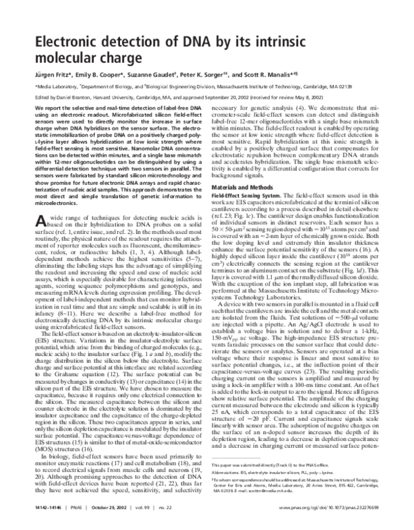
Fig. 1. Sensor schematics. (a and b) EIS interface of a n-type field-effect
sensor. DNA exhibits one intrinsic negative charge per base at its sugarphosphate backbone. Probe DNA is bound electrostatically to a layer of PLL
(gray) on the surface. (a) Binding of negatively charged target DNA (green) to
its complementary probe DNA (red) at the sensor surface (yellow) extends the
depletion region (black arrow) in the silicon portion of the sensor compared
with b, where no binding occurs to noncomplementary probe DNA (blue).
(c) Optical micrograph of a device consisting of field-effect sensors at the
terminus of two cantilevers. The cantilevers are 500 mm long, 75 mm wide, and
3 mm thick. (d) Cross section of a cantilever field-effect sensor. The sensing area
at the terminus of the cantilever is connected electrically to a metal contact on
the substrate by a layer of highly doped silicon inside the cantilever.
Fig. 2.
PLLyoligonucleotide multilayer growth. (a) Thickness of PLLy
oligonucleotide multilayer from ellipsometry measurements on silicon substrates that were prepared identically to the field-effect sensor surfaces. The
thickness increases linearly. (b) Surface potential signal of multilayer growth
in solution measured with a field-effect sensor. The signal alternates according to the charge of the adsorbed layer (positive for PLL and negative for
oligonucleotides). The same PLL and oligonucleotide concentrations as described for a were used. Each solution was injected twice and followed by an
injection of buffer before the next layer was adsorbed. Blue arrows indicate
PLL injections, and red arrows indicate oligonucleotide injections.
�Fig. 4. Concentration dependence and detection limit. Shown is the differential surface potential response for the hybridization of target oligonucleotides at concentrations of 2, 5, 20, and 80 nM.
Fig. 3. Field-effect detection of DNA hybridization. (a) Surface potential
response from sensor 1 (blue) functionalized with probe oligonucleotide A
and sensor 2 (red) functionalized with probe oligonucleotide B during a
hybridization experiment. Downward arrows indicate injections of oligonucleotides, and upward arrows indicate injections of buffer into the fluid cell.
(b) Differential signal obtained by subtracting the two sensor signals shown in
a (sensor 1-sensor 2). The order of injections was: buffer, B (80 nM), buffer, cA
(80 nM), cA (200 nM), buffer, cB (80 nM), cB (200 nM), and buffer. The second
injection of either cA or cB did not result in a change in hybridization signal,
indicating that saturation was reached.
to their thickness (24), we monitored the growth of polyelectrolyte multilayers consisting of positively charged PLL and
negatively charged oligonucleotides on the sensor surface (Fig.
2). Such layers bind to each other primarily by electrostatic
interactions and are known to overcompensate for the surface
charge of the previously adsorbed layer, which leads to a linear
growth in multilayer thickness as positively and negatively
charged molecules are successively applied to the surface (25).
Using ellipsometry, we determined the incremental thickness
increase of PLL-oligonucleotide multilayers to be '0.4 nm per
layer (Fig. 2a). In contrast, the field-effect sensor showed an
alternating response, over five successive cycles of PLL and DNA
adsorption, with an increase of '16 mV after the addition of
14144 u www.pnas.orgycgiydoiy10.1073ypnas.232276699
PLL and a subsequent decrease of '14 mV after the addition of
oligonucleotides (Fig. 2b). Thus, our device measures net charge
rather than layer thickness.
To explore the utility of our field-effect sensor for detecting
DNA in solution, two sensors were first functionalized with a
PLL layer. Next, the sensing area of one sensor was functionalized with the 12-mer oligonucleotide A (sensor 1), and the
adjacent sensor was functionalized with the unrelated 12-mer
oligonucleotide B (sensor 2). The sensors were then mounted in
a fluid cell. Solutions containing various target DNA oligonucleotides were injected in succession, and the surface potential
of the sensors was measured. The addition of control solutions
such as buffer or oligonucleotide B generated similar signals
from both sensors (Fig. 3a). These signals arose because the
surface potential is sensitive to thermal fluctuations, drifts,
nonspecific binding, and changes in electrolyte composition.
However, because these unwanted signals are similar for both
sensors, they can be eliminated by taking the differential response from the two sensors (sensor 1-sensor 2). When oligonucleotide cA, complementary to A, was injected, the surface
potentials of sensor 1 and sensor 2 diverged. The sensors showed
a differential response of 23 mV for oligonucleotide cA and of
13 mV for the subsequent addition of cB (Fig. 3b). These
observations demonstrate that a differential field-effect sensor
configuration is able to measure the sequence-specific formation
of A–cA and B–cB hybrids.
We observed that sensors can be reused more than 10 times
by regenerating their surface with a piranha-hydrofluoric acidpiranha cleaning. After a new round of functionalization with
PLL and oligonucleotides, the difference in signal strengths
between the original and regenerated surfaces was 10–20% for
hybridizing oligonucleotides at a concentration of 80 nM.
The ionic strength of the buffer used in our experiments (23
mM) is much lower than that commonly used for DNA hybridization (e.g., 825 mM for a 53 saline sodium citrate buffer). We
chose such a low ionic strength because field-effect detection is
most sensitive when counterion screening of the charged molecules is minimized (12, 26). However, at low ionic strength, the
electrostatic repulsion between two complementary DNA
strands strongly reduces their probability of hybridization and
extends the time required for annealing (27, 28). Still, the data
shown in Fig. 3 indicate that saturation is reached within a few
Fritz et al.
�minutes of the addition of target oligonucleotides to the fluid
cell, which is unusually rapid at this ionic strength. The positively
charged PLL layer seems to compensate the negative charge on
the probe DNA and reduce the electrostatic repulsion between
target and probe DNA. In addition, a positively charged surface
may increase the local concentration of target oligonucleotides
near the surface (29). The method of hybridizing nucleic acids on
a charge-compensated surface at low ionic strength has been
investigated recently for use in rapid microarray hybridization
(29). It might also be used for other field-effect sensing devices
such as silicon nanowires (30), which could lead to a dramatic
size reduction of nucleic acid sensors.
We used autoradiography to verify selective hybridization with
radiolabeled A, cA, and cB oligonucleotides. We found that the
surface coverage after functionalization was approximately 1
probe oligonucleotide per 2 nm2. Specific target oligonucleotides
hybridized with an efficiency of 5% at 80 nM, whereas binding
Fritz et al.
Fig. 6. Field-effect detection of a single base mismatch. Surface potentials
from two sensors that were functionalized with probe oligonucleotides A and
Am, which differ only in a single base. Control solutions with noncomplementary target oligonucleotide cB show no differential signal, whereas injection
of 80 nM of complementary sequences cA and cAm both show a distinct
hybridization signal. (a) Absolute surface potential. (b) Differential surface
potential.
by noncomplementary oligonucleotides was less than 0.6%.
These experiments with radiolabeled DNA showed that a change
in surface potential of '3 mV corresponds to the binding of
'3 3 104 12-mer oligonucleotides per mm2 or 15 ng of DNA per
cm2. The expected change in surface potential C0 for a given
change in surface charge density s0 can be calculated by using the
Grahame equation (12),
s 0 5 Î8« w«okTco sinh
S D
eC0
,
2kT
where k is the Boltzmann constant, T is absolute temperature, e
is elementary charge, «o is the permittivity of free space, «w is the
dielectric constant of water, and co is the buffer ionic strength.
Starting with a surface charge density of silicon dioxide of 0.8
Cym2 (31), we calculated that a change of the surface charge
PNAS u October 29, 2002 u vol. 99 u no. 22 u 14145
BIOPHYSICS
Fig. 5. Field-effect detection within a complex sample. Absolute surface
potential (a) and differential surface potential (b) for the hybridization of 20
nM oligonucleotides cA to A and cB to B within a 10-times-higher concentration of unrelated oligonucleotides. All injections were made at a constant 200
nM concentration. Arrows indicate injections of 200 nM C, then 20 nM cA 1
180 nM C, and then 20 nM cB 1 20 nM cA 1 160 nM C.
�density from the 3 3 104 hybridized 12-mer oligonucleotides per
mm2 leads to a change in surface potential of '3 mV, which is
in good agreement with the observed value. Here we assumed an
upper value of 12 charges per oligonucleotide, although their
effective charge may be less than that, which would reduce the
calculated change in surface potential. The surface charge
density of 0.8 Cym2 is an upper limit for silicon dioxide, and using
a lower effective surface charge density would increase the
calculated change in surface potential.
Fig. 4 shows the dependence of the hybridization signal on
target oligonucleotide concentration and demonstrates that a 2
nM concentration of 12-mer oligonucleotides or 8 ngyml DNA
can be detected easily. Although our detection method is less
sensitive than state-of-the-art label-dependent methods (5–7) or
typical microarray applications that have sensitivities in the tens
of picomolar range, our detection limit of 2 nM is one of the
lowest values reported thus far for label-independent methods
(8–11). Because the sensor response depends on the change in
surface charge during hybridization, the higher charge associated with longer DNA molecules will create a stronger signal per
molecule. Whether this will improve the detection limit remains
unclear, because the total number of duplexes created during
hybridization will also depend on the surface coverage of longer
probe oligonucleotides as well as the affinity and unspecific
binding of longer target oligonucleotides.
To demonstrate the specificity of our sensors, we investigated
two cases where unspecific binding of oligonucleotides to the
sensor surface can affect sensor response. First we successfully
detected a specific oligonucleotide within a high concentration
of other unrelated oligonucleotides. Fig. 5 shows the binding of
20 nM cA and cB to A and B, respectively, within a 10-timeshigher concentration (200 nM) of unrelated sequences. Comparing this signal with a signal from a pure oligonucleotide (Fig.
4) shows that the high unspecific background reduces the
hybridization signal by a factor of 3. Although conventional
microarrays are operated at an even higher background-totarget ratio (1), we anticipate that further improvement of
surface functionalization and hybridization conditions will improve the specificity as well as the sensitivity of our sensor (32).
Second, Fig. 6 shows that our field-effect device can identify a
single base mismatch in 12-mer oligonucleotides, i.e., it can
differentiate between a specific sequence and a sequence that
differs only by one base. This is particularly important, because
a potential application of DNA sensors is to detect DNA point
mutations associated with disease. A pair of sensors was functionalized with oligonucleotides A and Am, a sequence that
differs from A at a single base. When cA was injected, the
1.
2.
3.
4.
5.
6.
7.
8.
9.
10.
11.
12.
13.
14.
15.
16.
Lander, E. S. (1999) Nat. Genet. 21, Suppl. 1, 3–4.
Wang, J. (2000) Nucleic Acids Res. 28, 3011–3016.
Kricka, L. J. (1999) Clin. Chem. 45, 453–458.
Wang, J. (1999) Chem. Eur. J. 5, 1681–1685.
Castro, A. & Williams, J. G. K. (1997) Anal. Chem. 69, 3915–3920.
Budach, W., Abel, A. P., Bruno, A. E. & Neuschäfer, D. (1999) Anal. Chem.
71, 3347–3355.
Taton, T. A., Mirkin, C. A. & Letsinger, R. L. (2000) Science 289, 1757–1760.
Nelson, B. P., Grimsrud, T. E., Liles, M. R., Goodman, R. M. & Corn, R. M.
(2001) Anal. Chem. 73, 1–7.
Okahata, Y., Kawase, M., Niikura, K., Ohtake, F., Furusawa, H. & Ebara, Y.
(1998) Anal. Chem. 70, 1288–1296.
Fritz, J., Baller, M. K., Lang, H. P., Rothuizen, H., Vettiger, P., Meyer, E.,
Güntherodt, H.-J., Gerber, C. & Gimzewski, J. K. (2000) Science 288, 316–318.
Howorka, S., Cheley, S. & Bayley, H. (2001) Nat. Biotechnol. 19, 636–639.
Israelachvili, J. (1992) Intermolecular and Surface Forces (Academic, London),
2nd Ed.
Bergveld, P. (1972) IEEE Trans. Biomed. Eng. 19, 342–351.
Bousse, L. & Bergveld, P. (1983) J. Electroanal. Chem. 152, 25–39.
Bousse, L. (1982) J. Phys. Chem. 76, 5128–5133.
Nicollian, E. & Brews, J. (1982) MOS (Metal Oxide Semiconductor) Physics and
Technology (Wiley, New York).
14146 u www.pnas.orgycgiydoiy10.1073ypnas.232276699
differential signal decreased, showing specific hybridization to A.
When cAm was injected, the differential signal increased, showing hybridization to Am. The smaller differential signal from
binding of cAm to Am compared with that of cA to A could result
from a difference in surface functionalization of the two sensors
with probe oligonucleotides. However, this trend was subsequently verified by radiolabeling experiments, suggesting that
the difference could also result from the several degrees lower
melting temperature of the Am–cAm duplex relative to the A–cA
duplex. The smaller differential signal from cA in Fig. 6b
compared with that from cA in Fig. 3b suggests that cA also binds
unspecifically, to some extent, to Am, and this reduces the
differential signal. Nevertheless, the data in Fig. 6 demonstrate
that our device can distinguish between complementary and
mismatched DNA sequences.
Conclusions
In this report we have introduced an electronic method for the
direct detection of unlabeled nucleic acids. We have presented
its operating principle and investigated its concentration sensitivity and its specificity. We focused on real-time and label-free
DNA detection with sensors manufacturable by conventional,
high-yield fabrication processes that can produce hundreds of
sensors in parallel. In doing so, we target applications where
rapid, parallel DNA analysis is needed, e.g., for characterizing
pathogens, measuring mRNA levels during expression profiling,
or point-of-care applications. To approach these goals, we
anticipate further research toward improvement of concentration sensitivity and of specificity through optimization of experimental conditions, improved surface functionalization, and
integration in small-volume fluidic handling systems. The active
area of our sensor can be further miniaturized to approximately
1 mm2 by using standard photolithography. The geometry and
sensitivity of the sensor also implies the potential for novel
application to real-time, single-cell measurements. Given that an
individual gene can be expressed at concentrations greater than
1 nM inside a mammalian cell, a sensor at the terminus of a
silicon cantilever, similar to that shown in Fig. 1, might be able
to detect gene expression from a single cell.
Devices were fabricated in the Massachusetts Institute of Technology
Microsystems Technology Laboratories. We are thankful to M. Trulson
for a critical reading of the manuscript. This work was supported by the
Defense Advanced Research Projects Agency, the Air Force Office of
Scientific Research, and the Media Laboratory’s Things That Think
consortium. E.B.C. acknowledges support from a National Science
Foundation Graduate Research fellowship.
17.
18.
19.
20.
21.
22.
23.
24.
25.
26.
27.
28.
29.
30.
31.
32.
Hall, E. A. H. (1991) Biosensors (Prentice Hall, Englewood Cliffs, NJ).
Hafeman, D. G., Parce, J. W. & McConnell, H. M. (1988) Science 240, 1182–1185.
Offenhäusser, A. & Knoll, W. (2001) Trends Biotechnol. 19, 62–66.
Zeck, G. & Fromherz, P. (2001) Proc. Natl. Acad. Sci. USA 98, 10457–10462.
Souteyrand, E., Cloarec, J. P., Martin, J. R., Wilson, C., Lawrence, I.,
Mikkelsen, S. & Lawrence, M. F. (1997) J. Phys. Chem. B 101, 2980–2985.
Berney, H., West, J., Haefele, E., Alderman, J., Lane, W. & Collins, J. K. (2000)
Sens. Actuators B Chem. 68, 100–108.
Cooper, E. B., Fritz, J., Wiegand, G., Wagner, P. & Manalis, S. R. (2001) Appl.
Phys. Lett. 79, 3875–3877.
Berggren, C., Bjarnason, B. & Johansson, G. (2001) Electroanalysis (N.Y.) 13,
173–180.
Decher, G. (1997) Science 277, 1232–1237.
Bergveld, P. (1996) Sens. Actuators A Phys. 56, 65–73.
Wetmur, J. G. & Davidson, N. (1968) J. Mol. Biol. 31, 349–370.
Williams, A. P., Longfellow, C. E., Freier, S. M., Kierzek, R. & Turner, D. H.
(1989) Biochemistry 28, 4283–4291.
Belosludtsev, Y., Belosludtsev, I., Iverson, B., Lemeshko, S., Wiese, R., Hogan,
M. & Powdrill, T. (2001) Biochem. Biophys. Res. Commun. 282, 1263–1267.
Cui, Y., Wei, Q., Park, H. & Lieber, C. M. (2001) Science 293, 1289–1292.
Dong, Y., Pappu, S. V. & Xu, Z. (1998) Anal. Chem. 70, 4730–4735.
Andersen, M. L. M. (1999) Nucleic Acid Hybridization (Springer, New York).
Fritz et al.
�
 Suzanne Gaudet