408
BJID 2009; 13 (December)
Molecular Epidemiology of Acinetobacter baumannii in Central Intensive Care Unit in Kosova
Teaching Hospital
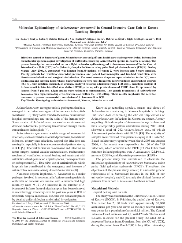
Lul Raka 1,2, Smilja Kalenc 3, Zrinka Bošnjak 3, Ana Budimir 3, Stjepan Katic 3, Dubravko Šijak 3, Gjyle Mulliqi-Osmani 1,2, Dick
Zoutman 4 and Arbëresha Jaka 1
1
Medical School, Prishtina University, Prishtina, Kosova; 2National Institute for Public Health of Kosova, Prishtina, Kosova
3
Department of Clinical and Molecular Microbiology, Clinical Hospital Centre Zagreb, Zagreb, Croatia; 4Queen’s University and Kingston
General Hospital, Kingston, Ontario, Canada
Infections caused by bacteria of genus Acinetobacter pose a significant health care challenge worldwide. Information
on molecular epidemiological investigation of outbreaks caused by Acinetobacter species in Kosova is lacking. The
present investigation was carried out to enlight molecular epidemiology of Acinetobacter baumannii in the Central
Intensive Care Unit (CICU) of a University hospital in Kosova using pulse field gel electrophoresis (PFGE). During
March - July 2006, A. baumannii was isolated from 30 patients, of whom 22 were infected and 8 were colonised.
Twenty patients had ventilator-associated pneumonia, one patient had meningitis, and two had coinfection with
bloodstream infection and surgical site infection. The most common diagnoses upon admission to the ICU were
politrauma and cerebral hemorrhage. Bacterial isolates were most frequently recovered from endotracheal aspirate
(86.7%). First isolation occurred, on average, on day 8 following admission (range 1–26 days). Genotype analysis of
A. baumannii isolates identified nine distinct PFGE patterns, with predominance of PFGE clone E represented by
isolates from 9 patients. Eight strains were resistant to carbapenems. The genetic relatedness of Acinetobacter
baumannii was high, indicating cross-transmission within the ICU setting. These results emphasize the need for
measures to prevent nosocomial transmission of A. baumannii in ICU.
Key-Words: Genotyping, Acinetobacter baumannii, Kosova, intensive care unit.
Acinetobacter spp. are opportunistic pathogens that have
emerged to an infectious agent of importance to hospitals
worldwide [1-3]. They can be found in the natural environment,
hospital surroundings and on the skin of the human body.
Some strains of Acinetobacter can survive environmental
desiccation for weeks, promoting transmission through fomite
contamination in hospitals [4].
Acinetobacter spp. cause a wide range of nosocomial
infections, such as ventilator-associated pneumonia, bloodstream
infections, urinary tract infections, surgical site infections and
meningitis, especially in immunocompromised patients staying
in ICU [5]. Other risk factors for colonization and infection are
recent surgery, central vascular catheterization, tracheostomy,
mechanical ventilation, enteral feeding and treatment with
antibiotics (third-generation cephalosporins, fluoroquinolones
or carbapenems)[6,7]. Extensive use of antimicrobials within
hospitals has contributed to the emergence and increase of
antimicrobial resistance among Acinetobacter strains [8].
Numerous reports implicates A. baumannii as a major
pathogen involved in nosocomial infections causing epidemic
outbreaks or endemic occurrence with a documented high
mortality rates [9-12]. An increase in the number of A.
baumannii isolates from clinical samples has been observed
in microbiology laboratory over the past few years in ICU of
university hospital in Kosova. But, this was not accompanied
by detailed epidemiological and clinical investigation.
Received on 6 May 2009; revised 20 November 2009.
Address for correspondence: Dr. Lul Raka. “Emin Duraku”, N=166, 71000
Kaçanik, Kosova; Phone: +3813829080666 and +37744368289. Email: lulraka@hotmail.com.
The Brazilian Journal of Infectious Diseases
2009;13(6):408-413.
© 2009 by The Brazilian Journal of Infectious Diseases and Contexto
Publishing. All rights reserved.
Knowledge regarding species, strains and clones of
Acinetobacter circulating in Kosova hospitals is lacking.
Published data concerning the clinical implications of
Acinetobacter spp. infections in Kosova are scarce. A study
regarding clinical samples of Acinetobacter spp. isolates and
their susceptibility pattern undertaken during 2001-2004,
showed a total of 242 Acinetobacter spp., of which
A.baumannii predominate with 81.2% [13]. The majority of
samples were revealed from patients staying in ICUs (62%).
Based on laboratory report between March 2005 and August
2006, A. baumannii was responsible for 100 of the 719
infections, which occurred in the CICU (13.9%). Other most
common isolated pathogens were P. aeruginosa (22.1%), S.
aureus (15.39%), and Klebsiella pneumoniae (12.9%).
The present study was undertaken to elucidate the
molecular epidemiology of Acinetobacter baumannii using
pulse field gel electrophoresis (PFGE). Therefore, the
objectives of the present study were (i) to assess the genetic
relatedness of A. baumannii isolates in the ICU of our
university hospital; and (ii) to study the clinical features of
patients from whom A. baumannii had been isolated.
Material and Methods
Hospital Setting and Patients
The study was conducted at the University Clinical Centre
of Kosova (UCCK), in Prishtina, the capital city of Kosova.
The center has 2,100 beds with approximately 60,000
admissions per year and serves as the only referral tertiary
care center for a population of around 2.1 million. The Central
Intensive Care Unit is a mixed ICU with 12 beds. The bacterial
isolates selected for the present study included 30 A.
baumannii isolates from 30 patients from the ICU of UCCK,
during the period from March 2006 to July 2006. Laboratory
www.bjid.com.br
�BJID 2009; 13 (December)
Epidemiology of Acinetobacter baumannii in an Hospital’s ICU in Kosova
diagnosis of microbiological samples and susceptibility testing
was done in the Department of Microbiology within the
National Institute for Public Health of Kosova. The genotyping
was performed in the Clinical Hospital Centre Zagreb,
Department of Clinical and Molecular Microbiology in Zagreb,
Croatia. Clinical specimens included cerebrospinal liquid,
endotracheal aspirate, thoracal drain and tracheostoma. The
following data were recorded from the medical charts of patients
with A. baumannii infection or colonisation: age, gender, number
of patient-days in hospital, underlying diseases or conditions,
susceptibility pattern and clinical outcome. Nosocomial
infections were classified according to standard CDC
definitions, whilst A. baumannii was considered to be a
colonising organism when it was isolated from clinical
specimens, but the criteria for infection were not met [14]. Only
one sample of A.baumanni per patient was enrolled in the study.
Microbiological Methods
A. baumannii strains were collected from clinical
specimens by using standard methods, isolated in pure
cultures on MacConkey agar plates. Organisms were identified
by using the API system for the identification (bioMerieux,
Marcy l’Etoile, France). From a fresh 18 hours plate culture of
A.baumanii, a heavy, cloudy suspension of the organism was
made in the CRYOBANK™ medium in the tube (COPAN
Diagnostics Inc., CA, USA). Tube was mixed by shaking and
inverting to allow the bacteria in the suspension to coat the
beads. Using a sterile pipet the CRYOBANK™ medium was
removed from the tube. Than, the tube was placed in a -70°C
freezer to store the culture. Afterwards the samples were
transported to Croatia where the bacteria were recovered
removing the cap of the CRYOBANK™ tube. Using forceps
one bead was rolled over the culture mediums (brain-heart
infusion) and Kaufman-Müller broth. Isolates were verified in
Croatia as A.baumannii using the Vitek 2 automatic system
(bioMerieux, Marcy l’Etoile, France).
Antimicrobial Susceptibility
Antimicrobial resistance was determined by the disk
diffusion method according to the Clinical and Laboratory
Standards Institute criteria, former NCCLS [15]. The following
antimicrobial drugs were tested: Ampicillin 10µg, Ceftriaxon
30µg, Gentamicin 10µg, Amikacin 30µg, Imipenem 10 µg,
Pipercilin + tazobactam 100µg, Cefoxitin 30µg, Ceftazidime
30µg, Tobramycin 10µg, Cotrimoxasole 1.25+23.75 µg and
Ciprofloxacin 5µg.
Molecular Typing by Pulsed-Field Gel Electrophoresis (PFGE)
and Dendrogram Analysis
The preparation of genomic DNA of A. baumannii isolates
was performed as described by Schwartz and Cantor with
minor modifications. Macrorestriction analysis of
chromosomal DNA with XbaI was carried out by PFGE
following published procedures [16]. PFGE was run in a CHEFDRIII apparatus (Bio-Rad Laboratories, CA, USA), with pulses
409
ranging from 5 to 50 seconds at a voltage of 6 V/cm at 10-12°C
for 20 h. Products were detected after staining with ethidium
bromide (50 mg/mL) and photographed with Polaroid type
667 film. A ladder of bacteriophage lambda concatemers (New
England Biolabs) was used as molecular weight markers.
Clusters of possibly related isolates were identified by
using the Dice coefficient of similarity and unweighted group
method with arithmetic averages at 80%, which indicates fourto six fragment differences in gels. The relationships between
all isolates were analysed using the GelComparII software
package and presented as a dendrogram (Applied Maths NV,
Belgium). DNA fingerprints were interpreted as recommended
by Tenover et al. [17].
Results
From March 16th to July 27th 2006, a total of 30 Acinetobacter
baumannii isolates were obtained from 30 patients (24 males,
6 females) admitted to the CICU. Their age range was from 2 to
82 years (mean age 47.5, median age 52.5 years). Based on
evaluation of clinical charts, 22 patients were classified as
infected and had nosocomial infections and eight of them
were considered colonized with A.baumannii.
Isolates were most frequently recovered from endotracheal
aspirate (n = 26); the other isolates were recovered from
tracheostoma (n=2), thoracal drain (n=1) and cerebrospinal
fluid (n = 1). Twenty patients developed nosocomial
pneumoniae; one patient had a diagnosis of meningitis, and
two had coinfection with bloodstream infection and surgical
site infection. The most common diagnoses upon admission
to the ICU were politrauma and cerebral hemorrhage. Other
pathogens were co-isolated from nine patients:
Staphylococcus aureus from two patients, P. aeruginosa from
4 patients, and Klebsiella pneumonaie from three patients.
The clinical characteristics of patients from whom A.
baumannii was isolated are shown in Table 1.
The length of stay in ICU ranged from 1-59 days with
median time of 17 days. The median time that had elapsed
between admission and isolation of A. baumannii was 8 days.
During the ICU stay, 16 patients died (crude mortality 53.3%)
and the A. baumannii-attributable mortality was 62.5% (10 /
16). The time-frame of admission, discharge and isolation of
A. baumanni from ICU patients is presented in Figure 1.
The length of ICU stay for non-survivors and survivors
was 14.5 and 18.5 days, respectively. The length of stay was
significant in comparison between infected and colonised
patients (19.5 vs. 9 days).
The length of ICU stay for epidemic strains and nonepidemic strains was 15.7 and 24.5 days, respectively.
Four patients yielded A. baumannii upon hospital
admission and were transferred from other hospitals to ICU,
while 22 patients yielded A. baumannii only following
hospitalization. Twelve patients were transferred to ICU from
other departments within the UCCK.
PFGE profiles of A. baumannii strains isolated from CICU
is shown in the Figure 2.
www.bjid.com.br
�410
Epidemiology of Acinetobacter baumannii in an Hospital’s ICU in Kosova
BJID 2009; 13 (December)
Figure 1. Time frame of admission, discharge and isolation of Acinetobacter baumanni from ICU patients.
Figure 2. Dendrogram depicting 30 representative isolates of Acinetobacter baumannii species obtained from CICU.
www.bjid.com.br
�BJID 2009; 13 (December)
Epidemiology of Acinetobacter baumannii in an Hospital’s ICU in Kosova
411
Table 1. Clinical and PFGE data of the patients with Acinetobacter baumannii isolates.
Nr Gender/ Day of Length of
age isolation ICU stay
1
2
3
4
5
6
7
8
9
10
11
12
13
14
15
16
17
18
19
20
21
22
23
24
25
26
27
28
29
30
M/ 66
M/ 33
M/ 62
M/ 17
M/ 11
M/ 62
M/41
F/33
M/ 51
M/ 2
M/ 21
M/ 72
M/ 43
M/ 55
M/ 74
M/ 71
M/ 57
M/ 46
F/ 20
M/ 22
M/ 50
F/ 24
M/ 82
F/ 54
F/ 72
M/ 60
M/ 74
F/ 71
M/ 61
M/ 20
17
4
3
1
12
4
4
10
14
1
8
3
1
4
18
9
2
14
2
1
5
15
8
26
12
10
8
10
16
8
18
28
8
14
37
6
28
14
59
10
10
5
13
8
23
34
8
20
15
1
11
30
32
44
34
14
17
19
19
11
Diagnosis
Tumor cerebri
Tumor cerebri
Tumor cerebri
Diabetes mellitus
Politrauma
Politrauma
Politrauma
Politrauma
Cerebral infarct
Politrauma
Politrauma
Cerebral infarct
cardiac arrest
Politrauma
Politrauma
Politrauma
Politrauma
Tumor cerebri
Pulmonal infiltrat
Politrauma
Tumor cerebri
Myocardiopathia
Peritonitis
Peritonitis
cardiac arrest
Cerebral infarct
Politrauma
Tumor cerebri
Cerebral infarct
Politrauma
Sensitive
AMI
TOB, AMI, IMI
AMI, IMI, TOB, GEN
AMI, CIP
TOB, AMI, IMI
AMI, IMI
IMI
AMI, IMI
TOB, AMI, CAZ
IMI
IMI, AMI
AMI, IMI
GEN, AMI, CAZ
AMI, IMI
TOB, AMI
AMI, CIP, IMI
GEN, TOB, AMI, IMI
AMI, IMI
AMI, IMI
IMI, CAZ, AMI
IMI
AMI
AMI, IMI
TOB, AMI,
AMI, IMI
AMI
IMI,CAZ.AMI
AMI,IMI
AMI
AMI, IMI
Outcome
Sample
Isolate PFGE type
Died
Died
Died
Recovered
Transferred
Died
Recovered
Transferred
Died
Died
Died
Died
Recovered
Recovered
Recovered
Recovered
Transfer
Recovered
Died
Transferred
Died
Died
Died
Transferred
Died
Died
Recovered
Transferred
Died
Died
EA
CLS
EA
EA
EA
EA
EA
EA
TRA
EA
EA
EA
EA
EA
EA
EA
EA
EA
TC
EA
EA
EA
EA
TRA
EA
EA
EA
EA
EA
EA
1277
1592
1007
930
1570
838
972
868
1622
1461
1517
768
1191
869
933
907
1574
943
929
1003
575
1438
931
1605
1188
815
1009
933
907
948
F4
G1
A3
E5
C1
E2
B2
E3
A2
C2
F6
E3
B1
E1
D1
E5
F5
D1
E5
A1
E4
F3
D1
H1
I1
F1
A1
D1
E5
F2
EA= endotracheal aspirate; AMI=amikacin, IMI=imipenem, TOB=tobramycin, CIP=ciprofloxacine, CAZ=cephtazidime, GEN=gentamycine;
TRA=tracheostoma; TC=Thoracal drain.
Genotypic analysis of A. baumannii isolates from ICU
patients identified nine major PFGE patterns, which we named
from A to I, that differed in migration of at least four DNA
fragments and showed a similarity of < 80% at dendrogram
analysis. Of these, PFGE pattern E predominate with isolates
from nine patients. Eight isolates were resistant to
carbapenems.
Discussion
Although only 5-10% of all hospitalized patients are
treated in ICUs, they account for approximately 25% of all
nosocomial infections [18]. The incidence of nosocomial
infections in ICUs is 5–10 times higher than that observed
in general hospital wards [19,20]. In developing countries
the occurrence of nosocomial infections is 12-20 fold
higher [21].
A. baumannii outbreaks have been reported previously,
particularly in ICU wards [22-26]. Severe underlying diseases,
invasive diagnostic and therapeutic procedures used in ICUs
have been demonstrated to predispose patients to severe
infections with A. baumannii [27-29]. Our results show that
nosocomial infections and colonizations by A. baumannii in
the ICU were prolonged for several months. The impact of A.
baumannii on ICU-acquired infections and colonization was
substantial from clinical samples received in our laboratory
from CICU. From March 2006 to August 2007 Acinetobacter
spp. were the second most prevalent identified microorganism
with 13.9% (100/719). Other most frequent isolates were P.
aeruginosa (22.1%), S. aureus (15.3%) and Klebsiella
spp.(12.9%). Acinetobacter strains (n=100) showed globally
high resistance pattern to cephalosporins (76.9%). Imipenem
and amikacine were the most effective drugs against
A.baumannii with sensitivity rate of 92.4% and 85.7%
respectively (unpublished data).
Previous prevalence studies in Kosova showed hight rates
of health care associated infections in UCCK (17.4%) and in
www.bjid.com.br
�412
Epidemiology of Acinetobacter baumannii in an Hospital’s ICU in Kosova
CICU with 68.7% of patients having nosocomial infections,
with a predominance of ventilator associated pneumonia
(72.7% of infections) [30,31].
There are many causes for high rate of nosocomial
infections in ICU and A. baumannii outbreaks. Main factor
remains the lack of support and implementation of prevention
and control policies. The proportion of health care workers
working in CICU to patients staying in ICU is only 5 HCW per
12 patients per shift. CICU is referent center for intensive care
for all 6 regional hospitals and other depratments within the
UCCK.
Single use devices were reused due to limited budget.
Suction catheters for aspiration of respiratory tract were
amongst most used equipment in this group. Audit in the
ward during the study period proved that these catheters
were placed in a containers containing diluted chlorhexidine.
The same catheters after “disinfection” were used for more
than one patient carrying a significant risk for cross-infection.
Some equipment used in ICU were outdated and their
maintenance services were not regular.
A study of compliance with hand hygiene in CICU showed
the alert rate of only 19% [32]. During the outbreak period
alcoholic hand rubs were not in used in ICU. There are three
washing sinks in the ward. Low number of wash sinks
contributed to high rate of infection in ICU. Gloves were not
changed after each contact with patients but they were used
and maintained for successive patients intervention.
For many years in Kosova, the cephalosporins are the
drugs of choice in empiric treatment in ICU and they have
been used without any restrictions not only in ICUs but also
in other hospital wards and ambulantory care. This could
explain the high resistance rates of Acinetobacter baumannii
to antimicrobials. For a decade in Kosova, all antimicrobials
have been available in pharmacies without a physician’s
prescription.
CICU is reference center for patients from other hospital
departments of CICU, from regional hospitals and also from
the private hospitals. Delay of referral to this unit contributed
to infections, severity of illness and poor outcome prognosis
for the patients.
Delay is related to patients who are previously treated at
the regional hospitals and they are not transferred on time to
the CICU, which is the only ICU reference center for six
regional hospitals and for 13 clinics within UCCK.
Genotypic analysis of A. baumannii isolates from ICU
patients identified nine major PFGE patterns. The most
predominant clones of A.baumannii (E and F) were related
with more than one outbreak during the study period occurring
sequentially. Case-control study was not performed in
epidemiological investigation. The data were recorded from
the medical charts of patients with A. baumannii infection or
colonization. Some genetically indistinguishable A.baumannii
isolates (931, 933 and 934) were isolated on the same day
(May 2, 2006) and had similar antimicrobial susceptibility
pattern, suggesting common source of infection. The median
BJID 2009; 13 (December)
time from admission to isolation of this bacteria revealed that
it’s shorter than in other publications [23-27].
In cases where the genetically same strains were not related
in timely manner, the only explanation would be poor hand
hygiene of health care workers (HCW). Another argument is
high endemic rate of MRSA, which is 61.3% of all S.aureus
isolates [13]. These facts suggests the horizontal transmission
of the epidemic strains from one patient to another through
the hospital staff.
As in other publications endotracheal aspirates were
predominant clinical samples received from ICU [33,34].
The length of stay was significant in comparison between
infected and colonised patients. This finding is consistent
with reports of other outbreaks [6,29,33,34]. But there was no
significant diference between non survivors and survivors.
This can be explained with a fact that some patients spent
some hospital days in regional hospitals before referral to
CICU and also six patients were sent for treatment in other
ICUs in neighbouring countries. As in previous studies, the
respiratory tract was the most frequent site of isolation of A.
baumannii in ICU patients [35,36]. Colonization with A.
baumannii in not performed routinely at admission to ICU.
In conclusion, we show here that A. baumannii strains
cause large and sustained hospital outbreak due to insufficient
preventive measures. These results emphasize the need for
preventive interventions in ICU.
References
1. Schreckenberger PC, Daneshvar MI, Weyant RS, Hollis DG.
Acinetobacter, Achromobacter, Chryseobacterium, Moraxella,
and other nonfermentative Gram-negative rods. In: Murray
PR, Baron EJ, Jorgensen JH, Landry ML, Pfaller MA, eds.
Manual of clinical microbiology. 9th ed. Washington, DC: ASM
Press, 2007:770-802.
2. Fournier PE, Richet H. The epidemiology and control of
Acinetobacter baumannii in health care facilities. Clin Infect
Dis 2006;42:692-699.
3. Munoz-Price LS, Weinstein RA. Acinetobacter infection. N Engl
J Med. 2008 Mar 20;358(12):1271-81.
4. Getchell-White SI, Donowitz LG, Gröschel DH. The inanimate
environment of an intensive care unit as a potential source of
nosocomial bacteria: evidence for long survival of Acinetobacter
calcoaceticus. Infect Control Hosp Epidemiol 1989;10:402407.
5. Garnacho-Montero J, Ortiz-Leyba C, Fernández-Hinojosa E, et
al. Acinetobacter baumannii ventilator-associated pneumonia:
epidemiological and clinical findings. Intensive Care Med
2005;31:649-655
6. Baran G, Erbay A, Bodur H, Ongürü P, Akinci E, Balaban N, Cevik
MA. Risk factors for nosocomial imipenem-resistant
Acinetobacter baumannii infections. Int J Infect Dis. 2008
Jan;12(1):16-21.
7. Playford EG, Craig JC, Iredell JR. Carbapenem-resistant
Acinetobacter baumannii in intensive care unit patients: risk
factors for acquisition, infection and their consequences. J Hosp
Infect. 2007 Mar;65(3):204-11
8. Murray CK, Hospenthal DR. Acinetobacter infection in the ICU.
Crit Care Clin. 2008 Apr;24(2):237-48.
9. Villegas MV, Hartstein AI. Acinetobacter outbreaks, 19772000. Infect Control Hosp Epidemiol 2003;24:284295
www.bjid.com.br
�BJID 2009; 13 (December)
Epidemiology of Acinetobacter baumannii in an Hospital’s ICU in Kosova
10. Meric M, Kasap M, Gacar G, Budak F, Dundar D, Kolayli F, Eroglu
C, Vahaboglu H. Emergence and spread of carbapenem-resistant
Acinetobacter baumannii in a tertiary care hospital in Turkey.
FEMS Microbiol Lett. 2008 May;282(2):214-8.
11. Kraniotaki E, Manganelli R, Platsouka E, Grossato A, Paniara O,
Palù G. Molecular investigation of an outbreak of multidrugresistant Acinetobacter baumannii, with characterisation of class
1 integrons. Int J Antimicrob Agents. 2006 Sep;28(3):193-9.
12. Zarrilli R, Crispino M, Bagattini M, Barretta E, Di Popolo A,
Triassi M, Villari P. Molecular epidemiology of sequential
outbreaks of Acinetobacter baumannii in an intensive care unit
shows the emergence of carbapenem resistance. J Clin Microbiol.
2004 Mar;42(3):946-53.
13. A.Kurti, L.Raka, Gj.Mulliqi: Acinetobacter species-clinical sample
isolates and antibiotic susceptibility in Kosova, 8th European
Congress of Chemotherapy and Infection, Budapest, Hungary,
25-28 October 2006.
14. Garner JS et al. CDC definitions for nosocomial infections. Am J
Infect Contr 1988;16:128-40.
15. Clinical and Laboratory Standards Institute. Methods for dilution
antimicrobial susceptibility tests for bacteria that grow
aerobically, 6th edn. Approved standard M7-A6. Wayne, PA:
CSLI, 2005.
16. Seifert H, Dolzani L, Bressan R et al. Standardization and
interlaboratory reproducibility assessment of pulsed-field gel
electrophoresis-generated fingerprints of Acinetobacter
baumannii. J Clin Microbiol 2005; 43: 4328–4335.
17. Tenover F, Arbeit R, Goering R. How to select and interpret
molecular strain typing methods for epidemiological studies of
bacterial infections: a review for healthcare epidemiologists.
Infect Control Hosp Epidemiol 1997; 18: 426–439.
18. Eggimann P., Pittet D. Infection Control in the ICU. Chest
2001;120:2059-2093.
19. Pittet D, Sax H. Health-care associated infections. In: Cohen J,
Powderly W, editors. Infectious diseases. 2nd ed. Mosby; 2003.
p. 881-92.
20. Ponce-de-Leon-Rosales S, Macias A. Global perspectives of infection
control. In: Wenzel: Prevention and control of nosocomial
infections. 4th ed. Philadelphia LWW, 2003:14-33.
21. Patricia Lynch P, Victor D. Rosenthal V, Michael A. Borg M, and
Eremin S. Infection Control in Developing Countries. In
Jarvis: Bennett and Brachman’s Hospital Infections. 4 th ed.
2007:255 -270.
22. Corbella X, Montero A, Pujol M et al. Emergence and rapid spread
of carbapenem resistance during a large and sustained hospital
outbreak of multiresistant Acinetobacter baumannii. J Clin
Microbiol 2000; 38: 4086–4095.
23. Rodriguez-Bano J, Cisneros JM, Fernandez-Cuenca F et al. Clinical
features and epidemiology of Acinetobacter baumannii
colonization and infection in Spanish hospitals. Infect Control
Hosp Epidemiol 2004; 25: 819–824.
24. Turton JF, Kaufmann ME, Warner M et al. A prevalent,
multiresistant clone of Acinetobacter baumannii in Southeast
England. J Hosp Infect 2004; 58: 170–179.
413
25. Marque´ S, Poirel L, He´ritier C et al. Regional occurrence of
plasmid-mediated carbapenem-hydrolyzing oxacillinase OXA58 in Acinetobacter spp. in Europe. J Clin Microbiol 2005; 43:
4885–4888.
26. Pournaras S, Markogiannakis A, Ikonomidis A et al. Outbreak of
multiple clones of imipenem-resistant Acinetobacter baumannii
isolates expressing OXA-58 carbapenemase in an intensive care
unit. J Antimicrob Chemother 2006; 57: 557–561.
27. Husni RN, Goldstein LS, Arroliga AC, Hall GS, Fatica C, Stoller JK,
Gordon SM. Risk factors for an outbreak of multi-drug-resistant
Acinetobacter nosocomial pneumonia among intubated patients.
Chest. 1999 May;115(5):1378-82.
28. Cardeñosa Cendrero JA, Solé-Violán J, Bordes Benítez A, Noguera
Catalán J, Arroyo Fernández J, Saavedra Santana P, Rodríguez
de Castro F. Role of different routes of tracheal colonization in
the development of pneumonia in patients receiving mechanical
ventilation. Chest. 1999 Aug;116(2):462-70.
29. Tejada Artigas A, Bello Dronda S, Chacón Vallés E, Muñoz Marco
J, Villuendas Usón MC, Figueras P, Suarez FJ, Hernández A. Risk
factors for nosocomial pneumonia in critically ill trauma patients.
Crit Care Med. 2001 Feb;29(2):304-9.
30. Raka L, Zoutman D, Mulliqi G, Krasniqi S, Dedushaj I, Raka N,
Ahmeti S, Shala M, Vishaj A, Elezi Y. Prevalence of nosocomial
infections in high-risk units in the university clinical center of
Kosova. Infect Control Hosp Epidemiol. 2006 Apr;27(4):4213.
31. Spahija G, Raka L, Mulliqi G, Spahija N, Bukoshi Z, Hoxha F,
Krasniqi A. Prevalence of Nosocomial Infections in Adult
Intensive Care Units at a Kosova Teaching Hospital. Infect
Control Hosp Epidemiol. 2008 May;29(5):475.
32. Rosenthal VD, Ajita Mehta, Luis Cuellar, Carlos Álvarez Moreno,
Hakan Leblebicioglu, Naoufel Madani, Lourdes Dueñas, Regina
Berba, Mitrev Z, Raka L, Souha Kanj-Sharara, FernandezHidalgo, Salisu Abubakar, Altaf Ahmed, Sanjeev Singh, Subhash
Kumar Todi, Nagamani Sen, Mónica Viegas, Nayide Barahona
Guzmán, A. Nevzat Yalcin, Teodora Atencio Espinoza, Alberto
Armas Ruiz, Gorki Grinberg. Hand Hygiene Compliance in 84
ICUs of 16 countries.Findings of the International Nosocomial
Infection Control Consortium (INICC). SHEA Annual
Conference, Orlando, Florida, 5-8 April, 2008.
33. Bergogne-Berezin E, Towner KJ. Acinetobacter spp. as nosocomial
pathogens: microbiological, clinical, and epidemiological features.
Clin Microbiol Rev 1996; 9: 148–165.
34. Webster CA, Crowe M, Humphreys H, Towner KJ. Surveillance of
an adult intensive care unit for long-term persistence of a multiresistant strain of Acinetobacter baumannii. Eur J Clin
Microbiol Infect Dis 1998; 17: 171–176.
35. Bergogne-Berezin E, Towner KJ. Acinetobacter spp. as nosocomial
pathogens: microbiological, clinical, and epidemiological features.
Clin Microbiol Rev 1996; 9: 148–165.
36. Corbella X, Montero A, Pujol M et al. Emergence and rapid spread
of carbapenem resistance during a large and sustained hospital
outbreak of multiresistant Acinetobacter baumannii. J Clin
Microbiol 2000; 38: 4086–4095.
www.bjid.com.br
�
 Aurora Aleo