Academia.edu no longer supports Internet Explorer.
To browse Academia.edu and the wider internet faster and more securely, please take a few seconds to upgrade your browser.
2013, Endocrine Abstracts
2011 •
Frontiers in Molecular Neuroscience
Iron entry in neurons and astrocytes: a link with synaptic activity2015 •
Free Radical Biology and Medicine
Progressive iron accumulation induces a biphasic change in the glutathione content of neuroblastoma cells2004 •
The Journal of Neuroscience
Neuronal Migration Depends on Intact Peroxisomal Function in Brain and in Extraneuronal Tissues2003 •
2005 •
Nuclear Instruments and Methods in Physics Research Section B: Beam Interactions with Materials and Atoms
Intracellular iron concentration of neurons with and without perineuronal nets2007 •
Journal of Neurochemistry
Expression of divalent metal transporter 1 in primary hippocampal neurons: reconsidering its role in non-transferrin-bound iron influx2012 •
Journal of Neuroscience
Molecular Mechanisms Controlling the Migration of Striatal Interneurons2015 •
2023 •
Korean Journal of Yoga Studies
Al-Bīrūnī’s Indian Philosophy: The Kitāb Sānk and the Kitāb Pātanǧal (Korean)2023 •
The Journal of Engineering Research
Ambient Air Quality Assessment of Al-Mansoriah Residential Area in the State of Kuwait2009 •
Slavonic Pedagogical Studies Journal
Implementation of Developmental Topics Into University Education2016 •
British Journal of Ophthalmology
Ocular signs associated with a rhodopsin mutation (Cys-167right-arrowArg) in a family with autosomal dominant retinitis pigmentosa1998 •
Journal of Medical Internet Research
Leveraging Virtual Reality and Augmented Reality to Combat Chronic Pain in Youth: Position Paper From the Interdisciplinary Network on Virtual and Augmented Technologies for Pain Management2021 •
Commagene journal of biology
Sivas ve Van’dan Toplanan Cüce Kertenkelelerin (Parvilacerta parva) Boulenger, 1887 Helmint Faunası2022 •
2017 •
Ri-vista. Ricerche per la progettazione del paesaggio
Paradossi dell’acqua. Un dialogo tra opposti2023 •
Journal of invertebrate pathology
Morphological, genetic and biological characterisation of a novel alphabaculovirus isolated from Cryptophlebia peltastica (Lepidoptera: Tortricidae)2018 •
Reviews in Mineralogy and Geochemistry
From Short to Medium Range Order in Glasses and Melts by Diffraction and Raman Spectroscopy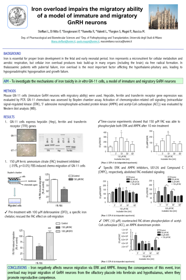
 Luca Valenti