JOURNAL OF BACTERIOLOGY, Dec. 1996, p. 7295–7303
0021-9193/96/$04.0010
Copyright q 1996, American Society for Microbiology
Vol. 178, No. 24
Analysis of the Region between Amino Acids 30 and 42 of
Intact UmuD by a Monocysteine Approach
ANGELINA GUZZO, MELISSA H. LEE, KAREN ODA,
AND
GRAHAM C. WALKER*
Department of Biology, Massachusetts Institute of Technology,
Cambridge, Massachusetts 02139
Received 3 June 1996/Accepted 3 October 1996
ticularly important during the shutdown of the SOS response
(3).
Several hypotheses have been proposed for the mechanism
of SOS mutagenesis, including (i) UmuD9 and UmuC affecting
the processivity of DNA polymerase III (2, 34); (ii) UmuD9
and UmuC binding to the RecA-ssDNA complex, causing it to
switch from a recombinational to a mutagenic bypass mode
(32); and (iii) UmuD9 and UmuC inhibiting the ε (proofreading) subunit of DNA polymerase III (9). Several experiments
have indicated an interaction between UmuD9 (10) or UmuC
(11) and a RecA–single-stranded DNA complex. Furthermore,
interactions between UmuC and UmuD or UmuC and UmuD9
have also been noted (16, 38). The different interactions between UmuD (or UmuD9), UmuC, and a RecA-ssDNA complex are consistent with the suggestion that these proteins are
targeted to the lesion.
In order to learn about the protein-protein interactions of
UmuD, we have initiated an approach based on the construction of a series of monocysteine derivatives of UmuD. The
mutant derivative of UmuD in which an alanine is substituted
for the single cysteine (designated CA24) is identical to wildtype UmuD in all properties that have been assessed. A series
of UmuD monocysteine mutants was then constructed from
CA24 that spanned the linear sequence of the protein (positions 19, 24, 34, 44, 57, 60, 67, 81, 89, 100, 112, and 126), and
several of their genetic and biochemical properties were characterized (19). Oxidation of the purified monocysteine UmuD
proteins with iodine revealed that derivatives having a single
cysteine at position 24, 34, or 44 are cross-linked into the
homodimer to a higher extent than derivatives having cysteines
at the other positions tested. This conclusion is further supported by the p-azidoiodoacetanilide cross-linking results described in one of the accompanying papers (20). UmuD shares
homology with LexA, l cI, and other phage repressors as well
The process of UV and chemical mutagenesis in Escherichia
coli requires the induction of cellular functions that facilitate
translesion DNA synthesis, a process that results in the introduction of mutations at the site of the lesion (35). Genetically,
this process was shown to require the expression of the products of three genes, umuC, umuD, and recA (17, 33), which are
regulated as part of the SOS regulon (12). After exposure to a
mutagen, RecA forms a nucleoprotein filament by binding to
single-stranded DNA (ssDNA) generated during a cell’s attempt to replicate its damaged DNA (30). This activates RecA
for its role in SOS induction, and such activated RecA is
referred to as RecA*. RecA* serves as a coprotease that facilitates the latent ability of LexA (23), l cI (27), and UmuD
(4) to autodigest. Cleavage of LexA is required for expression
of the numerous genes under the control of the SOS regulon
(12), including the umuDC operon. The 15-kDa UmuD protein is subsequently cleaved in a RecA-mediated fashion to
yield the 12-kDa carboxy-terminal derivative, designated
UmuD9 (4, 24, 31). Cleavage of UmuD to UmuD9 (24, 31)
activates the protein for its role in UV and chemical mutagenesis (1, 24). A reconstituted in vitro bypass assay using an
abasic site as a lesion showed that the proteins required for UV
mutability included UmuD9, UmuC, RecA, and DNA polymerase III (26). UmuD was found to inhibit the process (26).
UmuD and UmuD9 form homodimers as well as a heterodimer. The heterodimer was shown to be more stable in
vitro and has been postulated to play a posttranslational role in
negative regulation of UV mutagenesis which is perhaps par-
* Corresponding author. Mailing address: 68-633, Biology Department, Massachusetts Institute of Technology, 77 Massachusetts Ave.,
Cambridge, MA 02139. Phone: (617) 253-6716. Fax: (617) 253-2643.
7295
Downloaded from http://jb.asm.org/ on May 29, 2020 by guest
On the basis of characterizations of a set of UmuD monocysteine derivatives, we had suggested that positions
24, 34, and 44 are closer to the intact UmuD homodimer interface than other positions tested (M. H. Lee, T.
Ohta, and G. C. Walker, J. Bacteriol. 176:4825–4837, 1994). Because this region of UmuD also appeared to be
important for interactions with RecA, we followed up on our previous study by constructing a second set of
monocysteine UmuD derivatives with single cysteine substitutions at positions 30 to 42. We found that like the
VC34 mutant, UmuD derivatives with monocysteine substitutions at positions 32 and 35 showed deficiencies in
in vivo and in vitro RecA-mediated cleavage as well as in UV mutagenesis, suggesting that the position 32 to
35 region may be important for RecA-mediated cleavage of UmuD. Interestingly, UmuD with monocysteine
substitutions at residues 33 and 40 showed a reduction in UV mutagenesis while retaining the ability to be
cleaved by RecA in vivo, suggesting a deficiency in the subsequent role of the UmuD* derivatives in mutagenesis. All of the UmuD monocysteine derivatives in the position 30 to 42 series purified indistinguishably from
the wild-type protein. The observations that purified proteins of the UmuD derivatives RC37 and IC38 could
be disulfide cross-linked quantitatively upon addition of iodine and yet were poorly modified with iodoacetate
led us to suggest that the pairs of residues at positions 37 and 38 are extremely close to the UmuD2 homodimer
interface. These observations indicate that the structure of the UmuD2 homodimer in solution is very different
from the crystal structure of the UmuD*2 homodimer reported by Peat et al. (T. S. Peat, E. G. Frank, J. P.
McDonald, A. S. Levine, R. Woodgate, and W. A. Hendrickson, Nature [London] 380:727–730, 1996).
�7296
J. BACTERIOL.
GUZZO ET AL.
ments are discussed in light of the crystal structure of UmuD9
recently reported by Peat et al. (25).
MATERIALS AND METHODS
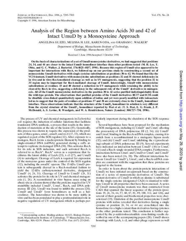
FIG. 1. Amino acid alignment of proteins that are homologous to UmuD.
Shown is the region between amino acids 24 and 44 of UmuD. This figure is
modified from reference 3. Positions of l cI (13), LexA (21, 22), and UmuD (3,
24) at which amino acid substitutions have been shown to yield stable proteins
that are defective in RecA-mediated cleavage are indicated by squares. Amino
acids that are identical in the three mutagenesis proteins but are not shared with
LexA or the three bacteriophage repressors are indicated by shading. Amino
acids that are identical in four or more members of the set are indicated by
boldfaced lettering.
TABLE 1. Bacterial strains and plasmids used in this study
Strain or plasmid
Relevant genotype or description
Reference or source
Strains
AB1157
GW3200
SG1161
RW82
GW8017
GW4212
GW8400
argE3
Same as AB1157 but umuD44
JM101 derivative; D(lac-pro) Dgal Dlon510 supE thi/(F9 traD36 proAB1 lacIq lacZDM15)
D(umuDC)595::cat
Same as AB1157 but D(umuDC)::cat
recA938::cat
Same as SG1161 but recA938::cat
24
14
37
AB1157XP1(RW82)
36
SG1161XP1(GW4212)
Plasmids
pGW6070
pGW6100
pGW7041
pGW7051
pGW7061
pGW7071
pGW7081
pGW7091
pGW7101
pGW7111
pGW7131
pGW7141
pGW7151
pGW7161
UmuD expressed from T7 promoter
70-TGT to GCC; Cys-24 to Ala; pGW6070 derivative; umuD131
103-GAA to TGT; Glu-35 to Cys; pGW6070 derivative; umuD142
106-CAG to TGT; Gln-36 to Cys; pGW6070 derivative; umuD143
109-CGC to TGC; Arg-37 to Cys; pGW6070 derivative; umuD144
112-ATC to TGC; Ile-38 to Cys; pGW6070 derivative; umuD145
115-GAT to TGT; Asp-39 to Cys; pGW6070 derivative; umuD146
118-CTG to TGC; Leu-40 to Cys; pGW6070 derivative; umuD147
121-AAT to TGT; Asn-41 to Cys; pGW6070 derivative; umuD148
124-CAA to TGT; Gln-42 to Cys; pGW6070 derivative; umuD149
88-GCA to TGC; Ala-30 to Cys; pGW6070 derivative; umuD150
91-GCA to TGC; Ala-31 to Cys; pGW6070 derivative; umuD151
94-GAT to TGT; Asp-32 to Cys; pGW6070 derivative; umuD152
97-TAC to TGT; Tyr-33 to Cys; pGW6070 derivative; umuD153
19
19
This
This
This
This
This
This
This
This
This
This
This
This
work
work
work
work
work
work
work
work
work
work
work
work
Downloaded from http://jb.asm.org/ on May 29, 2020 by guest
as with various UmuD-like proteins, including the region corresponding to amino acids 24 to 44 of UmuD (Fig. 1). Interestingly, mutations in the corresponding region in l cI (positions 111 to 132) were found to abolish RecA-mediated
cleavage but not self-cleavage (13), suggesting that this region
may be involved in interactions with RecA. Thus, we followed
up our previous study by constructing a second set of monocysteine UmuD derivatives, each containing a single cysteine
from amino acid 30 to 42. In this paper, we report our analyses
of the effects of the single cysteine changes on biological activity by assay of UV mutagenesis and the effects on UmuD
cleavage by RecA both in vivo and in vitro. We have also
assessed the relative proximity of the cysteines to each other in
the homodimer by measuring the ability of the cysteines to be
cross-linked after oxidation with iodine or copper phenanthroline. Moreover, the ability to spontaneously cross-link the
cysteines into a dimer after dialysis in a buffer lacking dithiothreitol (DTT) was assessed, and the results obtained by the
different methods of cross-linking were compared. Inferences
about the structure of intact UmuD made from these experi-
Bacterial strains and media. Bacterial strains and plasmids used in this study
are listed in Table 1. The antibiotics used (at the indicated concentrations) were
ampicillin (100 mg/ml), chloramphenicol (30 mg/ml), kanamycin (50 mg/ml), and
tetracycline (12.5 mg/ml).
Construction of monocysteine umuD mutant plasmids and overproduction
and purification of UmuD. Construction of monocysteine umuD mutant plasmids was described elsewhere (19). All of the umuD mutants are under the
control of the T7 promoter. E. coli SG1611 was used for the overproduction of
the UmuD derivatives EC35, QC36, DC39, and LC40, and strain GW8400 was
used for the overproduction of the UmuD derivatives AC30, AC31, DC32,
YC33, RC37, IC38, NC41, and QC42. The monocysteine mutant proteins were
purified to homogeneity as previously described (19) except that the buffer of the
UmuD-containing fractions eluted from the Mono Q column was not exchanged.
All the monocysteine UmuD derivatives purified indistinguishably from the
wild-type protein. UmuD protein concentrations were determined with respect
to the monomeric species.
UV mutability and RecA cleavage assays. UV mutagenesis was carried out
according to the procedure of Elledge and Walker (6) with strain GW3200. In
vivo RecA-mediated cleavage was performed with strain GW8017 by the following procedure. A saturated culture in minimal M9-glucose medium (28) supplemented with 0.1 mM CaCl2, 0.1 mM FeCl3, 0.1 mM ZnSO4, 0.4% glucose, 5 mg
of thiamine per ml, and antibiotics was diluted 1:10 into Luria broth containing
the appropriate antibiotics. At an A600 of 0.4 to 0.6, isopropyl-b-D-thiogalactopyranoside (IPTG) was added to a final concentration of 0.5 mM. After 1 h of
incubation at 378C, the culture was washed twice with an equal volume of 0.85%
saline. The cells were UV irradiated at 50 J/m2, centrifuged at 4,600 3 g for 10
min, resuspended in an equal volume of Luria broth containing antibiotics, and
incubated for 45 min at 378C. UmuD cleavage was assessed by centrifuging the
cells and resolving the protein from 0.05 A600 U of cells by electrophoresis on a
sodium dodecyl sulfate (SDS)-polyacrylamide gel, transferring the protein to a
polyvinylidene difluoride transfer membrane (Immobilon-P; Millipore, Bedford,
Mass.), and blotting with a 1:10,000 dilution of affinity-purified antibodies raised
against UmuD9. Cross-reacting material was visualized by chemiluminescence
(Tropix, Bedford, Mass.) which was quantitated with an LKB Bromma 2202
Ultroscan laser densitometer.
In vitro RecA-mediated cleavage was carried out according to the protocol of
Lee et al. (19) with some modifications. Reactions were conducted in 40 mM
Tris-HCl (pH 8.0)–6.8 mM MgCl2–30 mM NaCl–0.3 mM DTT with 42 ng of a
20-mer oligonucleotide per 20-ml sample volume and 0.68 mM ATPgS. UmuD at
a concentration of 10 mM was incubated with 3.15 mM RecA at 378C for 1 h.
Reactivity of mutant UmuD proteins to [3H]iodoacetate and cross-linking of
UmuD mutant derivatives with iodine and copper phenanthroline. Reactivity to
�VOL. 178, 1996
MONOCYSTEINE ANALYSIS OF AN N-TERMINAL REGION OF UmuD
7297
[3H]iodoacetate was conducted as previously described (19) except that 0.6 mM
DTT was present in the reaction mixture. Reactions with iodine and copper
phenanthroline were performed as previously described (19) with the following
exceptions: reactions with iodine were initiated by the addition of 1 mM aqueous
iodine to 10 mM UmuD in 50 mM HEPES (pH 8.1)–100 mM NaCl–0.3 mM
DTT. Oxidations with O2 catalyzed by copper phenanthroline were done by
reacting 10 mM UmuD with 1 mM Cu21 (as CuSO4) and 1.3 mM phenanthroline
for 10 min at 08C in 50 mM HEPES (pH 8.1)–100 mM NaCl–0.3 mM DTT.
Removal of reducing agent from UmuD solvent by dialysis. UmuD at a
concentration of 13 mM in 10 mM sodium phosphate (pH 6.8)–100 mM NaCl–
0.4 mM DTT was dialyzed against 10 mM sodium phosphate (pH 6.8)–100 mM
NaCl–5 mM EDTA with a System 100 microdialyzer (Pierce, Rockford, Ill.) at
48C for 2 h. SDS sample buffer was added after dialysis, and the proteins were
resolved by electrophoresis on an SDS–13% polyacrylamide gel. The Coomassie
blue-stained bands corresponding to the monomeric and dimeric forms of
UmuD were quantitated with an LKB Bromma 2202 Ultroscan laser densitometer.
RESULTS
Activity of the UmuD mutant proteins in UV mutagenesis
and RecA-mediated cleavage studies. In this study, our strategy
for choosing the sites for the single substitutions in this set of
derivatives differed from that described previously (19). In that
case we chose sites which would maximize the probability of
obtaining biologically active molecules spanning the entire
length of the UmuD protein (19), while in this study we chose
a particularly interesting region of UmuD to make successive
single cysteine substitutions. Thus, some derivatives have cysteine substitutions at sites which are conserved throughout the
set of analogous mutagenesis proteins. In addition, some derivatives have cysteine substitutions that do not necessarily
represent conservative substitutions; i.e., a cysteine may be
substituted for residues other than serine or alanine in many
cases. The effects that these mutations have on the ability of
UmuD to perform in various capacities, such as UV mutagenesis and RecA-mediated cleavage, have the potential to yield
insights into the significance of the residues in this particular
region of the UmuD protein. The ability of the mutant UmuD
proteins to participate in UV mutagenesis was determined by
expressing them in a umuD44 strain and measuring the reversion of an argE3 mutant to Arg1. Most of the UmuD monocysteine derivatives tested retained a substantial ability to participate in UV mutagenesis (40 to 90% of wild-type activity).
The UmuD monocysteine derivatives which were most substantially impaired by the cysteine substitution were DC32,
YC33, EC35, and LC40, which retained less than 30% of wildtype activity (Fig. 2), suggesting that the residues in these
positions are important, either directly or indirectly, for UmuD
to be able to participate in UV mutagenesis.
The ability of these monocysteine UmuD derivatives to productively interact with RecA in a manner which leads to
UmuD cleavage, both in vitro and in vivo, was assessed. The
UmuD monocysteine derivatives were purified to homogeneity, and their ability to be cleaved by RecA in vitro was measured. As mentioned in Materials and Methods, all the derivatives purified indistinguishably from wild-type UmuD. We
found that wild-type UmuD was cleaved to an extent of around
60% in 1 h under the conditions described in Materials and
Methods. Although cleavage of UmuD and related proteins
has been studied extensively in an in vitro reaction consisting of
RecA, ssDNA, and ATP (or a nonhydrolyzable analog) (5, 27),
it is possible that there is an additional factor that functions in
the in vivo reaction which is not present in our in vitro reaction.
To determine the extent of RecA-mediated cleavage in vivo, a
DumuDC strain carrying a UmuD mutant plasmid was induced
for UmuD production and irradiated with UV light at a dose
of 50 J/m2. After a 45-min incubation at 378C, the extent of
cleavage was determined by Western blotting (immunoblotting) with affinity-purified UmuD antiserum (3) and was found
to be around 75% for wild-type UmuD under these conditions.
Downloaded from http://jb.asm.org/ on May 29, 2020 by guest
FIG. 2. Relative mutation frequency and in vivo and in vitro RecA-mediated cleavage. Mutagenesis was determined for cells irradiated with a UV dose of 20 J/m2.
In vivo and in vitro RecA-mediated cleavage assays were conducted as described in Materials and Methods. Black bars, relative mutation frequency; hatched bars,
relative in vivo RecA-mediated cleavage; gray bars, relative in vitro RecA-mediated cleavage (all percentages of the wild-type level). The mean extent of in vivo
RecA-mediated cleavage of UmuD1 6 the standard deviation was 74.5% 6 5.5%. The extent of in vitro RecA-mediated cleavage of UmuD1 was 60.5% 6 3.0%. The
error bars signify the standard deviations. The numbers along the x axis correspond to the cysteine of the UmuD monocysteine derivative.
�7298
GUZZO ET AL.
study and subsequently diluted the protein stocks into the
appropriate reaction buffers. Thus, reaction buffers for this
study contained higher DTT concentrations (0.3 mM final concentration) than previously published reaction conditions
(,0.1 mM) (19). For this reason, the experiments using the
UmuD monocysteine derivatives VC34 and LC44 were repeated and included with the new series of monocysteine mutant derivatives.
When the monocysteine mutants were oxidized with iodine,
both RC37 and IC38 showed nearly quantitative cross-linking.
In contrast, oxidation of the remaining subset of monocysteine
derivatives resulted in a considerably lower level of cross-linking, ranging from 4.5 to 45%. This is a striking result considering the fact that most of the derivatives (with the exception
of RC37 and IC38) reacted quite well with iodoacetate, indicating that the sulfhydryl groups were reasonably exposed to
solvent. Even within this small region from amino acid 30 to 42,
the position of the cysteine substitution greatly affects the
ability of the UmuD derivatives to be cross-linked in the homodimer upon oxidation with iodine. As a control, this set of
monocysteine mutants was incubated in the presence of 0.3
mM DTT for 1 h at 378C without any oxidizing agent, and no
detectable disulfide bond formation occurred (data not
shown), ruling out the possibility that the observed cross-linking was due to spontaneous disulfide bond formation.
As observed previously (19), the copper phenanthrolinecatalyzed oxidation of the UmuD monocysteine mutant proteins in the homodimer resulted in cross-linking data that are
consistent with those obtained with iodine oxidation, but the
differences between the derivatives are less striking. RC37 and
IC38 are still the most efficiently cross-linked; however, all of
the other mutants were also able to cross-link with moderate
efficiency when this reagent was used. A simple explanation for
these observations is that during the more prolonged copper
phenanthroline-catalyzed air oxidation of the thiol groups, local flexibility within the monomer and the separation of the
homodimer into monomers allows two sulfhydryls to come
close enough for cross-linking to occur. No cross-linking was
observed when the UmuD derivative lacking cysteine, CA24,
was exposed to iodine or copper phenanthroline (data not
shown).
Spontaneous cross-linking of UmuD dimers after removal of
DTT by dialysis. Because certain UmuD monocysteine derivatives spontaneously cross-link via disulfide bond formation in
the presence of low concentrations of DTT, we thought it
would be interesting to survey the ability of our entire set of
UmuD monocysteine derivatives to spontaneously cross-link
upon removal of DTT by dialysis. The monocysteine derivatives in a buffer containing 0.4 mM DTT were dialyzed for 2 h
at 48C, and the resulting percentage of disulfide-cross-linked
dimers is plotted in Fig. 4. Dialysis of the derivatives with
cysteine substitutions within the region between positions 37
and 41 resulted in a high degree of cross-linking. Other derivatives which resulted in efficient dimer cross-linking are C24
(wild-type UmuD), AC30, AC31, and LC44. SC19, SC60,
SC112, DC126, and derivatives with cysteine substitutions
FIG. 3. Iodoacetate reactivities and cross-linking ability of UmuD monocysteine derivatives. (A) Reactivity of UmuD monocysteine mutant proteins to [3H]iodoacetate. The amount of total protein modified by [3H]iodoacetate (IAA) in 60 min was measured. UmuD at a concentration of 20 mM was incubated with a 65-fold molar
excess of [3H]iodoacetate in 50 mM HEPES (pH 8.1)–500 mM NaCl–0.6 mM DTT for 60 min in the dark at 378C. The counts determined for CA24 (UmuD without
a cysteine) were only slightly above background level and were subtracted as background. (B) Percentage of UmuD cross-linked by using iodine (I2). UmuD (10 mM)
was incubated with 1 mM iodine for 20 min at 228C as described in Materials and Methods. (C) Percentage of UmuD cross-linked by using copper phenanthroline
(CuP). Oxidations with O2 catalyzed by CuP were conducted by reacting 10 mM UmuD with 1 mM Cu21 and 1.3 mM phenanthroline for 10 min at 08C in 50 mM
HEPES (pH 8.1)–100 mM NaCl as described in Materials and Methods. The error bars in all three panels signify the standard deviations. The numbers along the x
axis correspond to the cysteine of the UmuD monocysteine derivative.
Downloaded from http://jb.asm.org/ on May 29, 2020 by guest
We found that for many of the monocysteine derivatives,
RecA-mediated cleavage roughly correlated with UV mutability. Since RecA-mediated cleavage is required to activate
UmuD for mutagenesis, it is not surprising that those derivatives which were defective in RecA-mediated cleavage should
also be defective in mutagenesis (3). One exception was the
AC31 derivative, which displayed a partial reduction in UV
mutability but a nearly wild-type ability to be cleaved by RecA,
both in vivo and in vitro. This observation suggests that this
mutant could be partially defective in a role in UV mutagenesis
that occurs after cleavage of UmuD to UmuD9, such as in the
interaction of UmuD9 with other proteins. Other exceptions
were YC33 and LC40, which, although defective in UV mutagenesis and in vitro RecA-mediated cleavage, were able to be
cleaved efficiently by RecA in vivo. There may be a factor
required in vivo that is not present in our in vitro reaction.
Additionally, these mutants may be like AC31 in that they are
partially impaired in their ability to perform in some capacity
in UV mutagenesis that occurs after the cleavage of UmuD to
UmuD9.
Solvent accessibility of the UmuD derivatives. In order to
test for the accessibility and reactivity of the unique cysteines in
UmuD, the purified UmuD derivatives were reacted with
[3H]iodoacetate. The results are expressed as the number of
nanomoles of [3H]iodoacetate that reacted with 0.20 nmol of
UmuD in 1 h, a time period in which a fully modified population of UmuD would have incorporated 0.20 nmol of [3H]
iodoacetate (Fig. 3A). Generally, the extent of reactivity for
each thiol group depends primarily on its exposure to solvent
and also on its particular local electrostatic environment (7).
Most of the mutants showed a moderate to high level of solvent accessibility. Two exceptions were RC37 and IC38, which
reacted poorly with [3H]iodoacetate. Moderate or high reactivity suggests that the thiol group of the UmuD derivative is
accessible to solvent, whereas low reactivity suggests that the
thiol group is buried within the interior of the protein or
possibly within the dimer interface.
Cross-linking of the UmuD monocysteine derivatives with
iodine or copper phenanthroline. In order to gain information
concerning the positions of the various monocysteine substitutions relative to the dimer interface, we examined the susceptibilities of the homodimers of the UmuD monocysteine derivatives to cross-linkage by disulfide bond formation. In order
for the UmuD monocysteine derivatives to cross-link, the two
cysteines must be proximal to each other and in the correct
orientation in the homodimer. This cross-linking was carried
out by the addition of iodine (Fig. 3B) or copper phenanthroline (Fig. 3C). During the course of the purification of these
monocysteine derivatives, we found that a significant proportion of the UmuD proteins (in particular, RC37 and IC38)
spontaneously cross-linked in 0.1 mM DTT in the absence of
any oxidizing agents. When we increased the DTT concentration to 1 mM, however, no disulfide bond formation was detectable. We therefore increased the final concentration of
DTT in the buffer of the stocks of all of the UmuD mutant
proteins to 1 mM in order to be consistent throughout the
J. BACTERIOL.
�7299
Downloaded from http://jb.asm.org/ on May 29, 2020 by guest
�7300
GUZZO ET AL.
J. BACTERIOL.
within the region between amino acids 32 and 36 all have a
moderate ability to spontaneously form disulfide cross-linkages
in the dimer. Finally, QC42, SC57, SC67, SC81, AC89, and
QC100 were able to cross-link poorly upon removal of the
reducing agent.
DISCUSSION
On the basis of the relative abilities of several monocysteine
derivatives of UmuD to cross-link via cysteine-specific crosslinking agents, we previously suggested that the region including the Cys-24–Gly-25 cleavage site, Val-34, and Leu-44 is
closer to the UmuD homodimer interface than the other residues tested (19). Other evidence also suggested that this region is important for UmuD interactions with RecA (3, 13, 20).
To further analyze the interactions in this region of UmuD, we
constructed a set of UmuD monocysteine derivatives in which
single cysteine substitutions were made in the region of UmuD
between amino acids 30 and 42 (inclusive) and characterized
them genetically and biochemically. Our studies of the structure of the homodimer of intact UmuD in solution strongly
indicate that Arg-37 and Ile-38 are very close to the dimer
interface of UmuD2. This inference is based on the striking
ease of disulfide cross-linking of the RC37 and IC38 monocysteine derivatives upon treatment with iodine. Nearly quantitative disulfide cross-linking of these derivatives occurred very
rapidly upon oxidation with iodine, in contrast to the crosslinking efficiencies of the other monocysteine UmuD derivatives tested, which ranged from approximately 10 to 50%. Interestingly, when the ability of each of the mutant UmuD
proteins to be modified by [3H]iodoacetate was assessed, we
found that all of the purified monocysteine derivatives except
RC37 and IC38 were quite reactive with iodoacetate (Fig. 3A).
A simple explanation for this observation is that these residues
are buried within the dimer interface and thus are less accessible for reaction with iodoacetate. The AC30 and VC34 proteins can be cross-linked by iodine relatively efficiently, although not as well as RC37 or IC38, but appear to be more
accessible to solvent as measured by reactivity to iodoacetate.
Thus, positions 30 and 34 may be relatively near the UmuD2
homodimer interface but are not as close to it as positions 37
and 38.
All of our solution studies on the intact UmuD2 homodimer
were performed in the absence of any structural data. However, after our studies were completed, Peat et al. reported the
crystal structure of the cleaved form of UmuD, UmuD9, to 2.5
Å (0.25 nm) (25). In the crystal structure of UmuD9, the
amino-terminal tail (including amino acids 30 to 42) extends
outward from a globular head. Residues Tyr-52, Val-54, Ile-87,
Phe-94, and Phe-128 of each UmuD9 monomer participate in
hydrophobic interactions at the UmuD92 homodimer interface.
In addition, a salt bridge is formed between Glu-93 of one
monomer and Lys-55 of the associating monomer. The aminoterminal tails in the UmuD92 homodimer protrude out in opposite directions and do not participate in dimer interactions.
It seems possible that the region from amino acid 32 to 40 is
seen as an extended terminal tail with a unique conformation
in the crystal as a consequence of crystal packing forces. As
shown in Fig. 5, residues 34, 37, and 38 are located in the
amino-terminal tails in the UmuD92 homodimer, whereas our
studies of the UmuD2 homodimer in solution indicate that
residues 30, 34, 37, and 38 (the region containing residue 30 is
disordered in the crystal) are very close to the UmuD2 dimer
interface. Thus, our results strongly suggest that the structures
of the UmuD2 homodimer in solution and the UmuD92 ho-
Downloaded from http://jb.asm.org/ on May 29, 2020 by guest
FIG. 4. Cross-linking of the monocysteine UmuD derivatives in the homodimer during dialysis. UmuD (13 mM) in a buffer containing 0.4 mM DTT was dialyzed
for 2 h at 48C against 10 mM sodium phosphate (pH 6.8)–100 mM NaCl–5 mM EDTA as described in Materials and Methods. The error bars signify the standard
deviations.
�VOL. 178, 1996
MONOCYSTEINE ANALYSIS OF AN N-TERMINAL REGION OF UmuD
7301
modimer (as determined from the crystal structure) are radically different.
To further study the structure and interactions of intact
UmuD by analyzing the region between residues 30 through 42
(which seems important not only for UmuD2 homodimer interactions but also for interactions with RecA [3, 13, 20]), our
strategy involved choosing this particularly interesting region
of UmuD as the site for successive single cysteine substitutions.
When the abilities of the mutants to perform in UV mutagenesis and to be cleaved by RecA both in vivo and in vitro were
assessed (Fig. 2), we observed that many of the monocysteine
derivatives retained a significant ability to perform in these
capacities. Although monocysteine derivatives DC32 and
EC35 were substantially impaired in all of the tested functions,
YC33 and LC40 were interesting because while they retained
significant in vivo RecA-mediated cleavage, their ability to
perform in UV mutagenesis was defective. We had previously
reported that the VC34 UmuD derivative was also impaired in
its ability to perform in these functions (19). In addition, as
discussed in one of the accompanying papers (20), we were
able to cross-link the VC34 derivative to RecA by using the
cysteine-specific photoactive cross-linker p-azidoiodoacetanilide. Taken together, these results suggest that amino acids
important for UmuD cleavage and its subsequent role in UV
mutagenesis overlap but are not identical, and they support the
theory that the amino acid 31 to 35 region is important for
interactions with RecA*. Such a conclusion is supported by
Gimble and Sauer’s isolation of l cI mutants that are deficient
in RecA*-mediated cleavage but not in the RecA-independent
cleavage under alkaline conditions (13). These mutations include EK117, TI122, GD124, DV125, DY125, DN125, and
EK127 of l cI, which correspond to Ala-30, Glu-35, Arg-37,
Ile-38, and Leu-40 of UmuD (Fig. 1). It is possible that residues in the region of UmuD9 that correspond to amino acids
30 to 40 of intact UmuD could play a role in the interaction
between UmuD9 and RecA* that has been reported (10), but
further work will be required to address this point.
The abilities of the UmuD monocysteine derivatives to
cross-link in the homodimer were determined by three different means: oxidation with iodine, oxidation with oxygen cata-
lyzed by copper phenanthroline, and spontaneous oxidation
with oxygen upon removal of the reducing agent by dialysis.
Of the three methods for discerning the relative proximity of
cysteine residues by disulfide cross-linking, iodine oxidation
seemed to be the most discriminatory. Interestingly, the oxidation reaction involving iodine occurs so rapidly that we were
unable to follow the kinetics of the cross-linking reaction. Although under our standard reaction conditions the proteins are
exposed to iodine at 228C for 20 min, we found no detectable
difference in the amount of disulfide bond formation even
when the reaction was carried out at 48C for 1 min (data not
shown). Furthermore, the amount of disulfide-cross-linked
dimers cannot be further increased by a second addition of
iodine. The explanation for this phenomenon lies in the mechanism of disulfide bond formation caused by iodine oxidation
(35). Iodine oxidation of the thiol group of a UmuD monocysteine derivative results in a sulfenyl iodide intermediate (35), a
reaction that apparently occurs very rapidly. A subsequent
reaction of the sulfenyl iodide with the thiol group of the
associating UmuD monomer is required to form a disulfide
bond and to cross-link the dimer. The sulfenyl iodide intermediate is very labile (35), and thus this reaction requires a close
proximal relationship between the sulfenyl iodide and the thiol
group (35). However, if a water molecule attacks the sulfenyl
iodide intermediate instead of an adjacent thiol group, a protein sulfenic acid is formed which is no longer available for
cross-linking. The sulfenic acid is possibly further oxidized to a
sulfinic or sulfonic acid (18). Thus, it seems likely that the
reaction mechanism can be summarized as shown in Fig. 6.
Because this reaction proceeds very rapidly (less than 1 min),
the extent of UmuD2 disulfide cross-linking promoted by iodine probably closely reflects the proportion of cysteines that
are in close proximity in the dimer within a small window of
time.
Our results suggest that iodine oxidation provides a good
assessment of the proximity of the cysteines to one another in
the homodimer interface. From the iodine cross-linking data it
is evident that RC37 and IC38 clearly cross-linked the most
efficiently of the derivatives that we tested. Oxidation with
copper phenanthroline yielded the same results; however,
Downloaded from http://jb.asm.org/ on May 29, 2020 by guest
FIG. 5. Ribbon diagram of the UmuD92 homodimer as determined by Peat et al. (25). Indicated are some of the amino acids that were changed in our study of
the intact UmuD protein. It is important to note that it is the structure of the UmuD9 protein which is depicted. Amino acids 30 to 42 are clearly not at the UmuD9
homodimer interface.
�7302
GUZZO ET AL.
J. BACTERIOL.
more, analysis of the UmuD92 structure by a monocysteine
approach will also provide insights into interactions within the
UmuD92 homodimer and into interactions of UmuD9 with
other proteins involved in mutagenesis. A better understanding of the mechanism of activation of UmuD to UmuD9 upon
RecA-mediated cleavage might also be gained by a comparison
of the structures and interactions of UmuD and UmuD9.
FIG. 6. Proposed reaction mechanism for disulfide bond formation via iodine
oxidation.
We thank Per Malkus for helping us to generate this set of mutants
and the members of the laboratory for many helpful suggestions. We
also thank Roger Woodgate, Wayne Hendrickson, and their collaborators for sharing information prior to publication.
This work was supported by Public Health Service grant CA21615
awarded by the National Cancer Institute. A.G. was supported by
a postdoctoral fellowship from the Medical Research Council of
Canada. M.H.L. was supported by predoctoral training grant
5T32GM07287 from the National Institutes of Health. K.O. carried
out her research as part of the Undergraduate Research Opportunities
Program (UROP) at the Massachusetts Institute of Technology.
REFERENCES
1. Bailone, A., S. Sommer, J. Knezevic, and R. Devoret. 1991. Substitution of
UmuD9 for UmuD does not affect SOS mutagenesis. Biochimie 73:471–478.
2. Battista, J. R., T. Nohmi, C. E. Donnelly, and G. C. Walker. 1988. Role of
UmuD and UmuC in UV and chemical mutagenesis, p. 455–459. In E. C.
Friedberg and P. C. Hanawalt (ed.), Mechanisms and consequences of DNA
damage processing. Alan R. Liss, Inc., New York.
3. Battista, J. R., T. Ohta, T. Nohmi, W. Sun, and G. C. Walker. 1990. Dominant negative umuD mutations decreasing RecA-mediated cleavage suggest
roles for intact UmuD in modulation of SOS mutagenesis. Proc. Natl. Acad.
Sci. USA 87:7190–7194.
4. Burckhardt, S. E., R. Woodgate, R. H. Scheuermann, and H. Echols. 1988.
UmuD mutagenesis protein of Escherichia coli: overproduction, purification,
and cleavage by RecA. Proc. Natl. Acad. Sci. USA 85:1811–1815.
5. Craig, N. L., and J. W. Roberts. 1980. E. coli recA protein-directed cleavage
of phage lambda repressor requires polynucleotide. Nature (London) 283:
26–30.
6. Elledge, S. J., and G. C. Walker. 1983. Proteins required for ultraviolet light
and chemical mutagenesis: identification of the products of the umuC locus
of Escherichia coli. J. Mol. Biol. 164:175–192.
7. Falke, J. J., A. F. Dernburg, D. A. Sternberg, N. Zalkin, D. L. Milligan, and
D. E. Koshland, Jr. 1988. Structure of a bacterial sensory receptor. A sitedirected sulfhydryl study. J. Biol. Chem. 263:14850–14858.
8. Falke, J. J., and D. E. Koshland, Jr. 1987. Global flexibility in a sensory
receptor: a site-directed cross-linking approach. Science 237:1596–1600.
9. Foster, P. L., and A. D. Sullivan. 1988. Interactions between epsilon, the
proofreading subunit of DNA polymerase III, and proteins involved in the
SOS response of Escherichia coli. Mol. Gen. Genet. 214:467–473.
10. Frank, E. G., J. Hauser, A. S. Levine, and R. Woodgate. 1993. Targeting of
the UmuD, UmuD9 and MucA9 mutagenesis proteins to DNA by RecA
protein. Proc. Natl. Acad. Sci. USA 90:8169–8173.
11. Freitag, N., and K. McEntee. 1989. “Activated”-RecA protein affinity chromatography of LexA repressor and other SOS-regulated proteins. Proc. Natl.
Acad. Sci. USA 86:8363–8367.
12. Friedberg, E. C., G. C. Walker, and W. Siede. 1995. DNA repair and mutagenesis. ASM Press, Washington, D.C.
13. Gimble, F. S., and R. T. Sauer. 1986. l repressor inactivation: properties of
purified ind2 proteins in the autodigestion and RecA-mediated cleavage
reactions. J. Mol. Biol. 192:39–47.
14. Gottesman, S. 1990. Minimizing proteolysis in Escherichia coli: genetic solutions. Methods Enzymol. 185:119–129.
15. Hughes, R. E., P. A. Rice, T. A. Steitz, and N. D. F. Grindley. 1993. Proteinprotein interactions directing resolvase site-specific recombination: a structure-function analysis. EMBO J. 12:1447–1458.
16. Jonczyk, P., and A. Nowicka. 1996. Specific in vivo protein-protein interactions between Escherichia coli SOS mutagenesis proteins. J. Bacteriol. 178:
2580–2585.
17. Kato, T., and Y. Shinoura. 1977. Isolation and characterization of mutants of
Escherichia coli deficient in induction of mutations by ultraviolet light. Mol.
Gen. Genet. 156:121–131.
18. Kharasch, N. 1961. Organic sulfur compounds. Pergamon Press, New York.
19. Lee, M. H., T. Ohta, and G. C. Walker. 1994. A monocysteine approach for
probing the structure and interactions of the UmuD protein. J. Bacteriol.
176:4825–4837.
20. Lee, M. H., and G. C. Walker. 1996. Interactions of Escherichia coli UmuD
with activated RecA analyzed by cross-linking UmuD monocysteine deriva-
Downloaded from http://jb.asm.org/ on May 29, 2020 by guest
UmuD derivatives with cysteines in other positions also
formed disulfide bonds in the dimer to a moderate degree
under these conditions. It is possible that this region (residues
30 to 42) is quite flexible in the intact UmuD proteins. If this
is the case, then the copper phenanthroline-catalyzed oxidation of the derivatives could involve the capturing of transient
intermediates in which the residues of associating monomers
are brought close enough to each other to be disulfide crosslinked (8, 19). Thus, we would expect all the derivatives with
cysteine substitutions in this region to react with about the
same efficiency unless the positions of the pairs of cysteines in
the dimer were particularly close in the native conformation, as
we suggest is the case for Arg-37 and Ile-38. Surprisingly, we
found that the spontaneous oxidation of thiols to form disulfide bonds during dialysis was not as discriminatory as the
oxidation of thiols by iodine. Since spontaneous air oxidation
of thiols appears to be a very mild method of oxidation, we had
initially expected this procedure to be highly selective for only
those pairs of cysteines which are optimally positioned in the
native structure.
Oxidation by dialysis is not as discriminatory (Fig. 4), but this
method has been used before in structure-function investigations of other proteins, such as the gd resolvase protein (15),
for which the crystal structure of the catalytic subunit is known
(29). In the case of the gd resolvase, inspection of the crystal
structure and computer modeling were employed to identify
positions for cysteine substitutions which would minimize distortion of the dimer interface in the disulfide-cross-linked
dimer. Two of the mutants cross-linked completely in the
dimer, and one mutant cross-linked to a level of 50% after 12 h
of dialysis. These observed results were interpreted to imply
that dialyzing away the reducing agent and then testing for
disulfide bond formation is a good method for identifying
those residues which are relatively close in the native conformation of the dimer. However, in our investigations of derivatives with cysteine substitutions within the region of amino
acids 30 to 44, we found that in addition to RC37 and IC38, the
UmuD derivatives AC30, AC31, DC39, LC40, NC41, and
LC44 also disulfide cross-linked quite efficiently during dialysis
(Fig. 4). In our experiments, we allowed dialysis to proceed for
only 2 h, although even an additional 2 h of dialysis resulted in
a higher yield of cross-linked dimers (data not shown). Moreover, we found that in addition to those derivatives in the first
set of monocysteine proteins which were expected to cross-link
efficiently (SC19, C24, VC34, and LC44), the UmuD derivatives SC60, SC112, and DC126 also appeared to cross-link
relatively more efficiently during dialysis than upon treatment
with iodine (determined previously [19]). These results suggest
that inferences about the solution structure made from the
spontaneous disulfide cross-linking of a single pair of cysteines
after dialyzing away the reducing agent should be made with
caution.
It will be interesting to evaluate the structure of UmuD with
results obtained from this monocysteine approach. Further-
ACKNOWLEDGMENTS
�VOL. 178, 1996
MONOCYSTEINE ANALYSIS OF AN N-TERMINAL REGION OF UmuD
30.
31.
32.
33.
34.
35.
36.
37.
38.
domain of the site-specific recombination enzyme gd resolvase at 2.7 Å. Cell
63:1323–1329.
Sassanfar, M., and J. W. Roberts. 1990. Nature of the SOS-inducing signal
in Escherichia coli: the involvement of DNA replication. J. Mol. Biol. 212:
79–96.
Shinagawa, H., H. Iwasaki, T. Kato, and A. Nakata. 1988. RecA proteindependent cleavage of UmuD protein and SOS mutagenesis. Proc. Natl.
Acad. Sci. USA 85:1806–1810.
Sommer, S., A. Bailone, and R. Devoret. 1993. The appearance of the
UmuD9C protein complex in Escherichia coli switches repair from homologous recombination to SOS mutagenesis. Mol. Microbiol. 10:963–971.
Steinborn, G. 1978. Uvm mutants of Escherichia coli K12 deficient in UV
mutagenesis. I. Isolation of uvm mutants and their phenotypical characterization in DNA repair and mutagenesis. Mol. Gen. Genet. 165:87–93.
Tadmor, Y., R. Ascarelli-Goell, R. Skaliter, and Z. Livneh. 1992. Overproduction of the b subunit of DNA polymerase III holoenzyme reduces UV
mutagenesis in Escherichia coli. J. Bacteriol. 174:2517–2524.
Trundle, D., and L. W. Cunningham. 1969. Iodine oxidation of the sulfhydryl
groups of creatine kinase. Biochemistry 8:1919–1925.
Winans, S. C., S. J. Elledge, J. H. Krueger, and G. C. Walker. 1985. Sitedirected insertion and deletion mutagenesis with cloned fragments in Escherichia coli. J. Bacteriol. 161:1219–1221.
Woodgate, R. 1992. Construction of a umuDC operon substitution mutation
in Escherichia coli. Mutat. Res. 281:221–225.
Woodgate, R., M. Rajagopalan, C. Lu, and H. Echols. 1989. UmuC mutagenesis protein of Escherichia coli: purification and interaction with UmuD and
UmuD9. Proc. Natl. Acad. Sci. USA 86:7301–7305.
Downloaded from http://jb.asm.org/ on May 29, 2020 by guest
tives. J. Bacteriol. 178:7285–7294.
21. Lin, L.-L., and J. W. Little. 1988. Isolation and characterization of noncleavable (Ind2) mutants of the LexA repressor of Escherichia coli K-12. J.
Bacteriol. 170:2163–2173.
22. Lin, L. L., and J. W. Little. 1989. Autodigestion and RecA-dependent cleavage of Ind2 mutant LexA proteins. J. Mol. Biol. 210:439–452.
23. Little, J. W. 1984. Autodigestion of LexA and phage l repressors. Proc. Natl.
Acad. Sci. USA 81:1375–1379.
24. Nohmi, T., J. R. Battista, L. A. Dodson, and G. C. Walker. 1988. RecAmediated cleavage activates UmuD for mutagenesis: mechanistic relationship between transcriptional derepression and posttranslational activation.
Proc. Natl. Acad. Sci. USA 85:1816–1820.
25. Peat, T. S., E. G. Frank, J. P. McDonald, A. S. Levine, R. Woodgate, and
W. A. Hendrickson. 1996. Structure of the UmuD9 protein and its regulation
in response to DNA damage. Nature (London) 380:727–730.
26. Rajagopalan, M., C. Lu, R. Woodgate, M. O’Donnell, M. F. Goodman, and
H. Echols. 1992. Activity of the purified mutagenesis proteins UmuC,
UmuD9, and RecA in replicative bypass of an abasic DNA lesion by DNA
polymerase III. Proc. Natl. Acad. Sci. USA 89:10777–10781.
27. Roberts, J. W., C. W. Roberts, and N. L. Craig. 1978. Escherichia coli recA
gene product inactivates phage lambda repressor. Proc. Natl. Acad. Sci. USA
75:4714–4718.
28. Sambrook, J., E. F. Fritsch, and T. Maniatis. 1989. Molecular cloning: a
laboratory manual, 2nd ed. Cold Spring Harbor Laboratory Press, Cold
Spring Harbor, N.Y.
29. Sanderson, M. R., P. S. Freemont, P. A. Rice, A. Goldman, G. F. Hatfull,
N. D. F. Grindley, and T. A. Steitz. 1990. The crystal structure of the catalytic
7303
�
 Graham Walker