(12) INTERNATIONAL APPLICATION PUBLISHED UNDER THE PATENT COOPERATION TREATY (PCT)
(19) World Intellectual Property Organization
International Bureau
(43) International Publication Date
(51) International Patent Classification:
A61K 36/20 (2006.01)
2 1 May 2008 (21.05.2008)
(25) Filing Language:
English
(26) Publication Language:
English
(30) Priority Data:
60/939,143
2 1 May 2007 (2 1.05 .2007)
WO 2008/144706 A2
(74) Agent: DAVIS, Bonnie, J.; TAYLOR, PORTER,
BROOKS & PHILLIPS L.L.P., P.O.BOX 2471, Baton
Rouge, PA 70821-2471 (US).
(21) International Application Number:
PCT/US2008/064303
(22) International Filing Date:
(10) International Publication Number
PCT
27 November 2008 (27.11.2008)
US
(71) Applicants (for all designated States except US):
BOARD OF SUPERVISORS OF LOUISIANA STATE
UNIVERSITY AND AGRICULTURAL AND ME¬
CHANICAL COLLEGE [US/US]; LSU AgCenter, 104
Efferson Hall, Baton Rouge, LA 70803 (US). BOARD OF
REGENTS, THE UNIVERSITY OF TEXAS SYSTEM
[US/US]; 201 West 7th Street, Austin, TX 78701 (US).
(72) Inventors; and
(75) Inventors/Applicants (for US only): LIU, Zhijun
[US/US]; 11142 South Lakeside Oaks Avenue, Baton
Rouge, LA 70810 (US). YANG, Peiying [US/US]; 5215
Riverstone Crossing Drive, Sugarland, TX 77479 (US).
NEWMAN, Robert, A. [US/US]; 1742- A Michigan
Street, Houston, TX 77006 (US).
(81) Designated States (unless otherwise indicated, for every
kind of national protection available): AE, AG, AL, AM,
AO, AT, AU, AZ, BA, BB, BG, BH, BR, BW, BY, BZ, CA,
CH, CN, CO, CR, CU, CZ, DE, DK, DM, DO, DZ, EC, EE,
EG, ES, FI, GB, GD, GE, GH, GM, GT, HN, HR, HU, ID,
IL, IN, IS, JP, KE, KG, KM, KN, KP, KR, KZ, LA, LC,
LK, LR, LS, LT, LU, LY, MA, MD, ME, MG, MK, MN,
MW, MX, MY, MZ, NA, NG, NI, NO, NZ, OM, PG, PH,
PL, PT, RO, RS, RU, SC, SD, SE, SG, SK, SL, SM, SV,
SY, TJ, TM, TN, TR, TT, TZ, UA, UG, US, UZ, VC, VN,
ZA, ZM, ZW
(84) Designated States (unless otherwise indicated, for every
kind of regional protection available): ARIPO (BW, GH,
GM, KE, LS, MW, MZ, NA, SD, SL, SZ, TZ, UG, ZM,
ZW), Eurasian (AM, AZ, BY, KG, KZ, MD, RU, TJ, TM),
European (AT, BE, BG, CH, CY, CZ, DE, DK, EE, ES, FI,
FR, GB, GR, HR, HU, IE, IS, IT, LT, LU, LV,MC, MT, NL,
NO, PL, PT, RO, SE, SI, SK, TR), OAPI (BF, BJ, CF, CG,
CI, CM, GA, GN, GQ, GW, ML, MR, NE, SN, TD, TG).
Published:
— without international search report and to be republished
upon receipt of that report
(54) Title: SWEET GUM FRUIT EXTRACT AS A THERAPEUTIC AGENT
Fig. 12
(57) Abstract: Sweet gum (Liquidambar styraciflua L., family Hamamelidaceae) fruit extract was discovered to possess potent ac
tivities against multiple targets of the PBK (phosphatidylinositide 3-kinase) pathway, especially the PI3K/Akt and mTOR pathways.
At a very low concentration of 1.85 µg/ml (IC50), sweet gun extract showed the ability of simultaneously blocking the pathways
of PI3K/Akt (upstream), mTOR (mammalian target of rapamycin) (downstream), as well as its downstream protein products S6K
and S6. It was also able to block 5-HETE, a lipoxygenase product that contributes to inflammation and activation of PI3K/Akt.
The sweet gum fruit extract was prepared with 50% methanol (47: 1; raw to extract) and concentrated to an organic fraction (210:
1 raw to extract) referred as LIS-100 via reverse-phase column chromatography using a bioassay directed fractionation approach.
The extract is a new targeted therapeutic agent for numerous disorders known to be treated by mTOR inhibitors, including cancer,
diabetes, obesity, and inflammation.
�SWEET GUM FRUIT EXTRACT A S A THERAPEUTIC AGENT
Zhijun Liu, Peiying Yang, and Robert A. Newman
Liu 07A10W
[0001]
The benefit of the filing date of provisional U.S. application Serial Number
60/939,143, filed 2 1 May 2007, is claimed under 35 U.S.C. § 119(e) in the United States, and is
claimed under applicable treaties and conventions in all countries.
TECHNICAL FIELD
[0002]
An
extract
from
sweet
gum
(Liquidambar
styraciflua
L.,
family
Hamamelidaceae) fruit was discovered to be a potent inhibitor against multiple targets of the
PI3K (phosphatidylinositide 3-kinase) pathway, especially the PI3K/Akt and mTOR pathways,
and to inhibit the proliferation of various cancer cells.
BACKGROUND ART
[0003]
Sweet Gum Tree:
Sweet gum {Liquidambar styraciflua L., family
Hamamelidaceae) is a large aromatic tree native to the Southeastern United States. The spiny
pendulous fruits are used as raw plant materials. Although there is no information available on
the use of the fruits as medicinal ingredients in the United States, the cousins of sweet gum are
used for medicinal purposes. For example, the dried fruit of Liquidambarformosana Hance is
an herbal ingredient that appears in the Chinese Herbal Pharmacopoeia as lulutong in Chinese.
[0004]
Traditionally, sweet gum was used to "dispel wind and remove obstruction from
the collaterals, to cause diuresis, and to stimulate menstrual discharge" (Chinese Meteria
Medica 1995). Another species of the same genus is Liquidambar orientalis Mill., which has
been used to clear mucus congestion and to relieve pain and congestion.
[0005]
Cinnamic acid and its derivatives as well as triterpenoids are reportedly the
main constituents of the bark of sweet gum tree. The sweet gum tree contains cinnamic acid
and betulonic acid, l-methoxy-9-caryolanol, and eudesm-4(14)-ene-l,6-diol.
Sweet gum
�fruits
(cones)
also
accumulate
the
following
triterpene
25-acetoxy-3 α-hydroxyolean-12-en-28-oic
carboxylic
acids:
3 β,
acid;
25-epoxy-3 α-hydroxylup-20(29)-en-28-oic acid, 3 β,25-epoxy-3 α-hydroxyolean-12-en-28-oic
acid;
oleanolic
butulinic
acid;
3-oxohydroxy-
(29)-en-28-oic
acid;
3α,
acid;
3-oxohydroxylup-20
olean-12-en-28-oic
acid;
25-dihydroxyolean-12-en-28-oic
acid;
6 β-hydroxy-3-oxolup-20 (29)-en-28-oic acid; 6 β-hydroxy-3-oxoolean-12-en-28- oic acid; and
3,l l-dioxoolean-12-en-28 oic acid. In examining the anti-tumor activity of sweet gum in a
two-stage carcinogenesis assay in mice, 25-acetoxy-3 α-hydroxyolean-12-en-28-oic acid and
3 β, 25-epoxy-3 α-hydroxylup-20(29)-en-28-oic acid in the fruits displayed moderate inhibitory
activities.
The
presence
6 β,
of
30-dihydroxy-3-oxolup-20
(29)-en-28-oic
acid,
3α-hydroxy-l l-oxoolean-12-en-28-oic acid, and massagenic acid G were found in the cones
(fruits) of sweet gum. Other compounds that have been isolated from the stem bark of sweet
gum
include
25-acetoxy-3 α-hydroxyolean-12-en-28-oic
acid,
3α,25-dihydroxyolean-12-en-28-oic acid, 6 β-hydroxy-3-oxolup-20 (29)-en-28-oic acid, and
3,l l-dioxoolean-12-en-28
oic
acid.
It
was
shown
that
25-acetoxy-3 α-hydroxyolean-12-en-28-oic acid has strong cytotoxicity against 39 human
cancer cell lines, while the other three showed weaker activity. The oral administration of
3 β,25-epoxy-3 α-hydroxylup-20(29)-en-28-oic acid led to an approximate halving of the tumor
incidence and tumor multiplicity in comparison with a control group, which indicated its
potential as an anti-tumor agent in UVB-radiation-induced photocarcinogenesis.
(Jordan,
1919; Nakajima et al, 1999; Fukuda et al, 2005; Fukuda et al, 2006; Sakai et al, 2004;
Fukuda et al., 2006).
[0006]
Cancer Therapy. Prostate cancer is the second most frequently diagnosed
malignancy in men, and prostate cancer ranks as having the highest incidence of any cancer
derived from a single organ. Despite the recent decline in the incidence of prostate cancer due
to the introduction of prostate-specific antigen (PSA) testing and improved diagnostic
modalities, mortality rates have remained unchanged.
This is primarily because recurrent
prostate cancers that have achieved androgen-independent growth are notoriously difficult to
treat. Single cytotoxic agents for the treatment of late-stage prostate cancer, if helpful at all,
usually provide no more than a palliative benefit. Thus, a new approach to the management of
�prostate cancers that have spread beyond the prostate and achieved androgen independence is
desperately needed.
[0007]
Malignant cells, including prostate cancer cells, are often characterized by the
up-regulation or constitutive activation of multiple signaling pathways that promote
proliferation, inhibit apoptosis, and enable cells to invade and migrate through tissues.
Therefore, targeting multiple signaling pathways that are prone to up-regulation in prostate
cancer would offer the best prospect for achieving long-term control of this particular tumor.
While studies have shown that combinations of chemotherapeutic agents and anti-angiogenic
agents at noncytotoxic levels can profoundly suppress tumor growth compared with individual
agents alone, an agent that could single-handedly target multiple signaling pathways would be
far superior to any single agent, alone or in combination, for the treatment of prostate cancer.
(Tortora et al, 2003)
[0008]
Multiple Signaling Pathways in Cancer: Abberrations of multiple signaling
pathways in cancer are common. For example, activation and mutations on PB kinase (PBK),
insulin-like growth factor (IGF-I), epidermal growth factor receptor (EGFR), NF- κB, HIF-l α,
HSP90, PKAl, COX-2, and 5- and 12-lipoxygenase pathways have been identified in multiple
forms of cancer. IGF-I related molecules are ligands for receptor tyrosine kinases (RTKs) and
initiate complex intracellular signaling pathways, such as the PBK pathway. Direct analysis of
cancer samples has led to the discovery of the tumor suppressor gene phosphatase and tensin
homologue (PTEN), which has been identified as a hot spot for mutations in glioblastomas,
breast and prostate cancer. Loss of PTEN has been correlated with up-regulation of AKT
phosphorylation, leading to activation of mTOR activity, and consequently increased activity
of ribosomal protein S6 kinase. (McCarty 2004; Chow et al., 2006; Cully et al., 2006)
[0009]
The PBKs are recognized as promising targets for small-molecule inhibitors
that have cancer curative potential. This is because increased PBK activity can be a significant
and even critical determinant of the oncogenic cellular phenotype. As demonstrated in Fig. 1,
the constitutively active PBK/ Akt signaling axis is critical for tumor cell growth, proliferation,
and survival in a variety of cancers, including prostate cancer. PBK is recruited and activated
by cell surface receptors, phosphorylates the D3 hydroxyl of phosphoinositides, and produces
phosphatidylinositol (PtdIns)-3 phosphates. Ptdlns (3,4,5,)p3 (PIP3) then binds to the PH
�domain of Akt, resulting in the translocation of cytosolic Akt to the membrane. The interaction
of Akt with its kinase (PDKl or PDK2) activates Akt kinase activity through the
phosphorylation of Thr308 (Aktl) or Ser473 (Aktl), respectively. Activation of Akt then leads
to the phosphorylation of multiple downstream substrates that are involved in a variety of
physiological events, including cell growth, apoptosis, proliferation, metastasis, and even cell
size. A recently identified Akt substrate is tuberin, also called TSC2, which is a tumor
suppressor that forms a heterodimeric complex with the TSCl protein. Akt can then
phosphorylate TSC2 at multiple sites, which prevents the substrate from acting as a GAP
toward Rheb within cells and allows Rheb-GTP to activate mTOR signaling (13). (Samuels,
2006; Testa et al, 2001; Sarbassov et al, 2005)
[0010]
mTOR,
a
289-kDa
serine/threonine
kinase
belonging
to
the
phosphatidylinositol kinase-related family, is a key regulator of cell growth and proliferation.
Its particular role is to regulate ribosomal and cap-dependent translation as well as to influence
cell size and autophagy. mTOR activation is a key element of the PI3K/Akt signaling pathway.
There are three main downstream messengers of the mTOR kinase, eukaryotic initiation factor
4E-binding protein- 1 (4E-BP1), the 4OS ribosomal protein S6 kinase (p70S6k, which controls
translation and cell cycle progression) and signal transducer and activator of transcription 3
(STAT3; which controls translation and cell cycle progression). It has been well documented
that mTOR functions downstream of the PI3K/Akt pathway and is phosphorylated (or
activated) in response to stimuli that activate the PI3K/Akt pathway. Interestingly, TSC2,
p70S6K, and S6 are also regulated through the Erk pathway. Therefore, mTOR activity can be
regulated through both PI3K/Akt and MAPK pathways. Because these are frequently
abnormally activated in numerous human maligancies, pathways involving mTOR have
become an attractive target for anticancer drug development. In fact, three mTOR inhibitors
that are rapamycin derivatives (including RadOOl, CCI779 and AP23573) are currently being
evaluated in clinical trials. However, several studies have demonstrated that mTOR-specific
inhibitors, such as rapamycin, enhance the cell survival pathway, PI3K-Akt activation and
elF4E, in many cell lines as well as tumor biopsy specimens from patients treated with
rapamycin derivatives, suggesting the existence of a basal mTOR-dependent feedback
mechanism. This feedback activation of Akt by mTOR inhibitors could markedly attenuate the
therapeutic effect of an mTOR inhibitor in treating malignant diseases. Activation of Akt
�kinase in multiple myeloma cells by the mTOR inhibitor was mediated by up-regulation of
insulin-like growth factor- I (IGF-I) receptor/insulin receptor substrate- 1 (IRS-I) and
PI3K/Akt cascade. mTOR activation by insulin initiates a feedback inhibition of PBK/ Akt
through p70S6K activation and reduces IRS-I expression, leading to decreased activity of
PBK/ Akt. The inhibition of mTOR was observed to enhance IRS-IGF-I receptor interactions,
inhibit IRS-I serine phosphorylation and lead to activate PBK/ Akt in multiple cell lines,
including multiple myeloma, prostate, breast, and lung cancer cells (Fig.l). Given the
complexity of PBK/ Akt and mTOR cell signaling, including the forward signaling from PBK
to Akt and to mTOR, and the negative feedback from mTOR, and the ability of cells to
compensate for loss of function, more than one kinase needs to be inhibited to achieve a
significant change in the malignant cell phenotype. On the basis of these observations, dual
mTOR and PBK/ Akt inhibitors appear to offer a promising combination therapy. In fact, dual
inhibition of PBK/ Akt and mTOR has been shown to be required for shutting down the
proliferation of glioma cells, suggesting that agents with a dual inhibitory effect on PBK and
mTOR cell signaling pathways would possess maximum therapeutic efficacy compared with
agents that inhibit only one of these kinases.
(Vignot et al., 2005; Fingar et al., 2004;
Smolewski, 2006; Chambard et al., 2006; Sun et al., 2005; Shi et al., 2005; O'Reilly et al.,
2006; Fan et al., 2006)
[0011]
Mechanistically, PBK regulates Gl cell cycle progression and cyclin
expression in prostate cancer cells through activation of the Akt/mTOR/p70S6k signaling
pathway. The growth of prostate tumors in the initial stages is dependent on androgens and is
effectively stopped by androgen ablation therapy. However, once the tumor eventually
progresses to an androgen-independent phenotype, it turned to be insensitive to hormone
withdrawal therapy. Most tumors still continue to express ARs and androgen-related genes,
indicating that the AR pathway is still active. Thus, these studies suggest that both PBK/ Akt
and downstream mTOR pathways are equally critical in controlling the proliferation of prostate
cancer cells, with the mTOR pathway more active in androgen-independent prostate cancer
cells, regardless of the status of AR expression. (Gao, N et al., 2003; Xu, Y et al., 2006)
[0012]
Role of Arachidonic Acid Metabolism in Cancer:
PBK/Akt/mTOR
In addition to the
pathways, alteration of arachidonic acid metabolism has also been
�recognized as being important to prostate cancer development. Dietary fat, especially n-6 fatty
acids, intake is among the most widely studied risk factor for prostate cancer. Therefore, the
most likely mechanism proposed for arachidonic acid-induced cell proliferation in prostate
cancer is generation of eicosanoids, which are key mediators of the inflammatory response.
Eicosanoids are produced both by tissue cells and by tumor-infiltrating leukocytes and may act
as autocrine or paracrine factors. They are synthesized from polyunsaturated fatty acids, with
predominantly arachidonic acid released from phospholipids of the cell membrane via the
action of phospho lipase A2. Eiconsanoids have potent biological activities in cell proliferation
and tissue repair, blood clotting, blood vessel permeability, inflammation, and immune cell
behavior. There are three known enzymatic pathways involved in eicosanoid metabolism,
including the cyclooxygenase (COX-I and COX-2), lipoxoygenase (LOX, at least 5 known
LOX enzymes), and cytochrome P450 enzymes. Eicosanoids fall into three general groups,
prostaglandins (PGs), leukotrienes (LTs), and thromboxanes. (Fig. 3).
These specific
eicosanoids are modulators of tumor cell interactions with certain host components within the
context of cancer growth, invasion, and metastasis. (Wang et al., [?]; Higgs et al., 1984;
Wallace, 2002; Jiang et al., 2004).
[0013]
In contrast to lung cancer, in which prostaglandin metabolism is a dominant
tumorigenic factor, LOX products appear to plan an important role in prostate cancer etiology.
For example, 12-HETE, a 12-LOX product, promotes the proliferation of human prostate cells,
as well as that of colon, pancreatic, and breast cancer cells, and plays an important role in cell
adhesion and promotion of metastasis.
12-LOX also appears to stimulate angiogenesis in
human prostate carcinoma cells. Inhibition of proliferation of PC3 prostate cancer cells was
shown to be associated with inhibition of 12-LOX. Additionally, the 5-LOX product, 5-HETE,
has been suggested to function as a potent survival factor in human prostate cancer cells. In
contrast, 15-LOX-2 and its arachidonic acid metabolite, 15-HETE, were shown to function as
tumor suppressors in prostate cancer cells. Thus 12-HETE and 5-HETE can both promote the
growth of cancer cells, but 15-HETE is considered a tumor suppressor. (Me et al., 2001; M e et
al., 2003; Yang et al., 2007; Ghosh et al., 1997; Yang et al., 2003; Subbarayan et al., 2001;
Tang et al., 2002)
�[0014]
Implication of mTOR pathway on other diseases. Over the past several years,
an increasing number of human diseases have been linked to the dysregulation of mTOR cell
signaling pathway, including cancer, diabetes, cardiovascular and neurological disorders.
Intriguingly, most of these diseases are due to aberrant hyperactivity of the mTOR pathway,
which makes an inhibitor of mTOR potentially effective therapeutics for the treatment of these
diseases. Inhibitor of mTOR have been proposed to be useful in treatment for the following
diseases -
including rejection
of allografts,
autoimmune
disorders
(e.g., allergic
encephalomyelitis, insulin-dependent diabetes mellitus, lupus, adjuvant arthritis, rheumatoid
arthritis, psoriasis, and multiple sclerosis), cancer(e.g., prostate cancer, renal cell carcinoma,
mantle cell lymphoma, endometric cancers, other cancers sensitive to mTOR inhibitors),
diabetes (e.g., both Type 1 and Type 2 diabetes), obesity (e.g., inhibition of fat accumulation),
cardiovascular diseases (ischemic disease, hypertension, valvular disease, and heart failure),
and neurological disorders (e.g., Huntington's, Alzheimer's and Parkinson's diseases). (See
Tsang et al, 2006; Hartford et al, 2007)
[0015]
There are two types of diabetes. Type 1 diabetes is caused by loss of insulin
production due to destruction of pancreatic β-cells. Type 2 diabetes is developed when insulin
secretion from pancreatic β-cells fails to compensate for the peripheral insulin resistance in
skeletal muscle, liver and fat cells. Clinical studies using immunosuppressive regimens
containing rapamycin to prevent the rejection of islet transplants have shown some significant
efficacy in type 1 diabetes patient. However, there is a special positive role of mTOR in the
growth of pancreatic β-cells. Thus, mTOR inhibition needs to be balanced carefully in islet
transplantation. (Shapiro et al., 2000)
[0016]
An mTOR inhibitor (e.g., sweet gum fruit extract) could be potentially useful in
management of Type 2 diabetes due to the following reasons: (1) Evidence indicates that
insulin resistance might be caused by inhibition of insulin-receptor substrate (IRS) proteins by
phosphorylation, which abolishes the signaling transduction pathway from insulin receptor to
PB kinase. Recent data suggest that sustained activation of mTOR signaling is a crucial event
that makes IRS not responsible to insulin. (2) An mTOR inhibitor, such as rapamycin, can
restore the sensitivity of IRS to insulin by negative feed-back loop due to mTOR being a
downstream of the IRS-PI3K-Akt pathway. (3) It has been demonstrated that S6kl, an effector
�of mTOR pathway, phosphorylates IRS directly, and resulted in the inhibition of the
association of IRS with insulin receptor. (4) More recent research indicated that chronic
hyperglycemia can lead to activation of mTOR in pancreatic β-cells. Chronic activation of
mTOR could trigger proteosomal degradation of IRS2, which would lead to an increase in
apoptosis of β-cells. (Saltie et al, 2001; Shah et al, 2004; Harrington et al, 2004; Briaud et al,
2005)
[0017]
Taken together, these studies have suggested that activation of mTOR in insulin
responsive cells reduces insulin signaling and leads to insulin resistance. On the other hand,
persistent activation of mTOR in pancreatic β-cells could contribute to reduction of cell mass
and insulin secretion. Therefore, the mTOR inhibitor would have great potential for the
treatment of Type 2 diabetes.
[0018]
In addition, renal enlargement occurs in early phases of human and
experimental diabetes and contributes to the later development of overt diabetic kidney
disease. The renal enlargement is mainly due to renal cellular hypertrophy. mTOR signaling
has been reported to regulate protein synthesis and cellular growth, specifically hypertrophy. It
has been suggested that activation of mTOR signaling, especially increased phosphorylation of
effector of mTOR, p70S6k, causes renal hypertrophy at early stage of diabetes in an animal
model.
Intraperitoneal injection of rapamycin markedly attenuated the enhanced
phosphorylation of p70S6k and subsequent renal enlargement without any changes in clinical
parameters in mice bearing streptozotocin-induced diabetes. Therefore, modulation of mTOR
activity might represent a novel approach for diabetic nephropathy. (Sakaguchi et al., 2006)
[0019]
The most common mode of treatment for early stage of prostate cancer, the
growth of which is dependent on androgens, is surgical or pharmacological castration to
decrease androgen action. However, this therapy often fails when tumor growth becomes
independent of androgen (late stage). Androgen independent prostate cancer tends to progress
and metastasize to a point where there is no consensus therapy. Insights into the epidemiology,
genetics, and molecular pathogenesis of prostate cancer strongly suggest that PBK/ Akt and
mTOR activation play important roles in the progression of prostate cancer to an
androgen-independent state. As such, mTOR inhibitors are showing promise as useful targeted
chemotherapeutic agents in many malignancies, including late stage prostate cancer. However,
�unexpected feedback activation of Akt by mTOR inhibitors has become a barrier for the
effective use of this class of drugs. Suppressing both Akt and mTOR pathways is highly
desirable but no such an agent has previously been identified.
DISCLOSURE OF INVENTION
[0020]
was
Sweet gum (Liquidambar styraciflua L., family Hamamelidaceae) fruit extract
discovered
to be a potent
inhibitor
against multiple
targets
of the PBK
(phosphatidylinositide 3-kinase) pathway, especially the PBK/ Akt and mTOR pathways. At a
very low concentration of 1.85 µg/ml (IC 50), a purified fraction of sweet gun extract (named
"LIS-100") simultaneously blocked the pathways of PBK/Akt (upstream) and mTOR
(mammalian target of rapamycin) (downstream), as well as the mTOR downstream protein
products S6K and S6.
This extract also blocked 5-HETE, a lipoxygenase product that
contributes to inflammation and activation of PBK/Akt.
The initial sweet gum fruit extract
was prepared with 50% methanol (47: 1; raw to extract) and concentrated to an organic fraction
(210:1 raw to extract). The extract was fractionated and the specific fraction, referred to as
LIS-100, was identified using reverse-phase column chromatography using a bioassay directed
fractionation approach. The LIS-100 extract was shown to be fifteen times more effective than
could be explained on the basis of a known active single component of sweet gum fruits. The
extract contains several components that appear to act synergistically.
LIS-100 is a good
candidate for a therapeutic agent in diseases, including cancer, diabetes, obesity, and
inflammation. At concentrations as low as 0.5 µg/ml, LIS-100 almost totally shut down the
expression of effectors of the mTOR pathway, yet did not cause activation of Akt. In fact, it
simultaneously inhibited phosphorylation of Akt in PC3 cells, suggesting that LIS-100 is dual
inhibitor of PBK/Akt and mTOR pathways. Because of this property sweet gum extract is
advantageous over the current therapy and presents a new opportunity in the treatments of
PBK mediated diseases including, but not limited to, late-stage prostate cancer.
[0021]
In particular, LIS-100 can be used for targeted cancer therapy for late stage
prostate cancer, since inhibiting both the PBK/Akt and mTOR pathway can control late stage
prostate cancer, while inhibiting a single target (e.g., an mTOR inhibitor) has not been a
successful approach.
�[0022]
In addition, the sweet gum extract (LIS- 100) as a potent inhibitor of mTOR will
be useful in treatment for diseases known to be affected by inhibitors of the mTOR pathway including rejection of allografts, autoimmune disorders (e.g., allergic encephalomyelitis,
insulin-dependent diabetes mellitus, lupus, adjuvant arthritis, rheumatoid arthritis, psoriasis,
and multiple sclerosis), cancer (e.g., prostate cancer, renal cell carcinoma, mantle cell
lymphoma, endometric cancers, other cancers sensitive to mTOR inhibitors), diabetes (e.g.,
both Type 1 and Type 2 diabetes), obesity (e.g., inhibition of fat accumulation), cardiovascular
diseases (ischemic disease, hypertension, valvular disease, and heart failure), and neurological
disorders (e.g., Huntington's, Alzheimer's and Parkinson's diseases). (See Tsang et al, 2006;
Hartford et al., 2007)
BRIEF DESCRIPTION OF DRAWINGS
[0023]
Fig. 1 illustrates a schematic representation of the PBK pathway and indicates
the possible target compounds of LIS-100, an active anticancer fraction of sweet gum. The
dashed line shows the most plausible mechanism by which the mTOR inhibitor rapamycin
activates Akt.
[0024]
Fig. 2 illustrates a schematic representation of the possible signaling
mechanisms leading to cell cycle progression in the two prostate cancer cell lines, LNCaP
(androgen dependent) and C4-2 (androgen independent) cells.
[0025]
Fig. 3 illustrates a schematic representation of the arachiodonate metabolism
cascade known to be important to prostate cancer development.
[0026]
Fig. 4A illustrates the antiproliferative activity of sweet gum extract (LIS-F)
against various human cancer cell lines, including human prostate (PC3), colon (HCTl 16),
pancreatic (BxPC3, Panc-1, AsPC-I), and non-small cell lung cancer cells (A549 cells).
[0027]
Fig 4B illustrates the antiproliferative activity of sweet gum extract (LIS-F) and
four fractions of LIS-F (LIS-00, LIS-20, LIS-50, and LIS 100) against the human prostate
cancer cell line (PC3 cells).
�[0028]
Fig. 4C illustrates the antiproliferative activity of the sweet gum extract
(LIS-IOO) on PC3 (prostate cancer), LNCaP (prostate cancer), HCTl 16 (pancreatic cancer),
DU145 (prostate cancer) and Panc-1 cells (pancreatic cancer).
[0029]
Fig. 5 illustrates the chromatographic fingerprint of sweet gum fruit extracts,
including the crude extract (LIS) and various fractions of the crude extract (LIS-00, LIS-20,
LIS-50, and LIS-100).
[0030]
Fig. 6A illustrates the effect of LIS-F on the cell cycle of prostate cancer (PC3)
cells.
[0031]
Fig 6B illustrates the effect of the sweet gum extract, LIS-100, on the cell cycle
of prostate cancer (PC3) cells.
[0032]
Fig 6C illustrates the effect of the sweet gum extract, LIS-100, on apoptosis of
prostate cancer (PC3) cells.
[0033]
Fig. 7 illustrates the change in eicosanoid metabolism in prostate cancer (PC3)
cells treated with different concentrations of LIS-F and LIS-100 (25 to 100 µg/ml).
[0034]
Fig.8A illustrates the change in cell signaling pathways in prostate cancer (PC3)
cells treated with several high concentrations of LIS-100 (0, 5, 10, 25, and 50 µg/ml).
[0035]
Fig.8B illustrates the change in cell signaling pathways in prostate cancer (PC3)
cells treated with several low concentrations of LIS-100 (0, 0.5, 1, 5, 10 µg/ml).
[0036]
Fig.8C illustrates the change in cell signaling pathways in prostate cancer (PC3)
cells treated with several high concentrations of LIS-F (0, 5, 10, 25, and 50 µg/ml).
[0037]
Fig. 9 illustrates the effect of sweet gum extract (LIS-100, 2 µg/ml) on
autophagic cell death in prostate cancer (PC3) cells for treatment at various time periods, as
measured by the expression of the hallmark autophagic protein, LC3-II.
[0038]
Fig. 10 illustrates the effect of sweet gum extract (LIS-100 at 1, 5, and 10 µg/ml)
and rapamycin and a control on activity of PI3-kinase in prostate cancer (PC3) cells, as
measured by the reduction in phosphorylation of the protein, pGSK-3.
�Fig. 11 illustrates the effect of sweet gum extract (LIS-100 at 5, 10, and 25
[0039]
µg/ml) and
rapamycin on activity of mTOR in prostate cancer (PC3) cells relative to the
control activity.
Fig. 12 illustrates the effect of treatment with sweet gum extract (LIS-100, at 50
[0040]
mg/kg (IP) or 250 mg/kg (oral)), rapamycin, or controls on the size of a mouse xenograft tumor
(human PC3) after three weeks of treatment.
Fig. 13A illustrates the chromatographic fingerprint of the crude sweet gum
[0041]
extract (LIS-F) developed at 254 nm.
Fig. 13B illustrates the chromatographic fingerprint of the sweet gum extract
[0042]
(LIS-100) developed at 254 nm.
Fig. 14A illustrates the chromatographic fingerprint of the crude sweet gum
[0043]
extract (LIS-F) developed at 203 nm, illustrating the presence of unknown triterpenoids.
Fig. 14B illustrates the chromatographic fingerprint of the sweet gum extract
[0044]
(LIS-100) developed at 203 nm, illustrating the presence of unknown triterpenoids.
Fig. 15 illustrates a comparison between the chromatographic fingerprint of the
[0045]
sweet gum extract (LIS-100) developed at 203 and 254 nm, and one of ellagic acid and
25-acetocy-3 α-hydroxyolean-12-en-28-oic acid, an oleanane-type triterpeneoid, developed at
254 nm.
MODES FOR CARRYING OUT THE INVENTION
Example 1
Materials and Methods
[0046]
Plant material extraction and fractionation.
Sweet gum (Liquidambar
styraciflua L.) fruit was collected in season in Louisiana, oven-dried, and ground to pass
through a 6-mm sieve. The ground particles were extracted using 50% aqueous methanol at a
raw:solvent ratio of 1:10 w/v at 60 0C for 4 hr. After filtration through Whatman # 4 filter
papers (>20 µm), the organic solvent was evaporated from the filtrate, and the filtrate was
�freeze-dried to powder. This powder is the crude extract LIS-F. LIS-F was then dissolved in
water at 1:33 ratio (w/v). The aqueous solution was subjected to column chromatographic
separation by C l 8 sorbent material using an Isco Companion Flash Chromatography unit with
online UV detection. Sequential elution with increasing solvent (0%, 20%, 50%, and 100%
aqueous methanol) yielded four fractions, named LIS-OO, LIS-20, LIS-50 and LIS-100.
[0047]
Chromatography of Extracts.
An analytical method for determining the
chromatographic fingerprint of the sweet gum extracts was been established. This method
showed good separation of major components and was used to monitor the sub-fractionation
and isolation processes. The method used an HPLC system (Waters Delta 600, Waters Co.,
Milford, MA) consisting of a solvent delivery pump unit, an autosampler (Waters 717 plus), a
UV- Vis diode array detector (Waters 2996 Photodiode Array Detector, 190 to 800 nm) coupled
with an EMD 1000 Mass Detector (Waters), and an evaporative light-scattering detector
(Waters 2420 ELSD). This system can analyze both analytical and preparative separations.
Separation of the fractions was performed using a Symmetry C l 8 column (5.0 µm, 150 x4.6
mm LD. ; Waters) with a guard cartridge (5.0 µm , 20 x3.9 mm I.D.), with a flow rate of 1.0
ml/min and a detection wavelength of 254, 230 or 203 nm, or any wavelength in the range of
200-400 nm. The mobile phase was as follows: 0-5 min, 10% MeOH; 5-28 min, gradient to
80% MeOH; 28-30 min, gradient to absolute MeOH until 40 min; 40 min, 10% MeOH to end.
[0048]
A chromatographic fingerprint for LIS-100 was generated using LC-MS
instrumentation to provide specific UV spectrum, MS spectra, and retention time information
for each peak. This chromatographic fingerprint was used as a template for controlling the
quality of LIS-100 extracts. (See Figs. 5, 13B, 14B, and 15)
Example 2
Antiproliferative Activity of Extracts of Sweet Gum
[0049]
The crude sweet gum extract (47:1, LIS-F) and the purified extract (LIS-100)
were prepared from the fruit with 50% methanol extraction as discussed above. LIS-F inhibited
the proliferation of multiple cell lines, including human prostate cancer (PC3, LNCaP, and
DU145), human colon HCTl 16, human pancreatic cancer (PANC-I, BxPC3, and AsPC-I),
and human non-small cell lunch cancer A549 cells. All of these human cancer cell lines were
�purchased from the American Type Culture Collection (Manassas, Virginia) and maintained in
a humidified atmosphere containing 5% CO at 37°C.
Cell lines derived from different
epithelial origins were routinely cultured in tissue culture medium (Invitrogen Corp., Grand
Island, New York) (Table 1) supplemented with 10% heat-inactivated fetal bovine serum
([FBS] Hyclone Laboratories Inc., Logan, Utah), 50 IU/ml penicillin and 50 µg/ml
streptomycin, and 2 mM L-glutamine from GIBCO (Invitrogen). (See Table 1).
Table 1. Description of human cancer cell phenotype and cell culture medium
[0050]
The human prostate LNCaP cancer cells (PTEN negative, PI3K/Akt and mTOR
active) and androgen independent PC3 (PTEN negative) cells were used as a model of
non-aggressive to aggressive prostate cancer. LNCaP cells are one of a few available
AR-expressing and androgen-responsive prostate cancer cells that represent prostate cancer at
an early stage. PC3 cells represent the late stage prostate cancer because they are androgen
independent.
[0051]
Anti-proliferative effect of sweet gum extract was assessed by MTT assay as
described by T. Mossman, 1983. Briefly, cells ( 1 x 10 5) were plated on 96-well plates and
�allowed to grow overnight. Cells were then treated with a series of concentrations of either
LIS-F or LIS-100 (0.04 to 500 µg/ml) and incubated for 72 hrs. The extracts were dissolved in
DMSO initially, then diluted with medium to reach a DMSO concentration of not more than
0 . 1% in the final treatment solution. The treated cells were then subjected to MTT assay at the
end of the experimental period.
[0052]
A s shown in Fig. 4A, among the responses to LIS-F of the above cell lines, the
inhibitory effect of LIS-F on the growth of prostate cancer cells was the most potent, with an
IC50 of around 7.1
µg/ml as compared with other cell lines, with
IC50S ranging from 80 to 122
µg/ml.
[0053]
LIS-F was further characterized.
One round of fractionation was performed
using column chromatography based on the polarity of the components as discussed above.
The effect on proliferation of human prostate PC3 cancer cells was then tested for the various
fractions. A s shown in Figure 4B, anti-proliferative activity was enhanced by over 4 fold by
further fractionation of LIS-F with 100% methanol (IC 50 , 1.85 µg/ml) (LIS-100) in comparison
with the activity of the crude extract (LIS-F) (IC 50 = 7.81 µg/ml).
A s shown in Fig. 4B,
fractions eluted with 50% (LIS-50) or 20% (LIS-20) methanol were much less potent in
inhibiting the proliferation of PC3 cells. LIS-00, which is a water-soluble fraction of LIS-F, did
not inhibit the proliferation of PC3 cells.
[0054]
When human prostate (PC3, LNCaP and DU145) cells, human colon cancer
HCTl 16 cells, and human pancreatic Panc-1 cells were treated with LIS-100 at various
concentrations, the inhibitory effect of this fraction on PC3 cells and LNCaP cells was similar,
with IC50S of 1.7 and 2.5 µg/ml, respectively. In comparison, LIS-100 inhibited the growth of
HCTl 16, Panc-1, and DU145 cells by 50% at a concentration of 1 1.1, 21.5, and 31.2 µg/ml,
respectively (Fig. 4C).
[0055]
Interestingly, although DU 145 prostate cancer cells are androgen independent,
but PTEN (the tumor suppressor gene, a phosphatase and tensin homologue on chromosome
10) positive, the inhibitory effect of LIS-100 on that particular cell line was much weaker than
that on the LNCaP and PC3 prostate cancer cells, which are PTEN negative. Without wishing
to be bound by this theory, it is believed that the anticancer efficacy of LIS-100 is associated
�with the expression and mutation of PTEN resulting in the activation of the PBK/ Akt/ mTOR
pathway. The above data consistently demonstrate that prostate cancer cells are more sensitive
to sweet gum extract and especially to the LIS-100 fraction than were the other malignant cells
tested.
Example 3
Characterization of the Sweet Gum Extracts
[0056]
The HPLC chromatographic fingerprints of the sweet gum fruit extracts were
performed as described above, and the results are shown in Fig. 5. LIS- 100, a purer fraction of
LIS-F, remains a complex mixture of components (Fig. 5). There are more than seventeen
major peaks in the potent fraction of LIS-100. Some peaks, however, may not represent a
single compound, but rather a small cluster of compounds. Compared with the crude extract
(LIS), LIS-100 obviously retains the least polar compounds whereas LIS-OO retains the most
polar ones, and LIS-20 and LIS-50 retain those in between.
Example 4
LIS-100 inhibited the growth of PCJ cells
[0057]
LIS-F and LIS-100 extract fractions were tested for the effect on cell cycle
regulation and apoptosis in PC3 cells. Cells ( 1 x 10 6) were treated with various concentrations
of either LIS-F or LIS-100 (25 to 250 µg/ml) for 24 and 48 hrs. Cells were trypsinized, washed
in PBS, and fixed overnight in 70% ethanol at 4 0 C . They were then washed and resuspended in
PBS containing 50 µg/ml of propidium iodide (PI) and 20 µg/ml of DNase-free RNase. Cells
were then incubated in the dark for 30 minutes at room temperature prior to analysis by
fluorescence-activated cell-sorting analysis (FACS) using a Coulter Epics-XL (Coulter Corp.,
Fullerton, California).
The percentage of cells in each phase of the cell cycle was then
estimated from the DNA histogram content.
[0058]
Intriguingly, when PC3 cells were treated with LIS-F (50-125 µg/ml) for 24 hrs,
the growth of the cells was inhibited through G l phase arrest in a concentration-dependent
manner. (Fig. 6A) In contrast, LIS-100 at lower concentrations (25-50 µg/ml), but not higher,
arrested cells in the G2/M phase, whereas apoptosis was induced in PC3 cells treated with a
�higher concentration (125 µg/ml) (Fig. 6B), suggesting that the two different concentrations of
LIS-100 inhibited PC3 cell proliferation through different mechanisms.
These findings were confirmed by staining the LIS-lOO-treated PC3 cells with
[0059]
Annexin IV (staining for early stage of apoptotic cells) and PI (staining for late stage and
necrotic cells) together (Fig. 6C), as described in Yang et al., 2003.
Example 5
Sweet Gum Extracts Inhibition of Arachiodonic Acid Metabolites
[0060]
Mediators associated with the development of prostate cancer include
arachidonic acid metabolites. For this reason, the effect of LIS-F and LIS-100 on levels of
eicosanoids were examined in PC3 cells as described in Yang et al., 2003. Briefly, PC3 cells
were harvested by trypsinization and washed with PBS. Cells (5 x 10 6) were then resuspended
in 500 µl of PBS with 1 mM CaCl 2 and incubated with either LIS-F or LIS-100 (25, 50, and 100
µg/ml) for 10 min, followed by the addition of A23187, a calcium ionophore. The reaction
mixture was then incubated for another 5 min, and a 5-µl aliquot of 1OmM arachidonic acid
solution was added and the mixture further incubated for 10 min. The reaction was stopped by
the addition of IN citric acid. Eicosanoids in the cells were then extracted and quantified with a
rapid, sensitive LC/MS/MS method as described in Kempen et al., 2001 and in Yang et al.,
2002. An Agilent HPLC coupled to a Quattro Ultima tandem mass spectrometer was used for
these analyses.
[0061]
Results are shown in Fig. 7 . Neither LIS-F nor LIS-100 alterned the formation
OfPGE in PC3 cells. The most significant changes were observed in metabolites representing
the lipoxygenase (LOX) pathway.
Both LIS-F and LIS-100 increased the formation of
15 -HETE (a 15-LOX-2 product), but inhibited the production of 5-HETE (a 5-LOX product) in
a concentration-dependent manner. The change in the eicosanoid balance seen with LIS-100
was much more significant that that seen with LIS-F. The 15-LOX-2 and its arachidonate
metabolite 15 -HETE have been shown to function as tumor suppressors in prostate cancer
cells, whereas 5-LOX and its metabolite 5-HETE are considered part of the survival pathway
for prostate cancer cells.
These results indicate that LIS-100 can shift the balance of
arachidonate metabolism by increasing the production of the tumor suppressor 15-HETE and
�reducing the levels of the tumor promoter gene product 5-HETE. These alterations may play a
critical role in LIS-100 mediated growth inhibition of PC3 cells.
Example 6
LIS-100 inhibits multiple cell cycle-associatedproteins and cell signalingproteins
[0062]
Studies have suggested that PTEN loss in human prostate cancer is highly
correlated with the subsequent activation of the tumor-promoting protein Akt, which in turn
activates mTOR and the subsequent ribosomal protein S6. Thus, the effect of LIS-F and
LIS-100 on the proteins associated with the PBK pathway, including both the PBK/ Akt and
mTOR pathways, was examined in PC3 cells. PC3 cells were treated in serum-free conditions
with LIS-F or LIS-100 (0.5 to 50 µg/ml) for 24 hr. Cells were then washed with cold phosphate
buffered saline (PBS) and scraped with a lysis buffer, containing 2OmM MOPS, 2mM EGTA,
5mM EDTA, 3OmM NaF, 4OmM b-glycerophosphate, 2OmM sodium pyruvate, 0.5% triton
X-100, and ImM sodium orthovanadate with Ix protease inhibitor cocktail (Sigma, Inc., St.
Louis, Missouri). Lysates were then sonicated on ice for 3 min, incubated for 10 min, and
centrifuged at (14,000 rpm) for 10 min at 4 0 C . Protein levels were quantified via the BioRad
Dc protein assay (BioRad, Inc., Hercules, California). Equal levels of protein (50 µg) were
fractionated on precast gels (BioRad, Inc., Hercules, California) and then transferred on
polyvinylidene diflouride membranes, according to standard methods. Following a 1-2 hr
incubation in 5% nonfat dry milk blocking buffer prepared in tris-buffered saline with 0.1%
Tween 20 (TBS-T), membranes were probed with primary antibodies (Table 2) diluted 1:2000
in blocking buffer. Protein bands were visualized via chemiluminesence, using the ECL+
detection kit and hyper- film (AmershamBiosciences, Piscataway, New Jersey). Equal loading
of samples was illustrated by Western blotting for β-Actin content.
�Table 2. Sources of primary antibodies of cell cycle and cell signaling proteins
[0063]
As demonstrated in Fig. 8A (Left), LIS-100 at a very low concentration (5
µg/ml) inhibited the phosphorylation of Akt, p70S6 kinase at ser389, and ribosomal S6 protein
at Ser235/236. In order to confirm whether the inhibitory effect of the above proteins were
concentration dependent, the levels of the above proteins in PC3 cells treated with lower
concentrations of LIS-100 (0.5 to 10 µg/ml) were measured by western blotting analysis (Fig.
8B). This analysis indicated that, indeed, LIS-100 inhibited Akt phosphorylation at 5 µg/ml
and the reduction was concentration dependent. In comparison to inhibition of Akt
phosphorylation, LIS-100, even at 0.5 µg/ml, dramatically inhibited the phosphorylation of
p70S6K and its downstream substrate S6, and this inhibition was also concentration dependent
�(Fig. 8B). Since the mTOR inhibitor, rapamycin, inhibits Gl cell cycle progression and the
expression of cyclin Dl, CDK4 and Rb phosphorylation, whether LIS-100 can cause similar
biological changes was examined. As shown in Figs. 8A and 8B, LIS-100 inhibited the
expression of cyclin D 1 and the phosphorylation of the Rb protein, and the concentration of
LIS-100 giving rise to those effects was very similar to the concentration that was able to
inhibit the phosphorylation of p70S6K and S6. This indicates that the inhibition of PC3
proliferation by LIS-100 might predominantly be mediated through inhibition of the mTOR
pathway. Because one of the pathways associated with the mTOR inhibitor, rapamycin,
induced activation of Akt was through induction of IGF-I/IRS-1, the expression of
phosphorylated IRS-I
in PC3 cells was examined. LIS-100 slightly reduced the
phosphorylation of IRS-I (Fig. 8A), which suggests that LIS-100 did not activate the insulin
receptor while inhibiting mTOR effector proteins.
LIS-F also inhibited the phosphorylation of Akt and p70S6K in PC3 cells, but at
[0064]
a much higher concentration (about 10 times higher) than that of LIS-100, respectively. (Fig.
8C).
Example 7
LIS-100 induces autophagic cell death
[0065]
autophagic
Classically, electron microscopy is used as the standard to demonstrate
cell
death.
More
recently,
the
autophagosome-associated
protein
microtubule-associated protein 1 light chain 3 (LC3) has been used as a marker for autophagy.
LC3 has two forms: type I is cytosolic and type II is membrane-bound. During autophagy,
LC3-II increases by conversion from LC3 type I .
Therefore, upregulation of LC3 type II
indicates that autophagy has occurred. In Fig. 9, cells were treated with LIS-100 (2 µg/ml) for
24, 48 and 72 hrs. At the relevant time period, cell lysates were collected and the expression of
the hallmark protein of autophagy, light chain 3 protein (LC3-II), was determined by western
blotting methods as described in Newman et al., 2007. Levels of LC3-II was increased after 24
hr of treatment with LIS-100 and the effect remained up to 72 hrs, suggesting LIS-100 may
induce autophagic cell death in PC3 cells.
�Example 8
LIS-100 Inhibition ofPB-kinase activity in PCJ cells
[0066]
The Akt activity assay was performed using the Akt Kinase Assay Kit (Cell
Signaling, Beverly, Massachusetts). PC3 cells were treated with various concentrations of
LIS-100 (1, 5, and 10 µg/ml) for 24 hrs. The whole cell lysates were immunoprecipitated with
immobilized anti-Akt antibody. The in vitro kinase assays were performed using GSK
(Glycogen Synthase Kinase-3)-fusion protein as a substrate according to the manufacturer's
instruction. Phosphorylation of GSK-3 was detected with anti-(phosphor-GSK-3). As shown
in Fig. 10, LIS-100 significantly inhibited the activity of PI3-kinase as shown by the marked
reduction of phosphorylated GSK-3. This inhibition of activity of PI3 -kinase was
concentration dependent.
Example 9
LIS-100 Inhibition of mTOR Pathway in PC3 cells
[0067]
In order to confirm that LIS- 100 not only inhibited the expression of the mTOR
effector protein, but also down-regulated the activity of mTOR protein, PC3 cells were treated
with LIS-100 at concentrations of 5, 10 and 25 µg/ml for 24 hrs. The mTOR protein was
subjected to immuno-precipitation, and activity of mTOR was analyzed by an ELISA kit
according to the manufacture's protocol (K-LISA mTOR Activity Kit, Calbiochem, San
Diego, California). A GST-fusion of P70S6K was used as a specific mTOR substrate for the in
vitro mTOR activity assay. As shown in Fig. 11, LIS-100 at 5 µg/ml inhibited the activity of
mTOR by almost 80%. At 10 µg/ml, the inhibitory effect of mTOR activity by LIS-100 was
comparable with that produced by rapamycin (10 µM), a known mTOR inhibitor. At 25 µg/ml,
the inhibition was even greater. Thus, LIS-100 inhibits the activity of mTOR and this effect is
concentration dependent.
Example 10
Acute Toxicity of LIS-100 in Mice
[0068]
To check for the toxicity of LIS-100, CD-I mice (5 per group) (Charles River
Laboratory, Wilmington, Massachusetts) were administered with LIS-100 (DMSO: PEG 400-
�1: 1) at the dose of 500, 100, and 50 mg/kg by either intraperitoneal (IP) injection (100 µl) or by
oral gavage once per day for 5 days.
Two different administration routes were selected to
determine the differential bioavailability of LIS-100 in mice. Mice were monitored very
carefully for the first 24 hrs, every 5 min for the first hour, every hour for the next 1lhrs and
then at 16 hr and 24 hr afterward. Their weights were measured every day and clinical signs of
toxicity recorded for 7 consecutive days. For the oral administration group, the body weights
and behavioral observations were similar in all three groups as compared to the control group.
(Data not shown) For the IP administration, an abnormal clinical observation was made with
the mice dosed at 500 mg/kg, but the mice dosed at 100 and 50 mg/kg were normal. Therefore,
in general, LIS-100 is considered to be relatively safe, especially if given orally.
Example 11
LIS-100 Inhibits Prostate Cancer Tumor Growth.
[0069]
To analyze the activity of LIS-100 on tumor growth, a human prostate cancer
PC3 xenograft model was used. Human prostate cancer PC3 cells (3 x 10 6) were suspended in
100 µl of PBS and mixed with Matrigel (Becton Dickinson, Bedford, Massachusetts) (1:1).
This suspension was injected subcutaneously into both flanks of Blbc/Nu/Nu mice (Charles
River Laboratory). When the tumor xenografts reached 5 mm in diameter, the animals were
assigned randomly into groups of 10 mice each, except for the positive group (5 mice per
group). Mice were treated with LIS-100 via either intraperitoneal route (50 mg/kg) or oral
gavage (250 mg/kg) once per day for 3 weeks. Control mice were given the similar amount of
vehicle (DMSO: PEG 400-1 : 1) used for the preparation of LIS-100 in the two different routes.
In addition, rapamycin (10 mg/kg per day for three weeks given by intraperitoneal injection),
was used as a positive control in this experiment. The largest tumor diameter (W) and its
perpendicular (L) were measured twice weekly, and the total volume was estimated by the
formula, L x W x W/2. At the end, the mice were killed, and tumor specimens were collected
and weighed. As shown in Fig. 12, LIS-100 significantly inhibited the growth of the tumor in
this human prostate cancer PC3 xenograft model administered by oral or IP by 66% and 54%,
respectively, as compared to that of the control group.
�Example 12
Further Characterization of the LIS-100 and LIS-F Extracts.
[0070]
To further characterize the LIS-F and LIS-100 fractions, additional HPLC
analyses were performed on a Waters 600E system with an auto sampler and a photodiode
array detector. The analyses used a LUNA C18 (5 µm) column of about 250mmx4.6mm. The
mobile phase A consisted of HPLC grade acetonitrile and mobile phase B consisted of
HPLC-grade water containing 0.3% phosphoric acid. The gradient of the eluting mobile phase
is described in Table 3 below. The mobile phase was pumped at 1.0 mL/min, the column
temperature was 25 0 C, and the injection volume was 10.0 µL . The wavelength of the PDA
detector ranged from 200 to 400 nm, and detections were made at 254 nm, 203 nm or 254+203
nm.
Table 3. Elution schemes of the HPLC analysis
[0071]
The results of the above chromatographic procedure are shown in Figs. 13 and
14, including fingerprints of both the crude extract (LIS-F) and the purified extract (LIS-100).
As shown in Fig. 13 A, using 254 nm, the crude extract contained both gallic acid and ellagic
acid. In contrast, Fig. 13B indicates that the purified extract (LIS- 100) only had ellagic acid. In
Figs. 14A and 14B, at 203 nm, more peaks were detected between 80 and 120 min than those at
254 nm, indicative of possible locations of terpenoids. Again, as shown in Fig. 14A, the polar
�components such as gallic acid were removed from LIS-F to result in the purified extract
(LIS-IOO). The purified extract contained more non-polar components such as tri-terpene
glucosides, but resulting compounds have not been identified.
Example 13
Comparison of LIS-100 and Known Components of Sweet Gum Extract
[0072]
Chromatographic fingerprints of the purified LIS-100 extract was compared to
a fingerprint of ellagic acid and a triterpeneoid 1 (25-acetoxy-3 α-hydroxyolean-12-2n-28-oic
acid, an oleanane-type triterpeneoid) that is known to occur in sweet gum extract ("Terpenoid
1"). The HPLC conditions included the use of a Prevail C l 8 column (about 4.6 mm x 250 mm,
size 5 µm).
Mobile phase A consisted of HPLC grade acetonitrile, and mobile phase B
consisted of HPLC-grade water containing 0.3% phosphoric acid. The gradient of the eluting
mobile phase was 0-65 min at 5-30% A , 65-85 min at 30-60% A , 85-90 min at 60-70% A , and
90-100 min at 70% A . The mobile phase was pumped at 1.0 mL/min, the column temperature
was 25 0 C, and the injection volume was 10.0 uL. The wavelength of the PDA detector ranged
from 200 to 400 nm, and the detections were made at 254 nm or 254+203 nm. The results are
shown in Fig. 15. Based on the results, the ellagic acid (retention time = 47.749 min) content in
LIS-100 is about 1.3% w/w, and the Terpeneoid 1 (retention time = 80.5) is about 8.3% w/w.
The peaks labeled A , B, C, and D are probably other major triterpeneoids.
[0073]
Based on published articles, several triterpenoids isolated from the cones of
Liquidαmbαr styrαcifluα were found to be active inhibitors of tumor growth.
The
concentrations that induced 50% cell death (IC 50 ) of various human cancer cells in vitro ranged
from 1.71 µg/ml to 17.9 µg/ml for various purified triterpenoids compounds. In comparison,
the concentration of LIS-100 that induced 50% cell death (IC 50 ) ranged from 1.85 µg/ml to
31.2 µg/ml for multiple cancer cell lines tested. In particular, the IC50 value of the reported
Terpenoid 1 against human prostate cancer PC3 cells was 5.9 µg/ml. This is in contrast to only
1.85 µg/ml of the sweet gum extract LIS-100 to exert the same effect against PC3 cells. It is
believed that the high potency of LIS-100 is due to the dual inhibition of PI3-kinase and mTOR
pathways, a synergistic approach to treating cancer. (Fukuda et al., 2006; Sakai et al., 2004)
�[0074]
LC-MS
analysis
of
LIS-100
(25-acetoxy-3 α-hydroxyolean-12-en-28-oic
extract
indicated
that
Terpeneoid
1
acid, an oleanane-type triterpeneoid) was present
in an amount of about 8.3% w/w. Based on this analysis and the observed IC50 in various
cancer cells treated with the Liquidambar styraciflua fruit extract (LIS-100) that ranged from
1.85 µg/ml to 31.25 µg/ml, at these IC50 concentration, Terpenoid 1 would be present at
concentrations about 0.15 µg/ml to 2.59 µg/ml. Clearly, the effectiveness of LIS-100 is greater
than Terpeneoid 1 alone, and shows additive and synergistic activity that make it 15 times more
effective than Terpeneoid 1.
[0075]
As shown above, the sweet gum extract, especially its methanolic fraction, has
been shown to inhibit the proliferation of multiple cell lines with IC50 values ranging from
1.85 to 3 1.25 µg/ml and was most potent against prostate cancer (IC50 of 1.85 and 2.75 µg/ml
for PC3 and LNCaP cells, respectively).
LIS-100 inhibited the proliferation of PC3 cells
through G2/M cell cycle arrest at low concentrations (10 to 50 µg/ml), but induced apoptosis at
higher concentrations (about 125 µg/ml). It inhibited cyclin D l expression, Stat3 and restored
the tumor suppressor Rb protein function in PC3 cells. Additionally, LIS-100 (at 5 µg/ml)
altered the phosphorylation of multiple cell signaling proteins, especially those associated with
PI3K/Akt/mTOR pathways.
LIS-100 not only inhibited phosphorylation of the effector of
mTOR, p70S6 kinase at ser389, and ribosomal S6 protein at Ser235/236, it also inhibited the
phosphorylation of Akt and eIF4E in both PC3 and LNCaP cells, yet increased the tumor
suppressor protein eIF4BPl.
These above effects were concentration
dependent.
In
comparison to a known mTOR inhibitor (a rapamycin derivative) which enchances cell
survival pathways, such as PI3K-Akt and 4IF4E, the LIS-100 extract has the ability to inhibit
both PI3K/Akt and mTOR pathways.
When the LIS-100 extract was administered orally to
CD-I mice, no signs of toxicity were observed even at does as high as 500 mg/kg.
Additionally, the LIS-100 extract inhibited the growth of PC3 xenograft by 66% and 54%,
when administered at 250 mg/kg orally and 50 mg/kg intraperitoneally, respectively. The most
striking result, is that LIS-100, as a plant extract, at a very low concentration can strongly
inhibit the mTOR pathway while simultaneously inhibiting the phosphorylation of Akt. This
dual inhibition is especially important for treatment of the particularly aggressive form of
prostate cancer.
�Miscellaneous
The term "active sweet gum fruit extract" is defined as an extract from the fruit
[0076]
of sweet gum (Liquidαmbαr styr αcifluα L., family Hamamelidacease) that possesses inhibitory
activity against multiple targets of the PBK (phosphatidylinositide 3-kinase) pathway,
especially the PI3K/Akt and mTOR pathways. An example of an active sweet gum fruit
extract is LIS-100 which has the chromatographic profile as in Figs. 5, 13B, 14B, and 15. For
convenience, in the Claims, the extract will be identified as the chromatogram in Fig. 15.
The term "effective amount" as used herein refers to an amount of "active sweet
[0077]
gum fruit extract" sufficient to inhibit the PI3K/Akt and mTOR pathway to a statistically
significant degree (p<0.05). The term "effective amount" therefore includes, for example, an
amount sufficient to decrease the growth of late stage prostate cancer or other forms of cancer,
preferably by at least 50%, and more preferably to by at least 90%. The dosage ranges for the
administration of active sweet gum fruit extract are those that produce the desired effect.
Generally, the dosage will vary with the age, weight, condition, sex of the patient, type of
tumor or other pathology, and the degree of tumor development. A person of ordinary skill in
the art, given the teachings of the present specification, may readily determine suitable dosage
ranges.
The dosage can be adjusted by the individual physician in the event of any
contraindications.
In any event, the effectiveness of treatment can be determined by
monitoring the growth of the tumor by methods well known to those in the field. Moreover,
active sweet gum fruit extract can be applied in pharmaceutically acceptable carriers known in
the art. Active sweet gum fruit extract can be used to treat cancers in animals and in humans in
vivo.
The application can be oral, by injection, or topical, providing that in an oral
administration active sweet gum fruit extract is preferably protected from digestion.
[0078]
Active sweet gum fruit extract may be administered to a patient by any suitable
means, including oral, parenteral, subcutaneous, intrapulmonary, topical, and intranasal
administration.
Parenteral infusions include intramuscular, intravenous, intraarterial,
intraperitoneal or intravitreal administration. Additionally, the infusion could be into an organ
or tumor or site of disease. Injection of active sweet gum fruit extract may include the above
infusions or may include intraperitonieal, intravitreal, or direct injection into a tumor. Active
sweet gum fruit extract may also be administered transdermally, for example in the form of a
�slow-release subcutaneous implant, or orally in the form of capsules, powders, or granules.
Although direct oral administration may cause some loss of anti-tumorigenic activity, active
sweet gum fruit extract could be packaged in such a way to protect the active ingredient(s) from
digestion by use of enteric coatings, capsules or other methods known in the art.
[0079]
Pharmaceutically acceptable carrier preparations for parenteral administration
include sterile, aqueous or non-aqueous solutions, suspensions, and emulsions. Examples of
non-aqueous solvents are propylene glycol, polyethylene glycol, vegetable oils such as olive
oil, and injectable organic esters such as ethyl oleate.
Aqueous carriers include water,
emulsions or suspensions, including saline and buffered media. Parenteral vehicles include
sodium chloride solution, Ringer's dextrose, dextrose and sodium chloride, lactated Ringer's, or
fixed oils.
The active therapeutic ingredient may be mixed with excipients that are
pharmaceutically acceptable and are compatible with the active ingredient. Suitable excipients
include water, saline, dextrose, and glycerol, or combinations thereof. Intravenous vehicles
include fluid and nutrient replenishers, electrolyte replenishers, such as those based on Ringer's
dextrose, and the like. Preservatives and other additives may also be present such as, for
example, antimicrobials, anti-oxidants, chelating agents, inert gases, and the like.
[0080]
The form may vary depending upon the route of administration. For example,
compositions for injection may be provided in the form of an ampule, each containing a unit
dose amount, or in the form of a container containing multiple doses. Direct injections into a
tumor tissue or fat mass would be the most direct way to deliver the active sweet gum fruit
extract to the target tissue.
[0081]
Active sweet gum fruit extract may be formulated into therapeutic compositions
as pharmaceutically acceptable salts. These salts include the acid addition salts formed with
inorganic acids such as, for example, hydrochloric or phosphoric acid, or organic acids such as
acetic, oxalic, or tartaric acid, and the like. Salts also include those formed from inorganic
bases such as, for example, sodium, potassium, ammonium, calcium or ferric hydroxides, and
organic bases such as isopropylamine, trimethylamine, histidine, procaine and the like.
[0082]
Controlled delivery may be achieved by admixing the active ingredient with
appropriate macromolecules, for example, polyesters, polyamino acids, polyvinyl pyrrolidone,
�ethylenevinylacetate,
methylcellulose,
carboxymethylcellulose,
prolamine
sulfate,
or
lactide/glycolide copolymers. The rate of release of active sweet gum fruit extract may be
controlled by altering the concentration of the macromolecule.
[0083]
Controlled delivery can also be achieved by conjugating active sweet gum fruit
extract with a known compound that targets cellular surface receptors that are known to be
unique to specific tumor types.
[0084]
Another method for controlling the duration of action comprises incorporating
active sweet gum fruit extract into particles of a polymeric substance such as a polyester,
peptide, hydrogel, polylactide/glycolide copolymer, or ethylenevinylacetate copolymers.
Alternatively, active sweet gum fruit extract may be encapsulated in microcapsules prepared,
for example, by coacervation techniques or by interfacial polymerization, for example, by the
use of hydroxymethylcellulose
or gelatin-microcapsules
or poly(methylmethacrylate)
microcapsules, respectively, or in a colloid drug delivery system. Colloidal dispersion systems
include macromolecule complexes, nanocapsules, microspheres, beads, and lipid-based
systems including oil-in-water emulsions, micelles, mixed micelles, and liposomes.
[0085]
The present invention provides a method of treating or ameliorating a disease
that would be inhibited by compounds with activity against the PBK/ Akt and mTOR pathways
such as late stage prostate cancer, other forms of cancer, diabetes, or obesity, comprising
administering to a subject at risk for a disease or displaying symptoms for such disease, a
therapeutically effective amount of active sweet gum fruit extract. The term "ameliorate"
refers to a decrease or lessening of the symptoms or signs of the disorder being treated. The
symptoms or signs that may be ameliorated include those associated with an increase in tumor
growth, or symptoms of diabetes (e.g., blood glucose), or change in body weight. The term
"substantially similar" is understood by a person skilled in the art to refer to the possibility that
small differences in factors and concentrations of factors may exist between two compositions
even after following the same extraction procedure, but that these small differences do not
affect the properties as measured in the original composition.
�References:
Briaud, L, et al. Insulin receptor substrate-2 proteasomal degradation mediated by a
mammalian target of rapamycin and amino acids in adipogenesis, Diabetes 53:
2748-2756, 2005.
Chambard, J .C , Lefloch, R., Pouyssegur, J., Lenormand, P. ERK implication in cell cycle
regulation. Biochim. Biophys. Acta 17, 2006.
Chow, L,.M, Baker, S.J. PETN function in normal and neoplastic growth. Cancer Lett. 241 :
184-196, 2006.
Cully, M . You, H., Levine, A.J., Mak, T.W. Beyond PTEN mutations: the PBK pathways
as an integrator of multiple inputs during tumorigenesis. Nat. Rev. cancer. 6 :
184-192, 2006.
Fan, Q., Knight, Z . A., Goldenberg, D . D., Yu, W., Mostov, K . E., Stokoe, D., Shokat, K .
M., and Weiss, W. A . A dual PI3-kinase/mTOR inhibitor reveals emergent efficacy
in glioma. Cancer Cell 9 : 341-349, 2006.
Fingar, D . C . and Blenis, J . Target of rapamycin (TOR): an integrator of nutrient and
growth factor signals and coordinator of cell growth and cell cycle progression.
Oncogene 23: 3151-3171, 2004.
Fukuda, Y., Sakai, K., Matsunaga, S., Tokuda, H . and Tanaka, R . Cancer chemopreventive
activity of lupine- and oleanane-type triterpenoids from the cones of Liquidambar
styraciflua. Chem Biodivers 2 : 421-8, 2005.
Fukuda, Y., Sakai, K., Matsunaga, S., Tokuda, H . and Tanaka, R . Cancer chemopreventive
effect of orally administrated lupine-type triterpenoid on ultraviolet light B induced
photocarcinogenesis of hairless mouse. Cancer Lett. 240 : 94-101, 2006.
Fukuda, Y., Yamada, T., Wada, S.I., Sakai, K., Matsunaga, S., and Tanaka, R . Lupane and
oleanane triterpenoids from the cones of Liquidambar styraciflua. J . Nat. Prod. 69:
142-4, 2006.
Gao, N., Zhang, Z., Jiang, B., and Shi, X . Role of PI3K/Akt/mTOR signaling in the cell
cycle progression of human prostate cancer. Biochem. Biophy. Res. Communi.
310: 1124-1 132, 2003.
Harrington, L .S., et al. The TSCl-2 tumor suppressor controls insulin-PI3K signaling via
regulation of IRS protein. J . Cell Biol. 166: 213-223, 2004.
�Hartford, CM. et al. Rapamycin: Something old, something new, sometimes borrowed and
now renewed. Clinical Pharmacology & Therapeutics, 82:381-388.
Higgs, G.A., Moncada, S., and Vane J.R. Eicosanoids in inflammation. Ann. Clin. Res. 16:
287-299, 1984.
Jiang., M., Shappell, S .B., and Hayward, S .W. Approaches to inderstanding the importance
and clinical implications of peroxisome proliferator-activated receptor Gamma
(PPARγ) signaling in prostate cancer. J . Cell Biol. 9 1 : 513-527, 2004.
Jordan, S . Comparison of American and oriental storax. Journal of Industrial and
Engineering Chemistry (Washington, D.C.), 9 : 770-1, 1919.
Kempen, E .C . et al., Simultaneous quantification of arachidonic acid metabolites in
cultured tumor cells using high performance liquid chromatography/electrospray
ionization tandem mass spectrometry. Anal. Biochem. 297:183-190, 2001.
Le Bacquer, O . et al. Elevated sensitivity to diet-induced obesity and insulin resistance to
mice lacking 4E-BPI and 4E-BP2. J . Clin. Invest. 117:387-396, 2007.
McCarty, M . F. Targeting multiple signaling pathways as a strategy for managing prostate
cancer : multifocal signal modulation therapy. Interg. Cancer Therap. 3 : 349-380,
2004.
Mossman, T. Rapid colorimetric assay for cellular growth and survuval: application to
proliferation and cytotoxicity assays. J . Immun. Methods. 65: 55-63, 1983.
Nakajima, S., Takeda, T., Nakahira, K . Nitoda, T., and Baba, N . Insect-neuroactive
substances in two species of the genus Liquidambar. Pesticide Science 55: 205-206,
1999.
Me, D., Che, M., Grignon, D., Tang, K., and Honn, K.V. Role of eicosaniods in prostate
cancer progression. Cancer Metastasis Rev. 20: 195-206, 2001.
Me, D., Nemeth, J., Qiao, Y., Zacharek, A., Li, L., Hanna, K., Tang, K., Hillman, G .G.,
Cher, M.L., Grigon, D .J., and Honn, K.V. Increased metastatic potential in human
prostate carcinoma cells by overexpression of arachidonate 12-lipoxygenase. Clin.
Exp. Metastasis 20: 657-663, 2003.
O'Reilly, K . E., Rojo, F., She, Q., Solit, D., Mills, D., Smith, D., Lane, H., Hofmann, F.,
Hicklin, D . J., Ludwig, D . L., Baselga, J., and Rosen, N . mTOR inhibition induces
�upstream receptor tyrosine kinase signaling and activates Akt. Cancer Res. 66:
1500-1508, 2006.
Sakaguchi, M., et al. Inhibition of mTOR signaling with rapamycin attenuates renal
hypertrophy in the early diabetic mice. Biochem. Biophys. Res. Comm. 340:
296-301, 2006.
Sakai, K., Fukuda, Y., Matsunaga, S., Tanaka, R., and Yamori, T. New cytotoxic
oleanane-type triterpenoids from the cones of Liquidambar styraciflua. J . Nat. Prod.
67: 1088-93, 2004.
Saltie, A . amd Kahn, C . Insulin signaling and the regulation of glucose and lipid
metabolism. Nature 414: 799-806, 2001.
Samuels, Y., Ericson, K . Oncogenic PI3K and its role in Cancer. Curr. Opin. Oncol. 18:
77-82, 2006.
Sarbassov, D.D, AIi, S .M., Sabatini, D.M. Growing roles for the mTOR pathways. Curr.
Opin. Cell Biol. 17: 596-603, 2005.
Shah, O., et al. Inappropriate activation of the TSC/Rheb/mTOR/S6K cassett induces
IRS 1/2 depletion, insulin resistance, and cell survival deficiencies. Curr. Biol. 14:
1650-1656, 2004.
Shapiro, A.M. J . et al. Islet transplantation in seven patients with type 1 diabetes mellitus
using a glucocoriticoid-free immunosuppressive regimen. N . Eng. J . Med. 343:
230-238, 2000.
Shi, Y., Yan, H., Frost, P., Gera, J . and Lichtenstein, A . Mammalian target of rapamycin
inhibitors activate the Akt kinase in multiple myeloma cells by upregulating the
insulin- like growth factor receptor/insulin receptor substrate- 1/phosphatidylinositol
3-kinase cascade. MoI. Cancer Ther. 4 : 1533-1540, 2005.
Smolewski, P. Investigating mammalian target of rapamycin inhibitors for their anticancer
properties. Expert. Opin. Investg. Drug 15: 1201-1227, 2006.
Subbarayan V., Sabichi, A.L. Llansa, Lippman, S .M . and Menter, D .G . Differential
expression of cyclooxygenase-2 and its regulation by tumor necrosis factor- α in
normal and malignant prostate cells. Cancer Res. 6 1 : 2720-2726, 2001.
�Sun, S., Rosenberg, L . M., Wang, X., Zhou, Z., Yue, P., Fu, H., and Khuri, F. R . Activation
of Akt and elF4E survival pathwys by rapamycin-mediated mammalian target of
rapamycin inhibition. Cancer Res. 65: 7052-7058, 2005.
Tang S, Bhatia B, Maldonado CJ, Yang P, Newman RA Et al. Evidence that arachidonic
acid 15-lipoxygenase-2, whose expression is lost in prostate cancer cells, is a
negative cell-cycle regulator in normal prostate epithelial cells. J Biol Chem 277:
16189-201, 2002.
Testa, J . R., and Bellacosa A., AKt plays a central role in tumorigenesis. Proc. Natl. Acad.
Sci. USA 98: 10983-10985. 2001.
Tortora, G, Caputo, R, Daminano, V, et al. Combinat ion of a selectives cyclooxygenase-2
inhibitor with epidermal growth factor receptor tyrosine kinase inhibitor ZD 1839
and protein kinase A antisense causes cooperative antitumor and antiangiogenic
effect. Clin. Cancer Res. 9 : 1380-1572, 2003.
Tsang, CK. et al. Targeting mammalian target of rapamycin (mTOR) for health and
diseases. Drug Discovery Today 12:1 12-124, 2007.
Vignot, S., Faivre, S., Aguirre, D., and Raymond, E . mTOR-targeted therapy of cancer
with rapamycin derivatives. Ann. Oncol. 1-13, 2005.
Wallace,
J.
M . Nutritional
and
botanical
modulation
of
the
inflammatory
cascade-eicosanoids, cyclooxygenases, lipoxygenases - as an adjunt in cancer
therapy. Inter. Cancer Therap. 1 : 7-37, 2002.
Wang, Y., Corr, J.G., Thaler, H.T., Tao, Y., Fair, W.R., and Heston, W.D. Decreased
growth of established human prostate LNCaP tumors in nude mice fed a low-fat
diet. J . Nat. Cancer Inst. 87: 1456-1462.
Xu, Y., Chen, S., Ross, K . N., and Balk, S . P. Androgens induce prostate cancer cell
proliferation
through
mammalian
target
of
rapamycin
activation
and
post-transcriptional increases in cyclin D proteins. Cancer Res. 66: 7783-7792,
2006.
Yang, P, Collin, P, Madden, T, Chan, D, Sweeney-Gotsch, B, McConkey, D, and Newman
R A . Inhibition of proliferation of PC3 cells by the branched-chain fatty acid,
12-methyltetradecanoic acid, is associated with inhibition of 5-lipoxygenase. The
prostate 55: 281-291, 2003.
�Yang, P. et al. Quantitative high performance liquid chromatography/electrospray
ionization tandem mass spectrometric analysis of 2- and 3-series prostaglandins in
cultured tumor cells. Anal. Biochem. 308:168-177, 2002.
Yang, P., Cartwright, C , Chan, D., V. M., Ding, J . and Newman, R.A. ZyflamendMediated Inhibition of Human Prostate Cancer PC3 Cell Proliferation: Effects on
12-LOX and Rb Protein Phosphorylation. Cancer Biol. Ther. 6 : 2007.
[0086]
The complete disclosures of all references cited in this application are hereby
incorporated by reference. Also incorporated by reference is the complete disclosure of the
following: P. Yang et al., "Sweet gum extract inhibits proliferation of prostate cancer through
dual inhibition of PBK/Akt and mTOR pathways," an abstract and presentation to the
American Association for Cancer Research Annual Meeting, April 12-16, 2008, San Diego,
California.
In the event of an otherwise irreconcilable conflict, however, the present
specification shall control.
�What is claimed:
1.
A method of treating a non-cancer disorder in a mammalian patient by inhibiting the
mTOR pathway, said method comprising administering to the patient an effective
amount of active sweet gum fruit extract.
2.
The method of Claim 1, wherein the non-cancer disorder is selected from the group
consisting of rejection of allografts, autoimmune disorders, allergic encephalomyelitis,
insulin-dependent diabetes mellitus, lupus, adjuvant arthritis, rheumatoid arthritis,
psoriasis, and multiple sclerosis, diabetes, obesity, cardiovascular disease, ischemic
disease, hypertension, valvular disease, and heart failure, and neurological disorders
Huntington's disease, Alzheimer's disease and Parkinson's disease.
3.
The method of Claim 1, wherein the non-cancer disorder is diabetes.
4.
The method of Claim 1, wherein the non-cancer disorder is obesity.
5.
A method of treating late-stage prostate cancer in a mammalian patient, said method
comprising administering to the patient an effective amount of an active sweet gum
fruit extract.
6.
A composition of a purified fraction of a sweet gum fruit extract, wherein said
composition is more soluble in alcohol than in water; wherein said composition
contains less than about 2% w/w ellagic acid; wherein said composition contains less
than about 10% w/w 25-acetoxy-3 α-hydroxyolean-12-en-28-oic acid; wherein said
composition contains at least one other triterpenoid; wherein said composition inhibits
mTOR; and wherein said composition has a chemical fingerprint on high performance
liquid chromatography at 203 and 254 nm substantially as shown in Fig. 15.
�7.
A method of treating a non-cancer disorder in a mammalian patient by inhibiting
mTOR, said method comprising administering to the patient an effective amount of the
active sweet gum fruit extract in Claim 6 .
8.
The method of Claim 7, wherein the non-cancer disorder is selected from the group
consisting of rejection of allografts, autoimmune disorders, allergic encephalomyelitis,
insulin-dependent diabetes mellitus, lupus, adjuvant arthritis, rheumatoid arthritis,
psoriasis, and multiple sclerosis, diabetes, obesity, cardiovascular disease, ischemic
disease, hypertension, valvular disease, and heart failure, and neurological disorders
Huntington's disease, Alzheimer's disease and Parkinson's disease.
9.
The method of Claim 8, wherein the non-cancer disorder is diabetes.
10.
The method of Claim 8, wherein the non-cancer disorder is obesity.
11.
A method of treating cancer in a mammalian patient, said method comprising
administering to the patient an effective amount of the active sweet gum fruit extract as
in Claim 6 .
12.
The method of Claim 11, wherein said cancer is selected from the group consisting of
prostate cancer, renal cell carcinoma, mantle cell lymphoma, endometric cancers, and
other cancers treatable by mTOR inhibitor.
13.
A method of Claim 11, wherein the cancer is prostate cancer.
14.
The method of Claim 13, wherein the prostate cancer is late-stage prostate cancer.
�15.
A method of treating in a mammal a disease that activates the PBK/ Akt and mTOR
pathways, said method comprising administering to the mammal a therapeutically
effective amount of active sweet gum fruit extract.
16.
The method of Claim 15, wherein said active sweet gum fruit extract is as in Claim 6 .
���������������������
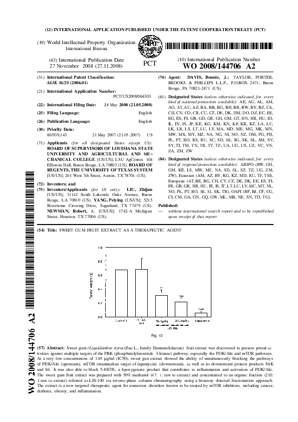
 Robert Newman