The effect of polarized light on wound healing
 K. Van Landuyt
2001, European Journal of Plastic Surgery
Free related PDFsRelated papers
Free PDF
Biomodulative Effects of Polarized Light on the Healing of Cutaneous Wounds on Nourished and Undernourished Wistar Rats
Photomedicine and Laser Surgery, 2006
Free PDF

Effects of a single near-infrared laser treatment on cutaneous wound healing: Biometrical and histological study in rats
Journal of Photochemistry and Photobiology B: Biology, 2007
Free PDF

Effects of laser irradiation (670-nm InGaP and 830-nm GaAlAs) on burn of second-degree in rats
Lasers in Medical Science, 2014
Free PDF

The Use of Polarized Light in Aesthetic Surgery
Aesthetic Plastic Surgery, 2004
Free PDF

The Nuts and Bolts of Low-level Laser (Light) Therapy
Annals of Biomedical Engineering, 2012
Free PDF
Combined reflectance confocal microscopy/optical coherence tomography imaging for skin burn assessment
Biomedical Optics Express, 2013
Free PDF
Polarized Light (λ400–2000 nm) on Third-Degree Burns in Diabetic Rats: Immunohistochemical Study
Photomedicine and Laser Surgery, 2010
Free PDF
International Journal of Biophysics 2015, 5(1): 18-23
DOI: 10.5923/j.biophysics.20150501.03
A Description of the Effect of Polarized Light as an
Adjuvant Therapy on Wound Healing Process in
Pediatrics
Samir M. Abdel-Mageed1,*, Ali Osman Selim2, Mohamed A. Abdel Ghafar3, Rania Reffat Ali4
1
Physics Department, Faculty of Science, Alexandria University
Physical Therapy for Surgery Department, Faculty of Physical Therapy, Cairo University
3
Physical Therapy for Pediatrics Department, Faculty of Physical Therapy, October 6 University
4
Basic science Department, Faculty of Physical Therapy, Cairo University
2
Abstract Back ground and purpose: The ability of light to penetrate a tissue and deposit energy via the optical
absorption properties of the tissue is the key to therapeutic applications. Birefringence which is an anisotropic property of
dermal layer of skin, the blood flow in the tissue capillaries and the multi-scattering from the tissue static components affect
the polarization of light. The goal of this study was to describe the healing process of deep partial thickness wound in
pediatrics submitted (or not) to the excitation property or the polarization property of light. Subjects: Thirty children who
suffered from deep partial thickness burn (20-30%) participated in this study (13 boys and 17 girls). Their ages ranged from
10 to18 years. They were classified randomly into two groups (study and control group) of equal numbers. Procedures:
Group A (study) was treated using polarized light as an adjuvant therapy to the regular wound care (debridement, Local
antimicrobial drug and beta dine) 5 sessions per week for 3 weeks. Group B (control) was treated using the regular wound
care (debridement, Local antimicrobial drug and beta dine). Wound surface areas were measured before and after the
treatment period by using wound tracing method. Results: The study showed a significant reduction in burn wound surface
areas in both groups (P < 0.05) while there was no significant difference between both groups. Conclusion: it was concluded
that polarized light has a limited effect as an adjuvant therapy on healing process of deep partial thickness burn wounds in
children.
Keywords Polarized light, Deep partial thickness burn, Wound healing, Paediatrics
1. Introduction
Burn is a coagulative necrosis of the skin caused by
contact with fire, chemicals such as acids, hot liquids, hot air
and hot gases, heated metals or electricity. Burn injuries
remain one of the major health problems that can happen
unexpectedly and have the potential to cause disability,
lifelong disfigurement, prolonged hospitalization and/or
death. [1, 2]
Unintentional and intentional burn injuries vary across age
groups, gender, income and global region. The worldwide
incidence of burn injuries was 1.3 per 100,000 populations in
low and moderate income countries. Children under 5 and
the elderly have the highest burn mortality worldwide. [3]
The repair of burn wound has long been a problem and this
has come more into focus in recent times. Wound closure is
undertaken as the most important advent preventing life
* Corresponding author:
samir75@hotmail.com (Samir M. Abdel-Mageed)
Published online at http://journal.sapub.org/biophysics
Copyright © 2015 Scientific & Academic Publishing. All Rights Reserved
threatening, infections, decreasing morbidity and mortality
[7].
Wound healing is an intricate process in which the skin or
any organ tissue repairs itself after injury [4]. Wound healing
is a complex and dynamic process characterized by
interaction of various cell types as lymphocytes, monocytes,
epithelial cells and fibroblasts. Three main overlapping
phases have been identified in tissue response to injury
include inflammation, formation of granulation tissue and
proliferation [5, 6].
Wound healing process is based on the vascular and
cellular activity. Vasomotion is the periodic constriction and
dilatation of small blood vessels. It is attributed to local
metabolic needs, vascular myogenic responses and
neurogenic controls. Besides the fibroblast and macrophage
activity, human wound associated lymphocyte populations
are modulated during a healing process. [4]
Polarized and non-polarized light from different sources in
the spectral ranges of visible and near infrared (NIR) are
widely used in clinical treatments and in medical research
especially in the therapeutic field, such as Low Level Laser
Therapy
(LLLT),
light
phototherapy
(lasers),
International Journal of Biophysics 2015, 5(1): 18-23
quasi-monochromatic radiation (LEDs) and broadband light
devices. [8] The most of laser types are polarized, where the
electric field oscillates in a certain direction perpendicular to
the propagation direction [9].
Light is a form of energy and has various colors with
different wavelength; it has been used as a healing tool since
ancient time. There is now better understanding of which
component of natural light are useful in the stimulation of
healing such as Biptron light therapy (BLT) which is a
device emits a polarized light containing a range of
wavelengths that correspond to visible light plus infrared
radiation which have been reported to stimulate the
biological reactions [10]. It was reported that polarized light
causes enhancement of the cell membrane activities,
acceleration of production of adenosine tri-phosphate (ATP)
in mitochondria, stimulation of regeneration processes and
acceleration of fibroblast proliferation and deposition of
collagen [11, 12].
It was considered that polarized light rearrange the polar
heads of a lipid bilayer in the cell membrane where enzyme
reactions take place lead to structural changes occur in cell
membranes in consequence the surface features and lipid
protein connection can be modified [11]. Schindl et al. [13]
stated that Biptron light therapy may significantly stimulate
the faster epithelialization of the damaged skin, reducing the
risk for the formation of the functionally and unacceptable
scars.
However, the polarization point has never been
experimentally proven and studies used polarized light in the
treatment of wounds revealed conflicting data and studies in
vivo are still scarce, with some studies reporting accelerated
wound closure and increased tensile strength of scars while
others have found no such improvement [14]. So, the
purpose of this study was to evaluate the effect of polarized
light in accelerating the healing rate in burn injuries in
children.
possible side effects had been explained under supervision of
their guardians.
Inclusion criteria
Children with deep partial thickness bun affecting 20 to
30% of total body surface area (TBSA) with no relative
contraindications for using of polarized light participated in
this study.
Exclusion criteria
Children suffering from medical conditions that might
affect burn healing were excluded. These conditions
included diabetes mellitus, malignancy, inhalation injuries or
any systemic diseases. Also children using drugs that can
affect the skin and delay in healing, especially steroids,
immunosuppressive agents, antineoplastic drugs and
anticoagulants were excluded.
Children were randomly divided into two equal groups.
Group A (study) was treated by polarized light as an adjuvant
therapy to the regular wound care (debridement, local
antimicrobial drug and beta dine) 5 sessions per week for 3
weeks. Group B (control) was treated by the regular wound
care (debridement, local antimicrobial drug and beta dine) 5
sessions per week for 3 weeks.
Equipments:
1. Wound tracing method used to measure burn surface
area (sterilized transparency film, fine tipped marker,
white paper, carbon paper and metric graph paper
1mm2). [15]
2. Bioptron Compact III Light Therapy System (PAG-860)
by (Bioptron AG, Switzerland) with the following
technical characteristics was used: Wavelength
480-3400 nm, degree of polarization >95% (590-1550
nm), specific power density 40 mW/cm2, light energy
per minute 2.4 J/cm2.
Procedures:
A. Measurement of wound surface area:
2. Materials and Methods
Table 1. Mean and Standard deviation of age, weight, height, and BMI
X±SD
X: Mean
19
Age(years)
15.2±2.6
Weight(Kg)
49.7±8.6
Height(cm)
160.6±7.6
BMI(kg/cm2)
24.7±4.2
SD: Standard deviation
The burn wound outline was traced on a transparent film
using a permanent marker. Each wound was traced twice to
establish measurement reliability. Then the wound tracing
was placed on a metric grid, and the number of square
millimeters of the traced area were counted to determine the
burn wound surface area, then the value was converted to
cm2. The mean of the two trials was calculated and
considered as a burn wound surface area (WSA). The wound
surface area was measured before starting the study and after
3 weeks of treatment.
Subjects:
B. Treatment procedures:
Thirty children (13 boys and17 girls) participated in this
study; their ages ranged from 10 to 18 years with a mean
value of 15.2±2.6 years. The mean values for their body
weight was 49.7±8.6 kg, and for their height was 160.6±7.6
cm, while it was 24.7±4.2 kg/cm2 for their body mass index
(Table 1). A consent form was signed after the procedure and
The patient was placed in suitable position. Before the
polarized light therapy all wounds were cleaned using 2%
hydrogen peroxide. Polarized light therapy was performed
for six min, at a distance of 10 cm, five times a week for three
weeks. Patient was asked to note the erythema (when it
begins and disappeared). If marked or painful erythema had
20
Samir M. Abdel-Mageed et al.: A Description of the Effect of Polarized Light
as an Adjuvant Therapy on Wound Healing Process in Pediatrics
occurred, therapy was stopped until erythema relieved and
then the session time was decreased to the previous level and
frequency.
Data analysis
Data was analyzed by using statistical package for social
sciences (SPSS). Unpaired t-test was used to compare
between the mean of wound surface area before and after the
treatment for both groups. Level of significance was set at
0.05.
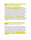
3. Results
Wound surface area (cm2)
The statistical analysis of the mean values of wound
surface area (WSA) in control group before treatment was
(105.7±20.96) and after treatment was (17.2±11.49) revealed
a significant statistical reduction of mean values of
WSA(P<0.05). Also the percentage of reduction was
120
110
100
90
80
70
60
50
40
30
20
10
0
105.7
calculated to be 88.3% after treatment, when compared to the
pretreatment value as shown in figure 1.
The statistical analysis of the mean values of wound
surface area (WSA) in study group before treatment
(104.9±24.22) and after treatment (12.3±9.45) revealed a
significant statistical reduction of mean values of WSA
(P<0.05). Also the percentage of reduction was calculated to
be 88.3% after treatment, when compared to the pretreatment
value as shown in figure 1.
The statistical analysis of the mean values of wound
surface area (WSA) pre treatment for control group and
study group revealed no statistical significant differences as
shown in figure 2.
The statistical analysis of the mean values of wound
surface area (WSA) post treatment for control group and
study group revealed no statistical significant differences as
shown in figure 3.
104.9
Pre Treatment
Post treatment
17.2
Control group
12.3
Study group
Wound surface area (cm2)
Figure 1. The statistical analysis of mean values of WSA pre treatment after post treatment in control and study groups
120
110
100
90
80
70
60
50
40
30
20
10
0
105.7
104.9
Control group
Study group
Pre treatment
Figure 2. The Mean values of WSA (cm2) post treatment for both control and study groups
International Journal of Biophysics 2015, 5(1): 18-23
21
Wound surface area (cm2)
17.2
18
16
14
12
10
8
6
4
2
0
12.3
Control group
Study group
Post treatment
Figure 3. The Mean values of WSA (cm2) post treatment for both control and study groups
4. Discussion
The analysis of the results indicated that both groups
showed significant statistical difference between pre and
post experiment measurements, but there was no statistically
significant difference between the study and the control
groups.
Results of the current study came in agreement with those
of Schlager et al. [16] who reported that irradiation of burns
with a 250-mW/670-nm laser light produced no beneficial
effects on wound-healing processes. Also it is supported by
Cambier et al. [17] who found no improvement in the
wound-healing process after irradiation with low-power
lasers or polarized light.
These results can be explained as the therapeutic effect of
polarized light may be due to either the polarization or the
excitation property of the light. Once the polarized lights
penetrate the tissues it starts to lose their polarization
direction. The polarization loss in the tissue may be mainly
due to many reasons; the blood flow in the tissue capillaries,
the birefringent properties of the medium and the
multi-scattering from the tissue static components [18].
First of all, according to the work of Fixler et al. [19] the
polarization loss is directly proportional to the volumetric
flow rate. Higher flow rate produces lower polarization
values especially when the polarization direction is
perpendicular to the direction of flow. The dermis layer of
the skin - first layer encountered by the polarized light during
second degree burn wound contains blood vessels and
capillaries where the blood is flowing in many directions
carrying its components. Accordingly, the polarized light
beam necessarily hits the fluid flow through the tissue
perpendicular to its polarization direction and this cause a
polarization loss.
Secondly, the randomization of linear polarization during
transmission through a tissue is more rapid in birefringent
tissues than in non-birefringent tissues [20]. Birefringence is
a polarization specific electromagnetic property of materials.
Collagen fiber is the main components of the dermis layer
(70% in dry weight) has a heterogeneous distribution in the
skin, and commonly refer to as birefringent tissue [21].
There are two types of birefringence have been reported for
collagen fibers, particularly intrinsic [22] and Form
birefringence which is caused by asymmetrical alignment of
chemical bonds or ions within the rod shaped triple chain
collagen molecules and the next arises from the nonlinear
optical property of the medium [23]. Therefore, Collagen’s
rod like triple helix conformation results in both linear and
circular anisotropic properties [24]. The most rapid
depolarization occurred in rich collagen tissues of the skin
[20].
Finally, on the cellular level, the mismatches of refractive
indices between cellular components result in scattering [25].
The scattering properties of the cellular level elements also
vary with their sizes. Therefore, the reduced scattering
coefficient of the dermis as a function of the light wavelength
can be described well as a combination of Mie and Rayleigh
scattering. The former occur when the size of the element is
comparable to the wavelength of the incident light such as
scattering by mitochondria, nuclei and collagen fibers can be
explained by Mie scattering. Contrary, when the size of the
element is much smaller than the wavelength, its scattering
property can be explained by Rayleigh scattering such as
scattering include membranes and the banded
ultra-structures of collagen fibrils fibers [26]. Therefore;
light with long wavelength has more penetration power in
skin. Rayleigh scattering dominates in the spectral range
below 650 nm while Mie scattering plays a major role above
650nm. As the main scatterers in the dermis are the collagen
fibers which are densely packed [27]. As a result, the
orientation of polarization becomes randomized by multiple
scattering events from these collagen fibers and the
polarization value reduced [28].
Based on the three facts discussed above ; the dermis static
22
Samir M. Abdel-Mageed et al.: A Description of the Effect of Polarized Light
as an Adjuvant Therapy on Wound Healing Process in Pediatrics
components such as collagen fibers exhibit optical
birefringent properties which acts as a strong multiple
scatterers for linear polarized light, also the blood flow in the
tissue capillaries and the blood dynamic components act as a
scatterers. Consequently, linear polarization of radiation
randomized through the dermis layer and polarization lost.
So there are no additional benefits in using polarized light as
an excitation source without maintaining the excitation
direction.
The slight improvement in the group treated by polarized
light compared to the control group comes in agreement with
Ribeiro [29] who reported that the relative direction of the
laser polarization plays an important role in the wound
healing process when highly coherent He-Ne laser is used.
Also these results confirmed by the clinical results of
Monstrey et al. [30] that polarized light therapy was effective
in the treatment of burn wounds.
These results may be attributed to the excitation property
of the light despite of its polarization property. The light
interaction with the biological tissue is not an intrinsic
property of the light. Where the first law of photochemistry
states that light must be absorbed before photochemistry can
occur.
It was stated that mitochondria thought to be a likely site
for the initial effects of light; absorption of photons by
chromosphere molecules such as cytochrome c that proposed
as the primary photoacceptor for the red-NIR range in
mammalian cells leads to electronically excited states and
consequently acceleration of electron transfer reactions in
the mitochondria membrane respiratory chain [31]. A further
electron transport automatically leads to increasing of ATP
production rate [32], alteration of reactive oxygen species
and induction of transcription factors which increasing the
activity of the Na+ /H+ and Ca2+/Na+ antiporters and of all the
ATP driven carriers for ions, such as Na+ /K+ ATPase and
Ca2+ pumps. ATP is the substrate for adenyl cyclase, and
therefore the ATP level controls the level of cAMP. Both
Ca2+ and cyclic adenosine mono-phosphate cAMP are very
important second messengers. Ca2+ adjusts most of the
biological process in the human body [31].
The positive effect of light on wound healing can be
explained by many biological mechanisms including
modulation in levels of cytokines and growth factors which
are responsible for the different stages of wound healing. It
was reported that cytokines responsible for the initial
inflammatory phase, fibroblast proliferation and migration in
wound healing [33]. Also it was confirmed that light can
increase
growth
factors
responsible
for
the
neovascularization and inducing collagen synthesis from
fibroblasts which necessary for wound healing [34]. Light
can incite fibroblasts transformation to myofibloblasts which
has the phenotype of contractile cells that accelerate wound
contraction [35].
5. Conclusions
The effects of polarized light as an adjuvant therapy for
deep partial thickness second degree burn were not
satisfactory and statistically non- significant. But it showed
a very slight improvement in the wound healing process.
REFERENCES
[1]
Peck MD, Kruger GE, Van Der Merwe AE, Godakumbura W,
Ahuja RB. Burns and fires from non electric domestic
appliances in low and middle income countries Part1. The
scope of the problem. Burns 2008; 34(3):303-311.
[2]
Peck M and Pressman MA. The correlation between burn
mortality rates from the flame and economic status of
countries. Burns 2013; 39(6):1054-9.
[3]
World health Organization (WHO) Global Burden of Disease
2008. www.who.int/healthinfo/global_burden_disease/estim
ates_regional/en/index.html (Accessed on September 20,
2011).
[4]
Nguyen DT, Orgill DP and Murphy GF. The
Pathophysiologic Basis for Wound Healing and Cutaneous
Regeneration. Biomaterials for Treating Skin Loss. CRC
Press (US) & Woodhead Publishing (UK/Europe), Boca
Raton/Cambridge 2009, 25-57.
[5]
Morison MJ, Ovington LG and Wilki K. Chronic wound care:
a problem based learning approach. Edinburgh: Mosby, 2004.
[6]
Nienartowicz A, Sobaniec-Lotowska ME, Jarocka-Cytra E
and Lemancewicz D. Mast cells in neoangiogenesis. Med Sci
Monit 2006; 12(3): 53-56.
[7]
Desanti L. Pathophysiology and current managment of burn
injury. Adv skin wound care. 2005; 18(6):323-332.
[8]
Sankaran V, Everett MJ, Maitland DJ et al .Comparison of
polarized light propagation in biological tissue and phantoms.
Optics Letters 2003; 24(15): 1044-46.
[9]
Lin JF, Lo YL. Measurement of optical rotation and phase
retardance of optical samples with depolarization effects
using linearly and circularly polarized probe lights. Opt Laser
Eng 2009; 47(9):948–55.
[10] Depuydl K, Monstrey S and Hoeksema H. The use of
polarized light in the treatment of burn wounds. Abstract.
Presented at the 10th annual EURAPS meeting. Madrid. Spain
2001.
[11] Monstrey SA, Hoeksema HG and Depuydt KA. The effect of
polarized light on wound healing. European Jornal of plastic
surgery; 2004, 24(8): 304-310.
[12] Medenica LA and Lens MA.the use of polarized
polychromatic non coherent light alone as a therapy for
venous leg ulceration. Journal of wound care; 2004, 12(1),
37-40.
[13] Schindl A, Schindl M, Pernerstorfer-Schon H and Schindl L.
Low intensity laser therapy: a review. J Investing Med.2000,
48(5): 312-326.
[14] Samoilova KA, Obolenskaya KD and Vologdina AV.
Proceedings of EUROPTO conference on low power light on
biological systems. Stockholm Sweden, Sep 2008: 90-103.
[15] Bohannon RE and Pfaller BV. "Documentation of wound
International Journal of Biophysics 2015, 5(1): 18-23
surface area from tracing of wound perimeters: Clinical report
of three techniques". Phys.there 1983; 63(10): 1622-1628.
[16] Schlager A, Oehler K, Huebner K-U, Schmuth M, Spoetl L.
Healing of burns after treatment with 670-nanometer
low-power laser light. Plast Reconstr Surg 2000; 105(5):
1635–9.
[17] Cambier DC, Vanderstraeten GG, Mussen MJ, van der Spank
JT. Low-power laser and healing of burns: a preliminary
assay. Plast Reconstr Surg 1996; 97(3): 555–8.
[18] Hoeksema HG, Monstrey SA and Saelens HU. Efficacy of
polarized light therapy in the conservative treatment of deep
dermal burns. Br J Plastic Surg; 2002; 55(5):420-426.
[19] Fixler D, Ankri R, Duadi H, Lubart R, and Zalevsky Z,
Depolarization of light in biological tissues, Optics and
Lasers in Engineering 2012; 50(6): 850–854
[20] Steven L. Jacques, Jessica R. Roman, and Ken Lee, Imaging
Superficial Tissues With Polarized Light, Lasers in Surgery
and Medicine 2000; 26:119–129.
[21] Maitland DJ and Walsh JT. Quantitative Measurements of
Linear Birefringence during Heating of Native Collagen
Lasers in Surgery and Medicine1997; 20(3):310–318.
[22] Bennett HS. The microscopical investigation of biological
materials with polarized light. In: Jones RM (ed.) McClung’s
Handbook of Microscopical Technique. New York: Hafner
Publ. Co. 1967; 591–766.
[23] Vidal BC. Form birefringence as applied to biopolymer and
inorganic material supraorganization. Biotech Histochem
2010; 85(6): 365–378.
[24] Yoshioka K, O’Konski CT. Electric properties of
macromolecules. IX. Dipole moment, polarizability, and
optical anisotropy factor of collagen in solution from electric
birefringence. Biopolymers 1966; 4(5):499–507.
[25] Guzlksu N, Federici J.F, Lim H.C, Chauhdry H.R, Ritter A.B,
and Findley T. Measurement of skin stretch via light
reflection. Journal of Biomedical Optics 2003; 8:80–86.
[26] Mobley J and Dinh TV. Biomedical Photonics Handbook.
23
CRC Press, 2003.
[27] Jacques S.L. Origins of tissue optical properties in the UVA,
visible, and NIR regions. Trends in Optics and Photonics:
Advances in Optical Imaging and Photon Migration. 1996;
2:364–371.
[28] Drezek R, Dunn A, and Kortum R.R. Light scattering from
cells: finite-difference time-domain simulations and
goniometric measurements. Applied Optics 1999,
38(16):3651–3661.
[29] Ribeiro MS1, Da Silva Dde F, De Araújo CE, De Oliveira SF,
Pelegrini CM, Zorn TM, Zezell DM. Effects of low-intensity
polarized visible laser radiation on skin burns: a light
microscopy study. J Clin Laser Med Surg. 2004; 22(1):59-66
[30] Monstrey S, Hoeksema H, Saelens H, Depuydt K, Hamdi M,
Van Landuyt K, et al. A conservative approach for deep
dermal burn wounds using polarised-light therapy. Br J Plast
Surg 2002; 55(5): 420–6.
[31] W. Yu, J.O. Naim, M. McGowan, K. Ippolito and R.J.
Lanzafame. Photomodulation of oxidative metabolism and
electron chain enzymes in rat liver mitochondria, Photochem
Photobiol 1997; 66(6): 866-71.
[32] Passarella S. He-Ne laser irradiation of isolated mitochondria,
J Photochem Photobiol B 1989; 3(16): 642-3.
[33] Poon VK, Huang L, and Burd A. Biostimulation of dermal
fibroblast by sublethal Q-switched Nd: YAG 532 nm laser:
collagen remodeling and pigmentation, J Photochem
Photobiol B 2005; 81(1): 1-8.
[34] Kipshidze N, Nikolaychik V, Keelan MH, Shankar LR,
Khanna A, Kornowski R, Leon M and Moses J. Low-power
helium: neon laser irradiation enhances production of
vascular endothelial growth factor and promotes growth of
endothelial cells in vitro, Lasers Surg Med 2001; 28(4):
355-64.
[35] Medrado AR, Pugliese LS, Reis SR and Andrade ZA,
Influence of low level laser therapy on wound healing and its
biological action upon myofibroblasts, Lasers Surg Med 2003;
32(3): 239-44.
Free related PDFsRelated papers
Photobiomodulation and bacterial cellulose membrane in the treatment of third-degree burns in rats
Journal of Tissue Viability, 2018
Free PDF


Noninvasive imaging technologies for cutaneous wound assessment: A review
Wound repair and regeneration : official publication of the Wound Healing Society [and] the European Tissue Repair Society, 2015
Free PDF
Quantification and the Underlying Mechanism of Skin Thermal Damage: A Review
Journal of Mechanics in Medicine and Biology, 2010
Free PDF

Determination of burn depth by polarization-sensitive optical coherence tomography
Journal of Biomedical Optics, 2004
Free PDF


Review of near-infrared methods for wound assessment
Journal of biomedical optics, 2016
Free PDF


Low-level laser therapy for spinal cord injury in rats: effects of polarization
Journal of Biomedical Optics, 2013
Free PDF

<title>Effects of a polarized light source (λ400-2000nm) on H.Ep.2 and L929 cell lines: a spectroscopic in vitro study</title>
Mechanisms for Low-Light Therapy IV, 2009
Free PDF
A systematic review of objective burn scar measurements
Burns & Trauma, 2016
Free PDF
Nanoceutical Adjuvants as Wound Healing Material: Precepts and Prospects
International Journal of Molecular Sciences, 2021
Free PDF
Effect of orange polarized light on post burn pediatric scar: a single blind randomized clinical trial
Journal of physical therapy science, 2018
Free PDF
Green Light Emitting Diode Irradiation Enhances Fibroblast Growth Impaired by High Glucose Level
Photomedicine and Laser Surgery, 2005
Free PDF

Efficacy of low-level laser therapy on scar tissue
Journal of Cosmetic and Laser Therapy, 2013
Free PDF

Effect of low-level helium–neon laser therapy on histological and ultrastructural features of immobilized rabbit articular cartilage
Journal of Photochemistry and Photobiology B-biology, 2007
Free PDF


Good, better, best? The effects of polarization on photobiomodulation therapy
Journal of Biophotonics
Free PDF
Effectiveness of Laser Photobiomodulation at 660 or 780 Nanometers on the Repair of Third-Degree Burns in Diabetic Rats
Photomedicine and Laser Surgery, 2008
Free PDF

Electromagnetic Stimulation Combined with Aloe vera Increases Collagen Reorganization in Burn Repair
Journal of Pharmacy and Pharmacology, 2018
Free PDF
Biophotonic methods in microcirculation imaging
Medical Laser Application, 2007
Free PDF
Collagen-Based Films Containing Liposome-Loaded Usnic Acid as Dressing for Dermal Burn Healing
Journal of Biomedicine and Biotechnology, 2011
Free PDF
Birefringence and Second Harmonic Generation on Tendon Collagen Following Red Linearly Polarized Laser Irradiation
Annals of Biomedical Engineering, 2013
Free PDF

Enhancement of bone formation in rat calvarial bone defects using low-level laser therapy
Oral Surgery, Oral Medicine, Oral Pathology, Oral Radiology, and Endodontology, 2004
Free PDF

Effect of photobiomodulation associated with cell therapy in the process of cutaneous regeneration in third degree burns in rats
Journal of Tissue Engineering and Regenerative Medicine, 2020
Free PDF

Intradermally focused infrared laser pulses: Thermal effects at defined tissue depths
Lasers in Surgery and Medicine, 2005
Free PDF
A Review of Scar Treatment Related to Acne and Burn
Journal of critical reviews, 2020
Free PDF


- Find new research papers in:
- Physics
- Chemistry
- Biology
- Health Sciences
- Ecology
- Earth Sciences
- Cognitive Science
- Mathematics
- Computer Science