History
A directed history is vital to the proper care of a patient with cellulitis. The patient may or may not relate an episode of trauma that preceded symptoms; when cellulitis develops, it is usually several days after the inciting trauma. Rapid progression or significant pain is a concerning sign that may indicate a severe problem, such as necrotizing fasciitis, which should be managed promptly. [1]
If the patient recalls an episode of trauma, the clinician should ask about circumstances surrounding the incident that may elicit clues to a particular etiology. For example, exposure to standing or brackish water could mean that Aeromonas or Vibrio is the cause of infection; or a cut that occurred while butchering may be an important clue to consider Erysipelothrix rhusiopathiae. Identifying the specific inciting cause helps the clinician identify the most likely pathogen, choose appropriate antibiotic therapy, and offer appropriate immunization, such as tetanus toxoid (Td or Tdap), if indicated. [1]
The patient should also be questioned about the presence of other skin disorders, including various types of dermatitis and especially any preceding fungal infection, which may serve as a portal of entry for bacterial pathogens. [21]
The past medical history should focus on the presence of comorbid conditions that may increase the risk for cellulitis, with the most common ones being diabetes mellitus, human immunodeficiency virus (HIV) infection/acquired immunodeficiency syndrome (AIDS), chronic kidney disease, and chronic liver disease. [1]
The surgical history may include a recent procedure that resulted in wound infection. For example, severe bacterial cellulitis may occur as a postsurgical complication following hip replacement [60] or liposuction. Alternatively, a remote surgical history involving lymph node dissection (eg, following either radical mastectomy or conservative breast surgery) may predispose to cellulitis, even years after the surgery, because of lymphatic occlusion. [61, 62, 63, 64] Impaired lymphatic drainage and edema are also considered predisposing factors to leg cellulitis following saphenous vein resection for coronary artery bypass. [26] In addition, the presence of foreign bodies, including indwelling IV catheters, external orthopedic pins, and other surgical devices, may predispose to infection. [1]

Physical Examination
The physical examination should first focus on the area of concern. Nonpurulent cellulitis is associated with four cardinal signs of infection: erythema, pain, swelling, and warmth. Several physical examination findings may help the clinician identify the most likely pathogen and assess the severity of the infection, thereby facilitating appropriate treatment. Those findings include the following [1] :
-
The involved site(s)is/are red, hot, swollen, and tender
-
Unlike erysipelas, the borders are not elevated or sharply demarcated
-
Regional lymphadenopathy is present
-
Malaise, chills, fever, and toxicity are present
-
Skin infection without underlying drainage, penetrating trauma, eschar, or abscess is most likely caused by streptococci; on the other hand, S aureus, often community-acquired methicillin-resistant S aureus (CA-MRSA), is the most likely pathogen when these factors are present [3]
-
Perianal cellulitis is usually observed in children with perianal fissures; it is characterized by perianal erythema and pruritus, purulent secretions, painful defecation, and blood in the stools [66]
-
Cellulitis characterized by violaceous color and bullae suggests more serious or systemic infection with organisms such as V vulnificus or S pneumoniae
 Cellulitis due to documented Vibrio vulnificus infection. (Image courtesy of Kepler Davis.)
-
Lymphangitic spread (red lines streaking away from the area of infection), crepitus, and hemodynamic instability are indications of severe infection, requiring more aggressive treatment
-
Circumferential cellulitis or pain that is disproportional to examination findings should prompt consideration of severe SSTI
The IDSA indicates that the following are also signs/symptoms of potentially severe deep SSTI (Note: these frequently appear later in the course of necrotizing infections), which necessitate emergent surgical evaluation [4] :
-
Violaceous bullae
-
Cutaneous hemorrhage
-
Skin sloughing
-
Skin anesthesia
-
Rapid progression
-
Gas in the tissue
-
Mild cellulitis with a fine lacelike pattern of erythema. This lesion was only slightly warm and caused minimal pain, which is typical for the initial presentation of mild cellulitis.
-
Swelling seen in cellulitis involving the hand. In a situation with hand cellulitis, always rule out deep infection by imaging studies or by obtaining surgical consultation.
-
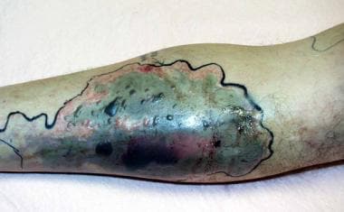
Severe cellulitis of the leg in a woman aged 80 years. The cellulitis developed beneath a cast and was painful and warm to the touch. Significant erythema is evident. The margins are irregular but not raised. An ulcerated area is visible in the center of the photograph.
-
Burns complicated by cellulitis. The larger lesion is a second-degree burn (left), and the smaller lesion is a first-degree burn (right), each with an expanding zone of erythema consistent with cellulitis.
-
Cellulitis due to documented Vibrio vulnificus infection. (Image courtesy of Kepler Davis.)
-
A case of cellulitis without associated purulence in an infant. Note the presence of lymphedema, a risk factor for cellulitis.(Photo courtesy of Amy Williams.)
-
Patient with cellulitis of the left ankle. This cellulitis was caused by community-acquired methicillin-resistant Staphylococcus aureus (CA-MRSA). (Photo courtesy of Texas Dept. of Public Health.)
-
Abscess and associated cellulitis caused by community-acquired methicillin-resistant Staphylococcus aureus (CA-MRSA). (Photo courtesy of Texas Dept. of Public Health.)
-
Guidelines for the management of patients who require hospitalization for cellulitis or cutaneous abscess. AFB = acid-fast bacilli; BID = twice daily; CRP = C reactive protein; CT = computed tomography scanning; DS = double strength; DM = diabetes mellitus; ESR = erythrocyte sedimentation rate; ESRD = end-stage renal disease; HIV = human immunodeficiency virus; ICU = intensive care unit; I&D = incision and drainage; ID = infectious disease; IDU = injection drug user; IV = intravenous; LRINEC = Laboratory Risk Indicator for Necrotizing Fasciitis; MRI = magnetic resonance imaging; MSRA = methicillin-resistant Staphylococcus aureus; NSAIDS = nonsteroidal anti-inflammatory drugs; PO = by mouth; SSTI = skin and soft-tissue infections; TID = 3 times daily. Adapted from Jenkins TC, Knepper BC, Sabel AL, et al. Decreased antibiotic utilization after implementation of a guideline for inpatient cellulitis and cutaneous abscess. Arch Intern Med. 2011;171(12):1072-9.
-
A male patient with orbital cellulitis with proptosis, ophthalmoplegia, and edema and erythema of the eyelids. The patient also exhibited pain on eye movement, fever, headache, and malaise.
-
A male patient with orbital cellulitis with proptosis, ophthalmoplegia, and edema and erythema of the eyelids. The patient also exhibited chemosis and resistance to retropulsion of the globe.
-
Gross photograph of complicated cellulitis. Instead of the presence of yellow fat, the tissue is hemorrhagic and necrotic.
-
Hematoxylin and eosin (H&E) stain, high power. This image shows deeper subcutaneous tissue involved in a case of cellulitis, with acute inflammatory cells and fat necrosis.
-
Hematoxylin and eosin (H&E) stain, high power. This image shows cellulitis caused by herpes simplex virus, with the multinucleated organism in the center of the picture.
Tables
Location |
Likely Organisms |
Other Organisms |
Complication/ Discussion |
Antibiotic Regimen -- Oral/ Outpatient |
Indication for Hospitalization |
Antibiotic Regimen -- Parenteral/ Hospitalized |
Uncomplicated cellulitis |
Group A streptococci much more likely than Staphylococcus aureus |
|
|
Cephalexin or dicloxacillin or clindamycin |
|
Cefazolin or oxacillin or nafcillin |
Cellulitis, concern for methicillin-resistant S aureus is a concern |
Group A streptococci and S aureus |
|
|
[(Cephalexin or dicloxacillin or clindamycin) plus trimethoprim/ sulfamethoxazole] or Clindamycin |
|
Vancomycin Daptomycin Ceftaroline |
Dog bite |
Pasteurella species (50% of wounds) S aureus Streptococcus pyogenes |
Staphylococci, streptococci Aerobes --Moraxella and Neisseria Anaerobes --Fusobacterium, Bacteroides, Porphyromonas, and Prevotella |
Capnocytophaga canimorsus may cause sepsis in patients with asplenia/hepatic disease. Avoid first-generation cephalosporins/ erythromycin/ dicloxacillin. High likelihood of infection – Prophylactic antibiotics indicated for the following wounds: deep puncture, hands, requiring surgical repair, immunocompromised host, venous or lymphatic compromise, crush injury. Requires close follow-up care within 24-48 h. |
Amoxicillin/ clavulanate Penicillin allergic: Moxifloxacin |
Deep wounds or severe wounds; infections not responding to oral antibiotics |
Third-generation cephalosporin (ceftriaxone [Rocephin]) plus metronidazole or beta-lactam/beta-lactamase inhibitor (eg, ampicillin/sulbactam) or fluoroquinolone plus metronidazole or carbapenem (ertapenem) |
Human bite |
Eikenella corrodens (gram-negative facultative anaerobe, 29% of wounds) Aerobic gram-positive cocci, anaerobes |
|
Clenched fist lacerations over metacarpophalangeal joints should be considered human bites; anesthetize wounds and irrigate; reevaluate within 24-48 h. Intercanine distance >3 cm is likely bite from adult; if wound to child, consider abuse. |
Amoxicillin/ clavulanate Penicillin allergic: Moxifloxacin or (Clindamycin or metronidazole) plus (doxycycline or cefuroxime or trimethoprim/ sulfamethoxazole) |
|
Third-generation cephalosporin (Rocephin) plus metronidazole or beta-lactam/beta-lactamase inhibitor (eg, ampicillin/sulbactam) or fluoroquinolone plus metronidazole or carbapenem (ertapenem) |
Cat bite |
Pasteurella multocida and P septica (75% of wounds) |
Staphylococci, streptococci, Bacteroides, Peptostreptococcus, Actinomyces, Fusobacterium, Porphyromonas, and Veillonella parvula |
Avoid first-generation cephalosporins/ erythromycin/ dicloxacillin High likelihood of infection -- Prophylactic antibiotics indicated for the following wounds: deep puncture, hands, requiring surgical repair, immunocompromised host, venous or lymphatic compromise. Requires close follow-up care within 24-48 h. |
Amoxicillin/ clavulanate Penicillin allergic -- Moxifloxacin or (Clindamycin or metronidazole) plus (doxycycline or cefuroxime or trimethoprim/ sulfamethoxazole) |
Deep wounds or severe wounds; infections not responding to oral antibiotics |
Third-generation cephalosporin (Rocephin) plus metronidazole or beta-lactam/beta-lactamase inhibitor (eg, ampicillin/sulbactam) or fluoroquinolone plus metronidazole or carbapenem (ertapenem) |
Preseptal (periorbital) cellulitis |
Haemophilus influenzae type b, Streptococcus pneumoniae, S aureus, other streptococcal species, and anaerobes |
Nocardia brasiliensis, Bacillus anthracis, Pseudomonas aeruginosa, Neisseria gonorrhoeae, Proteus species, Pasteurella multocida, Mycobacterium tuberculosis |
Largest study indicates that H influenzae type b and S pneumoniae not diminished in facial cellulitis as a result of immunizations [29] |
Amoxicillin-clavulanate, cefpodoxime, cefdinir |
Age < 1 y/ more severe disease require intravenous antibiotic |
Third-generation cephalosporin (Rocephin) |
Lower extremity -- Complicating saphenous venectomy site after coronary bypass grafting |
No pathogen identifiable in most infections, but it is likely to be streptococcal (> staphylococcal) Non-group A beta-hemolytic streptococci most likely organism; S aureus less common |
|
Recurrent episodes common; may be associated with rigors, extreme fatigue, myalgias, and hypotension; some associated with tinea pedis (toe web cultures may be useful in establishing probable pathogen) |
Dicloxacillin or cephalexin. Add trimethoprim/ sulfamethoxazole or tetracycline or clindamycin if concern for methicillin-resistant S aureus |
|
First-generation cephalosporin (cefazolin); clindamycin; vancomycin |
Breast/arm - - (not mastitis) Complicating breast cancer surgery/lymph node dissection |
No pathogen identifiable in most infections Group A or Non-group A beta-hemolytic streptococci most likely organisms |
|
|
Dicloxacillin, cephalexin. Add trimethoprim/ sulfamethoxazole or tetracycline or clindamycin if concern for methicillin-resistant S aureus |
Fever, recent chemotherapy, neutropenia |
Multiple regimens, none clearly superior –Piperacillin/tazobactam or ceftazidime plus aminoglycoside; or ciprofloxacin plus beta-lactam or monotherapy with piperacillin/tazobactam or cefepime |
Aquatic environment -- Fresh water/ salt water/ brackish water/ swimming pools/ aquarium Puncture/ laceration |
Aeromonas hydrophila, Pseudomonas and Plesiomonas species, Vibrio species, Erysipelothrix rhusiopathiae, Mycobacterium marinum, and others |
|
A hydrophila and Vibrio vulnificus may produce rapidly progressive soft tissue infection and sepsis |
Fluoroquinolone (eg, ciprofloxacin or levofloxacin) Note: For M marinum infection, use clarithromycin plus either ethambutol or rifampin |
|
Third- or fourth-generation cephalosporin (eg, ceftazidime or cefepime) or fluoroquinolone (eg, ciprofloxacin or levofloxacin) |
Clenched-fist injury |
E corrodens (gram-negative anaerobe, 29 % of wounds); aerobic gram-positive cocci, anaerobes |
|
Lacerations over metacarpophalangeal joints should be considered human bites; anesthetize wounds and irrigate; reevaluate within 24-48 h Lacerations of extensor tendon |
Amoxicillin/ clavulanate; penicillin allergic: Moxifloxacin or (clindamycin or metronidazole) plus (doxycycline or cefuroxime or trimethoprim/ sulfamethoxazole) |
Failure to respond to oral therapy marked by increasing pain and swelling or purulent drainage |
Beta-lactam/beta-lactamase inhibitor (eg, ampicillin/sulbactam) |
Odontogenic facial cellulitis |
Aerobic and facultative organisms: group A beta-hemolytic streptococci, Neisseria and Eikenella species Anaerobes: Prevotella and Peptostreptococcus species |
|
Require extraction or root canal |
Amoxicillin-clavulanate or clindamycin |
|
Beta-lactam/beta-lactamase inhibitor (eg, ampicillin/sulbactam) or clindamycin |